 ムービー
ムービー コントローラー


+ データを開く

- 基本情報
基本情報
| 登録情報 | データベース: EMDB / ID: EMD-1862 | |||||||||
|---|---|---|---|---|---|---|---|---|---|---|
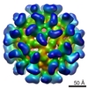
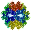
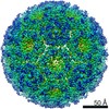
| タイトル | Visualization of a missing link in retrovirus capsid assembly | |||||||||
 マップデータ マップデータ | Icosahedral assembly of Rous sarcoma virus capsid proteins | |||||||||
 試料 試料 |
| |||||||||
 キーワード キーワード | Retrovirus / Capsid / Icosahedral | |||||||||
| 生物種 |  Avian leukosis virus (ニワトリ白血病ウイルス) Avian leukosis virus (ニワトリ白血病ウイルス) | |||||||||
| 手法 | 単粒子再構成法 / クライオ電子顕微鏡法 / 解像度: 10.4 Å | |||||||||
 データ登録者 データ登録者 | Cardone G / Purdy JG / Cheng N / Craven RC / Steven AC | |||||||||
 引用 引用 | ジャーナル: Nature / 年: 2009 タイトル: Visualization of a missing link in retrovirus capsid assembly. 著者: Giovanni Cardone / John G Purdy / Naiqian Cheng / Rebecca C Craven / Alasdair C Steven /  要旨: For a retrovirus such as HIV to be infectious, a properly formed capsid is needed; however, unusually among viruses, retrovirus capsids are highly variable in structure. According to the fullerene ...For a retrovirus such as HIV to be infectious, a properly formed capsid is needed; however, unusually among viruses, retrovirus capsids are highly variable in structure. According to the fullerene conjecture, they are composed of hexamers and pentamers of capsid protein (CA), with the shape of a capsid varying according to how the twelve pentamers are distributed and its size depending on the number of hexamers. Hexamers have been studied in planar and tubular arrays, but the predicted pentamers have not been observed. Here we report cryo-electron microscopic analyses of two in-vitro-assembled capsids of Rous sarcoma virus. Both are icosahedrally symmetric: one is composed of 12 pentamers, and the other of 12 pentamers and 20 hexamers. Fitting of atomic models of the two CA domains into the reconstructions shows three distinct inter-subunit interactions. These observations substantiate the fullerene conjecture, show how pentamers are accommodated at vertices, support the inference that nucleation is a crucial morphologic determinant, and imply that electrostatic interactions govern the differential assembly of pentamers and hexamers. | |||||||||
| 履歴 |
|
- 構造の表示
構造の表示
| ムービー |
 ムービービューア ムービービューア |
|---|---|
| 構造ビューア | EMマップ: SurfViewMolmilJmol/JSmol |
| 添付画像 |


- ダウンロードとリンク
-EMDBアーカイブ
| マップデータ | emd_1862.map.gz | 23.5 MB | EMDBマップデータ形式 | |
|---|---|---|---|---|
| ヘッダ (付随情報) | emd-1862-v30.xmlemd-1862.xml | 11 KB 11 KB | 表示 表示 | EMDBヘッダ |
| 画像 | 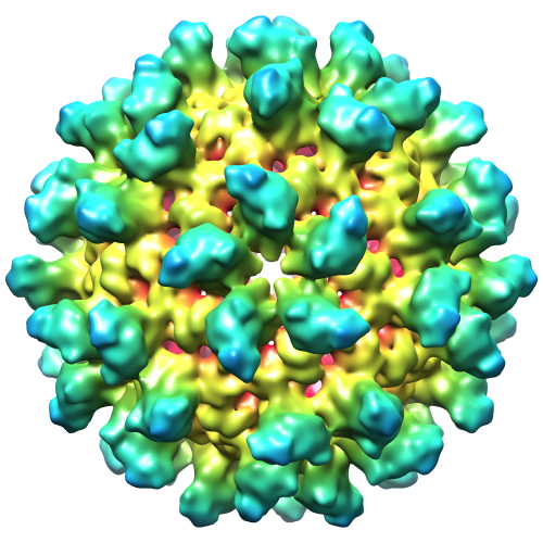 1862.png 1862.png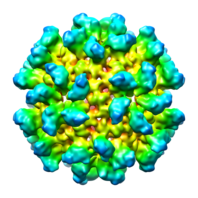 emd_1862.png emd_1862.png | 285.5 KB 473.4 KB | ||
| アーカイブディレクトリ |  http://ftp.pdbj.org/pub/emdb/structures/EMD-1862ftp://ftp.pdbj.org/pub/emdb/structures/EMD-1862 http://ftp.pdbj.org/pub/emdb/structures/EMD-1862ftp://ftp.pdbj.org/pub/emdb/structures/EMD-1862 | HTTPS FTP |
-検証レポート
| 文書・要旨 | emd_1862_validation.pdf.gz | 250.1 KB | 表示 | EMDB検証レポート |
|---|---|---|---|---|
| 文書・詳細版 | emd_1862_full_validation.pdf.gz | 249.3 KB | 表示 | |
| XML形式データ | emd_1862_validation.xml.gz | 5.9 KB | 表示 | |
| アーカイブディレクトリ | https://ftp.pdbj.org/pub/emdb/validation_reports/EMD-1862ftp://ftp.pdbj.org/pub/emdb/validation_reports/EMD-1862 | HTTPS FTP |
-関連構造データ
-リンク
| EMDBのページ | EMDB (EBI/PDBe) / EMDataResource |
|---|
-マップ
| ファイル | ダウンロード / ファイル: emd_1862.map.gz / 形式: CCP4 / 大きさ: 30.9 MB / タイプ: IMAGE STORED AS SIGNED INTEGER (2 BYTES) | ||||||||||||||||||||||||||||||||||||||||||||||||||||||||||||||||||||
|---|---|---|---|---|---|---|---|---|---|---|---|---|---|---|---|---|---|---|---|---|---|---|---|---|---|---|---|---|---|---|---|---|---|---|---|---|---|---|---|---|---|---|---|---|---|---|---|---|---|---|---|---|---|---|---|---|---|---|---|---|---|---|---|---|---|---|---|---|---|
| 注釈 | Icosahedral assembly of Rous sarcoma virus capsid proteins | ||||||||||||||||||||||||||||||||||||||||||||||||||||||||||||||||||||
| 投影像・断面図 | 画像のコントロール
画像は Spider により作成 | ||||||||||||||||||||||||||||||||||||||||||||||||||||||||||||||||||||
| ボクセルのサイズ | X=Y=Z: 1.27 Å | ||||||||||||||||||||||||||||||||||||||||||||||||||||||||||||||||||||
| 密度 |
| ||||||||||||||||||||||||||||||||||||||||||||||||||||||||||||||||||||
| 対称性 | 空間群: 1 | ||||||||||||||||||||||||||||||||||||||||||||||||||||||||||||||||||||
| 詳細 | EMDB XML:
CCP4マップ ヘッダ情報:
| ||||||||||||||||||||||||||||||||||||||||||||||||||||||||||||||||||||
 Z (Sec.)
 Y (Row.)
 X (Col.)


-添付データ
- 試料の構成要素
-全体 : Icosahedral assembly of Rous sarcoma virus capsid proteins
| 全体 | 名称: Icosahedral assembly of Rous sarcoma virus capsid proteins |
|---|---|
| 要素 |
|
-超分子 #1000: Icosahedral assembly of Rous sarcoma virus capsid proteins
| 超分子 | 名称: Icosahedral assembly of Rous sarcoma virus capsid proteins タイプ: sample / ID: 1000 / 集合状態: 60 capsid protein subunits form 12 pentamers / Number unique components: 1 |
|---|---|
| 分子量 | 理論値: 1.53 MDa |
-分子 #1: Avian leukosis virus capsid protein
| 分子 | 名称: Avian leukosis virus capsid protein / タイプ: protein_or_peptide / ID: 1 / Name.synonym: RSV CA protein / コピー数: 60 / 集合状態: Icosahedral composed of 12 pentamers / 組換発現: Yes |
|---|---|
| 由来(天然) | 生物種: Avian leukosis virus (ニワトリ白血病ウイルス) 株: Prague C |
| 分子量 | 理論値: 1.53 MDa |
| 組換発現 | 生物種:  |
-実験情報
-構造解析
| 手法 | クライオ電子顕微鏡法 |
|---|---|
 解析 解析 | 単粒子再構成法 |
| 試料の集合状態 | particle |
-試料調製
| 濃度 | 2 mg/mL |
|---|---|
| 緩衝液 | pH: 7.5 詳細: 10 mM Tris-HCl, 75 mM NaCl, 0.05 mM ETDA, 0.5 M NaPO4 |
| グリッド | 詳細: Holey carbon film on 400 mesh gold grid |
| 凍結 | 凍結剤: ETHANE / チャンバー内温度: 93.15 K / 装置: LEICA KF80 詳細: Vitrification instrument: Reichert-Jung KF80 plunger. Vitrification carried out in nitrogen atmosphere 手法: 4.5 microliter sample dropped onto grid, blotted on one side for 1 second, then plunged |
- 電子顕微鏡法
| 顕微鏡 | FEI/PHILIPS CM200FEG |
|---|---|
| 温度 | 最低: 88.15 K / 最高: 98.15 K / 平均: 93.15 K |
| 撮影 | カテゴリ: FILM / フィルム・検出器のモデル: KODAK SO-163 FILM デジタル化 - スキャナー: NIKON SUPER COOLSCAN 9000 デジタル化 - サンプリング間隔: 6.35 µm / 実像数: 6 / 平均電子線量: 14 e/Å2 / Od range: 4.8 / ビット/ピクセル: 16 |
| 電子線 | 加速電圧: 120 kV / 電子線源:  FIELD EMISSION GUN FIELD EMISSION GUN |
| 電子光学系 | 照射モード: FLOOD BEAM / 撮影モード: BRIGHT FIELD / Cs: 2.0 mm / 最大 デフォーカス(公称値): 1.5 µm / 最小 デフォーカス(公称値): 1.0 µm / 倍率(公称値): 50000 |
| 試料ステージ | 試料ホルダー: Eucentric / 試料ホルダーモデル: GATAN LIQUID NITROGEN |
-画像解析
| CTF補正 | 詳細: Each particle, phase reversal and baseline subtraction |
|---|---|
| 最終 再構成 | 想定した対称性 - 点群: I (正20面体型対称) / 解像度のタイプ: BY AUTHOR / 解像度: 10.4 Å / 解像度の算出法: FSC 0.5 CUT-OFF / ソフトウェア - 名称: EMAN, Bsoft, PFT2, EM3DR2 詳細: Particles were classified using EMAN and initial template generated using 3-fold class average. Preprocessing was done using Bsoft. For orientation assignment and 3D reconstruction PFT2 and ...詳細: Particles were classified using EMAN and initial template generated using 3-fold class average. Preprocessing was done using Bsoft. For orientation assignment and 3D reconstruction PFT2 and EM3DR2 were used, repsectively 使用した粒子像数: 1478 |
-原子モデル構築 1
| 初期モデル | PDB ID: Chain - Chain ID: A |
|---|---|
| ソフトウェア | 名称: SITUS |
| 詳細 | PDBEntryID_givenInChain. Protocol: Rigid Body. The NTD domain was fitted automatically using colores |
| 精密化 | 空間: RECIPROCAL / プロトコル: RIGID BODY FIT / 当てはまり具合の基準: Laplacian correlation |
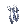
-原子モデル構築 2
| 初期モデル | PDB ID: Chain - Chain ID: A |
|---|---|
| ソフトウェア | 名称: SITUS |
| 詳細 | PDBEntryID_givenInChain. Protocol: Rigid Body. The CTD domain (aa 152-230) was extracted and fitted using colacor. Model 14 was selected as the best fit. |
| 精密化 | 空間: RECIPROCAL / プロトコル: RIGID BODY FIT / 当てはまり具合の基準: Laplacian correlation |

