 Movie
Movie Controller

[English] 日本語
 Yorodumi
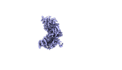
- EMDB-18501: Structure of the plastid-encoded RNA polymerase complex (PEP) fro... -
+ Open data

- Basic information
| Entry |  | ||||||||||||||||||
|---|---|---|---|---|---|---|---|---|---|---|---|---|---|---|---|---|---|---|---|
| Title | Structure of the plastid-encoded RNA polymerase complex (PEP) from Sinapis alba - Map C | ||||||||||||||||||
 Map data Map data | Map C, sharpened. | ||||||||||||||||||
 Sample Sample |
| ||||||||||||||||||
 Keywords Keywords | Transcription / Chloroplasts / Gene Expression / RNA / Polymerase | ||||||||||||||||||
| Biological species |  Sinapis alba (white mustard) Sinapis alba (white mustard) | ||||||||||||||||||
| Method | single particle reconstruction / cryo EM / Resolution: 3.52 Å | ||||||||||||||||||
 Authors Authors | do Prado PFV / Ahrens FM / Pfannschmidt T / Hillen HS | ||||||||||||||||||
| Funding support |  Germany, 5 items Germany, 5 items
| ||||||||||||||||||
 Citation Citation | Journal: Mol Cell / Year: 2024 Title: Structure of the multi-subunit chloroplast RNA polymerase. Authors: Paula F V do Prado / Frederik M Ahrens / Monique Liebers / Noah Ditz / Hans-Peter Braun / Thomas Pfannschmidt / Hauke S Hillen / Abstract: Chloroplasts contain a dedicated genome that encodes subunits of the photosynthesis machinery. Transcription of photosynthesis genes is predominantly carried out by a plastid-encoded RNA polymerase ...Chloroplasts contain a dedicated genome that encodes subunits of the photosynthesis machinery. Transcription of photosynthesis genes is predominantly carried out by a plastid-encoded RNA polymerase (PEP), a nearly 1 MDa complex composed of core subunits with homology to eubacterial RNA polymerases (RNAPs) and at least 12 additional chloroplast-specific PEP-associated proteins (PAPs). However, the architecture of this complex and the functions of the PAPs remain unknown. Here, we report the cryo-EM structure of a 19-subunit PEP complex from Sinapis alba (white mustard). The structure reveals that the PEP core resembles prokaryotic and nuclear RNAPs but contains chloroplast-specific features that mediate interactions with the PAPs. The PAPs are unrelated to known transcription factors and arrange around the core in a unique fashion. Their structures suggest potential functions during transcription in the chemical environment of chloroplasts. These results reveal structural insights into chloroplast transcription and provide a framework for understanding photosynthesis gene expression. | ||||||||||||||||||
| History |
|
- Structure visualization
| Supplemental images |
|---|
- Downloads & links
-EMDB archive
| Map data | emd_18501.map.gz | 306.7 MB |  EMDB map data format EMDB map data format | |
|---|---|---|---|---|
| Header (meta data) | emd-18501-v30.xmlemd-18501.xml | 19.7 KB 19.7 KB | Display Display | EMDB header |
| Images |  emd_18501.png emd_18501.png | 18.4 KB | ||
| Masks | emd_18501_msk_1.map | 325 MB | Mask map | |
| Filedesc metadata | emd-18501.cif.gz | 4.2 KB | ||
| Others | emd_18501_additional_1.map.gzemd_18501_half_map_1.map.gzemd_18501_half_map_2.map.gz | 163.3 MB 301.3 MB 301.3 MB | ||
| Archive directory |  http://ftp.pdbj.org/pub/emdb/structures/EMD-18501ftp://ftp.pdbj.org/pub/emdb/structures/EMD-18501 http://ftp.pdbj.org/pub/emdb/structures/EMD-18501ftp://ftp.pdbj.org/pub/emdb/structures/EMD-18501 | HTTPS FTP |
-Related structure data


-Links
| EMDB pages | EMDB (EBI/PDBe) / EMDataResource |
|---|
-Map
| File | Download / File: emd_18501.map.gz / Format: CCP4 / Size: 325 MB / Type: IMAGE STORED AS FLOATING POINT NUMBER (4 BYTES) | ||||||||||||||||||||||||||||||||||||
|---|---|---|---|---|---|---|---|---|---|---|---|---|---|---|---|---|---|---|---|---|---|---|---|---|---|---|---|---|---|---|---|---|---|---|---|---|---|
| Annotation | Map C, sharpened. | ||||||||||||||||||||||||||||||||||||
| Projections & slices | Image control
Images are generated by Spider. | ||||||||||||||||||||||||||||||||||||
| Voxel size | X=Y=Z: 1.05 Å | ||||||||||||||||||||||||||||||||||||
| Density |
| ||||||||||||||||||||||||||||||||||||
| Symmetry | Space group: 1 | ||||||||||||||||||||||||||||||||||||
| Details | EMDB XML:
|
 Z (Sec.)
 Y (Row.)
 X (Col.)


-Supplemental data
-Mask #1
| File | emd_18501_msk_1.map | ||||||||||||
|---|---|---|---|---|---|---|---|---|---|---|---|---|---|
| Projections & Slices |
| ||||||||||||
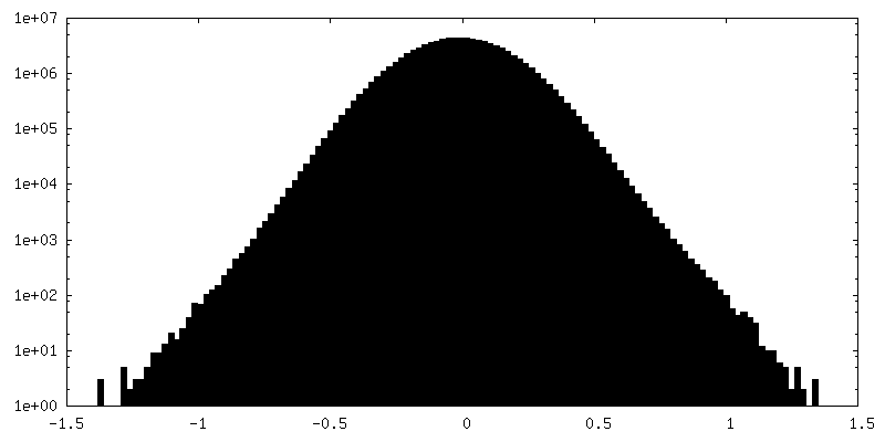
| Density Histograms |


-Additional map: Map C, unsharpened.
| File | emd_18501_additional_1.map | ||||||||||||
|---|---|---|---|---|---|---|---|---|---|---|---|---|---|
| Annotation | Map C, unsharpened. | ||||||||||||
| Projections & Slices |
| ||||||||||||
| Density Histograms |


-Half map: Map C, half map A.
| File | emd_18501_half_map_1.map | ||||||||||||
|---|---|---|---|---|---|---|---|---|---|---|---|---|---|
| Annotation | Map C, half map A. | ||||||||||||
| Projections & Slices |
| ||||||||||||
| Density Histograms |


- Sample components
-Entire : Plastid-encoded DNA-dependent RNA polymerase (PEP)
| Entire | Name: Plastid-encoded DNA-dependent RNA polymerase (PEP) |
|---|---|
| Components |
|
-Supramolecule #1: Plastid-encoded DNA-dependent RNA polymerase (PEP)
| Supramolecule | Name: Plastid-encoded DNA-dependent RNA polymerase (PEP) / type: complex / ID: 1 / Parent: 0 / Macromolecule list: #1-#17 |
|---|---|
| Source (natural) | Organism: Sinapis alba (white mustard) |
| Molecular weight | Theoretical: 1 MDa |
-Experimental details
-Structure determination
| Method | cryo EM |
|---|---|
 Processing Processing | single particle reconstruction |
| Aggregation state | particle |
-Sample preparation
| Buffer | pH: 7.6 Component:
| |||||||||||||||||||||
|---|---|---|---|---|---|---|---|---|---|---|---|---|---|---|---|---|---|---|---|---|---|---|
| Vitrification | Cryogen name: ETHANE / Chamber humidity: 95 % / Chamber temperature: 277.15 K / Instrument: FEI VITROBOT MARK IV |
- Electron microscopy
| Microscope | FEI TITAN KRIOS |
|---|---|
| Image recording | Film or detector model: GATAN K3 BIOQUANTUM (6k x 4k) / Average exposure time: 2.7 sec. / Average electron dose: 40.0 e/Å2 |
| Electron beam | Acceleration voltage: 300 kV / Electron source:  FIELD EMISSION GUN FIELD EMISSION GUN |
| Electron optics | Illumination mode: OTHER / Imaging mode: BRIGHT FIELD / Cs: 2.7 mm / Nominal defocus max: 2.0 µm / Nominal defocus min: 0.5 µm / Nominal magnification: 81000 |
| Sample stage | Specimen holder model: FEI TITAN KRIOS AUTOGRID HOLDER / Cooling holder cryogen: NITROGEN |
| Experimental equipment |  Model: Titan Krios / Image courtesy: FEI Company |
-Image processing
| Startup model | Type of model: INSILICO MODEL |
|---|---|
| Final reconstruction | Resolution.type: BY AUTHOR / Resolution: 3.52 Å / Resolution method: FSC 0.143 CUT-OFF / Number images used: 46170 |
| Initial angle assignment | Type: MAXIMUM LIKELIHOOD |
| Final angle assignment | Type: MAXIMUM LIKELIHOOD |
