 ムービー
ムービー コントローラー


+ データを開く

- 基本情報
基本情報
| 登録情報 |  | |||||||||
|---|---|---|---|---|---|---|---|---|---|---|
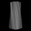
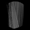
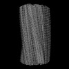
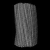
| タイトル | CTE Tau intermediate amyloid (MIA-19) | |||||||||
 マップデータ マップデータ | Sharpened map | |||||||||
 試料 試料 |
| |||||||||
 キーワード キーワード | Amyloid / tau / PROTEIN FIBRIL | |||||||||
| 生物種 |  Homo sapiens (ヒト) Homo sapiens (ヒト) | |||||||||
| 手法 | らせん対称体再構成法 / クライオ電子顕微鏡法 / 解像度: 6.73 Å | |||||||||
 データ登録者 データ登録者 | Lovestam S / Li D / Scheres SHW / Goedert M | |||||||||
| 資金援助 |  英国, 1件 英国, 1件
| |||||||||
 引用 引用 | ジャーナル: Nature / 年: 2024 タイトル: Disease-specific tau filaments assemble via polymorphic intermediates. 著者: Sofia Lövestam / David Li / Jane L Wagstaff / Abhay Kotecha / Dari Kimanius / Stephen H McLaughlin / Alexey G Murzin / Stefan M V Freund / Michel Goedert / Sjors H W Scheres /  要旨: Intermediate species in the assembly of amyloid filaments are believed to play a central role in neurodegenerative diseases and may constitute important targets for therapeutic intervention. However, ...Intermediate species in the assembly of amyloid filaments are believed to play a central role in neurodegenerative diseases and may constitute important targets for therapeutic intervention. However, structural information about intermediate species has been scarce and the molecular mechanisms by which amyloids assemble remain largely unknown. Here we use time-resolved cryogenic electron microscopy to study the in vitro assembly of recombinant truncated tau (amino acid residues 297-391) into paired helical filaments of Alzheimer's disease or into filaments of chronic traumatic encephalopathy. We report the formation of a shared first intermediate amyloid filament, with an ordered core comprising residues 302-316. Nuclear magnetic resonance indicates that the same residues adopt rigid, β-strand-like conformations in monomeric tau. At later time points, the first intermediate amyloid disappears and we observe many different intermediate amyloid filaments, with structures that depend on the reaction conditions. At the end of both assembly reactions, most intermediate amyloids disappear and filaments with the same ordered cores as those from human brains remain. Our results provide structural insights into the processes of primary and secondary nucleation of amyloid assembly, with implications for the design of new therapies. | |||||||||
| 履歴 |
|
- 構造の表示
構造の表示
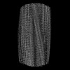
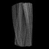
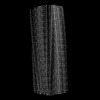
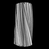
| 添付画像 |
|---|

- ダウンロードとリンク
-EMDBアーカイブ
| マップデータ | emd_18356.map.gz | 55.5 MB |  EMDBマップデータ形式 EMDBマップデータ形式 | |
|---|---|---|---|---|
| ヘッダ (付随情報) | emd-18356-v30.xmlemd-18356.xml | 14.6 KB 14.6 KB | 表示 表示 | EMDBヘッダ |
| FSC (解像度算出) | emd_18356_fsc.xml | 13.7 KB | 表示 | FSCデータファイル |
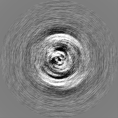
| 画像 |  emd_18356.png emd_18356.png | 65.7 KB | ||
| Filedesc metadata | emd-18356.cif.gz | 3.9 KB | ||
| その他 | emd_18356_additional_1.map.gzemd_18356_half_map_1.map.gzemd_18356_half_map_2.map.gz | 172.7 MB 172.3 MB 172.4 MB | ||
| アーカイブディレクトリ |  http://ftp.pdbj.org/pub/emdb/structures/EMD-18356ftp://ftp.pdbj.org/pub/emdb/structures/EMD-18356 http://ftp.pdbj.org/pub/emdb/structures/EMD-18356ftp://ftp.pdbj.org/pub/emdb/structures/EMD-18356 | HTTPS FTP |
-検証レポート
| 文書・要旨 | emd_18356_validation.pdf.gz | 1.1 MB | 表示 | EMDB検証レポート |
|---|---|---|---|---|
| 文書・詳細版 | emd_18356_full_validation.pdf.gz | 1.1 MB | 表示 | |
| XML形式データ | emd_18356_validation.xml.gz | 21.4 KB | 表示 | |
| CIF形式データ | emd_18356_validation.cif.gz | 28 KB | 表示 | |
| アーカイブディレクトリ | https://ftp.pdbj.org/pub/emdb/validation_reports/EMD-18356ftp://ftp.pdbj.org/pub/emdb/validation_reports/EMD-18356 | HTTPS FTP |
-関連構造データ
| 関連構造データ |  8ppoC  8q27C  8q2jC  8q2kC 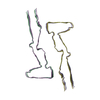 8q2lC 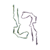 8q7fC 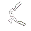 8q7lC 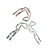 8q7mC 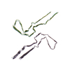 8q7pC 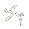 8q7tC 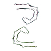 8q88C 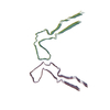 8q8cC 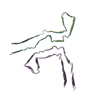 8q8dC  8q8eC 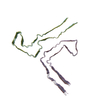 8q8fC 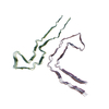 8q8lC 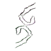 8q8mC  8q8rC 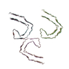 8q8sC 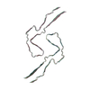 8q8uC 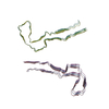 8q8vC 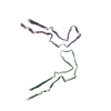 8q8wC 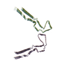 8q8xC 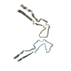 8q8yC 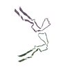 8q8zC 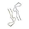 8q97C  8q98C  8q99C  8q9aC  8q9bC  8q9cC 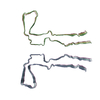 8q9dC 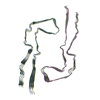 8q9eC  8q9fC  8q9gC  8q9hC 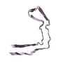 8q9iC 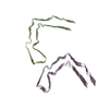 8q9jC 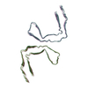 8q9kC 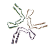 8q9lC  8q9mC 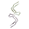 8q9oC 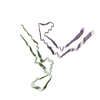 8qcpC  8qcrC  8qjjC C: 同じ文献を引用 ( |
|---|


-リンク
| EMDBのページ | EMDB (EBI/PDBe) / EMDataResource |
|---|---|
| 「今月の分子」の関連する項目 |

-マップ
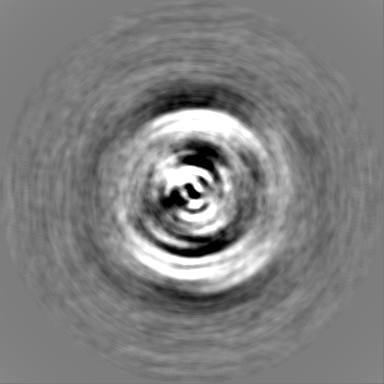
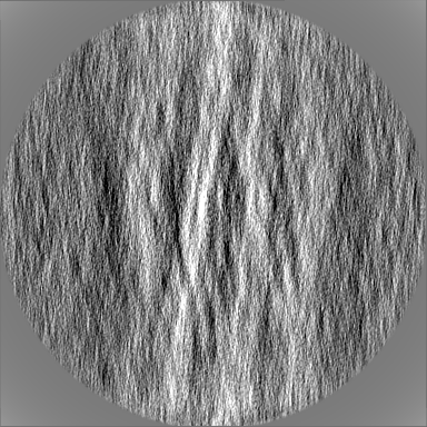
| ファイル | ダウンロード / ファイル: emd_18356.map.gz / 形式: CCP4 / 大きさ: 216 MB / タイプ: IMAGE STORED AS FLOATING POINT NUMBER (4 BYTES) | ||||||||||||||||||||||||||||||||||||
|---|---|---|---|---|---|---|---|---|---|---|---|---|---|---|---|---|---|---|---|---|---|---|---|---|---|---|---|---|---|---|---|---|---|---|---|---|---|
| 注釈 | Sharpened map | ||||||||||||||||||||||||||||||||||||
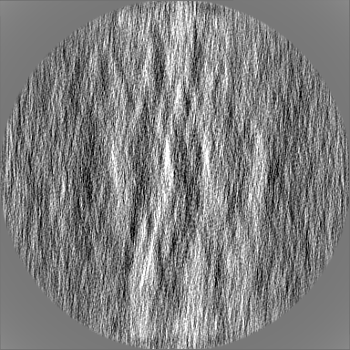
| 投影像・断面図 | 画像のコントロール
画像は Spider により作成 | ||||||||||||||||||||||||||||||||||||
| ボクセルのサイズ | X=Y=Z: 0.824 Å | ||||||||||||||||||||||||||||||||||||
| 密度 |
| ||||||||||||||||||||||||||||||||||||
| 対称性 | 空間群: 1 | ||||||||||||||||||||||||||||||||||||
| 詳細 | EMDB XML:
|
 Z (Sec.)
 Y (Row.)
 X (Col.)


-添付データ
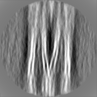
-追加マップ: Unsharpened map
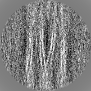
| ファイル | emd_18356_additional_1.map | ||||||||||||
|---|---|---|---|---|---|---|---|---|---|---|---|---|---|
| 注釈 | Unsharpened map | ||||||||||||
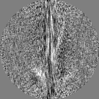
| 投影像・断面図 |
| ||||||||||||
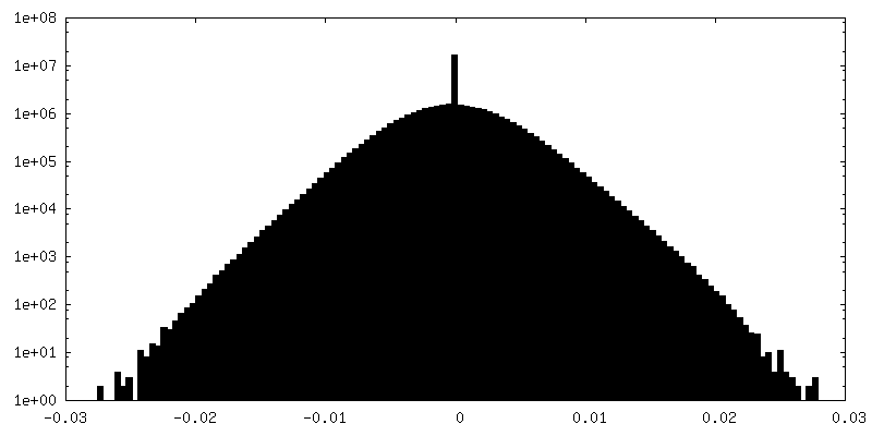
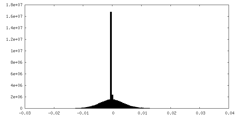
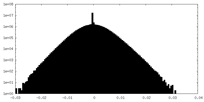
| 密度ヒストグラム |

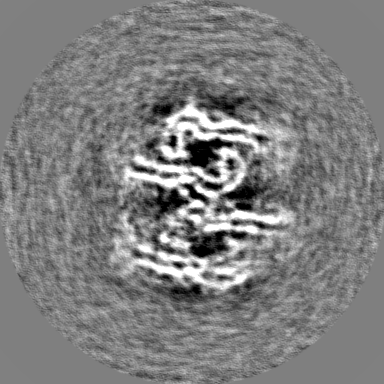


-ハーフマップ: Half map 1
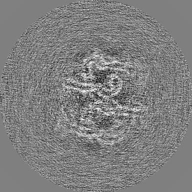
| ファイル | emd_18356_half_map_1.map | ||||||||||||
|---|---|---|---|---|---|---|---|---|---|---|---|---|---|
| 注釈 | Half map 1 | ||||||||||||
| 投影像・断面図 |
| ||||||||||||
| 密度ヒストグラム |


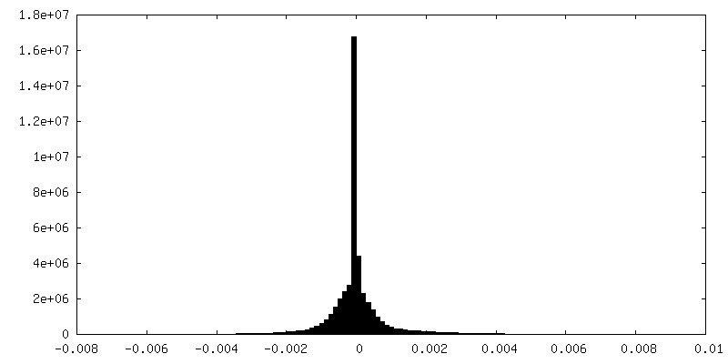
-ハーフマップ: Half map 2
| ファイル | emd_18356_half_map_2.map | ||||||||||||
|---|---|---|---|---|---|---|---|---|---|---|---|---|---|
| 注釈 | Half map 2 | ||||||||||||
| 投影像・断面図 |
| ||||||||||||
| 密度ヒストグラム |


- 試料の構成要素
-全体 : Amyloid
| 全体 | 名称: Amyloid |
|---|---|
| 要素 |
|
-超分子 #1: Amyloid
| 超分子 | 名称: Amyloid / タイプ: complex / ID: 1 / 親要素: 0 / 含まれる分子: #1 |
|---|---|
| 由来(天然) | 生物種: Homo sapiens (ヒト) |
-実験情報
-構造解析
| 手法 | クライオ電子顕微鏡法 |
|---|---|
 解析 解析 | らせん対称体再構成法 |
| 試料の集合状態 | filament |
-試料調製
| 緩衝液 | pH: 7.2 |
|---|---|
| 凍結 | 凍結剤: ETHANE |
- 電子顕微鏡法
| 顕微鏡 | FEI TITAN KRIOS |
|---|---|
| 撮影 | フィルム・検出器のモデル: FEI FALCON IV (4k x 4k) 平均電子線量: 40.0 e/Å2 |
| 電子線 | 加速電圧: 300 kV / 電子線源:  FIELD EMISSION GUN FIELD EMISSION GUN |
| 電子光学系 | 照射モード: FLOOD BEAM / 撮影モード: BRIGHT FIELD / 最大 デフォーカス(公称値): 2.5 µm / 最小 デフォーカス(公称値): 0.5 µm |
| 実験機器 |  モデル: Titan Krios / 画像提供: FEI Company |
-画像解析
| 最終 再構成 | 想定した対称性 - らせんパラメータ - Δz: 4.76 Å 想定した対称性 - らせんパラメータ - ΔΦ: -1.20 ° 想定した対称性 - らせんパラメータ - 軸対称性: C1 (非対称) 解像度のタイプ: BY AUTHOR / 解像度: 6.73 Å / 解像度の算出法: FSC 0.143 CUT-OFF / ソフトウェア - 名称: RELION (ver. 4) / 使用した粒子像数: 3787 |
|---|---|
| 初期モデル | モデルのタイプ: OTHER / 詳細: de novo |
| 最終 角度割当 | タイプ: NOT APPLICABLE |
| FSC曲線 (解像度の算出) | 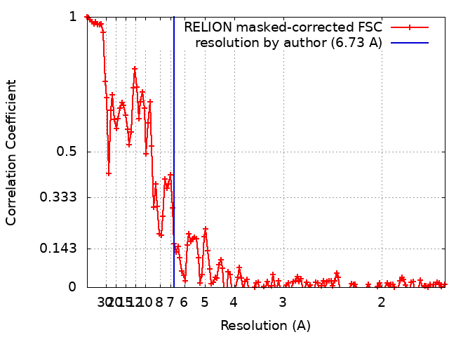 |
