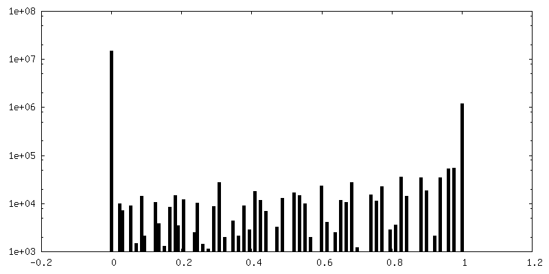
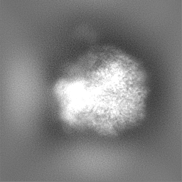
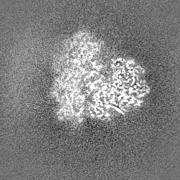
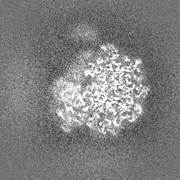
ジャーナル: Nat Methods / 年: 2024 タイトル: CryoDRGN-ET: deep reconstructing generative networks for visualizing dynamic biomolecules inside cells. 著者: Ramya Rangan / Ryan Feathers / Sagar Khavnekar / Adam Lerer / Jake D Johnston / Ron Kelley / Martin Obr / Abhay Kotecha / Ellen D Zhong / 要旨: Advances in cryo-electron tomography (cryo-ET) have produced new opportunities to visualize the structures of dynamic macromolecules in native cellular environments. While cryo-ET can reveal ...Advances in cryo-electron tomography (cryo-ET) have produced new opportunities to visualize the structures of dynamic macromolecules in native cellular environments. While cryo-ET can reveal structures at molecular resolution, image processing algorithms remain a bottleneck in resolving the heterogeneity of biomolecular structures in situ. Here, we introduce cryoDRGN-ET for heterogeneous reconstruction of cryo-ET subtomograms. CryoDRGN-ET learns a deep generative model of three-dimensional density maps directly from subtomogram tilt-series images and can capture states diverse in both composition and conformation. We validate this approach by recovering the known translational states in Mycoplasma pneumoniae ribosomes in situ. We then perform cryo-ET on cryogenic focused ion beam-milled Saccharomyces cerevisiae cells. CryoDRGN-ET reveals the structural landscape of S. cerevisiae ribosomes during translation and captures continuous motions of fatty acid synthase complexes inside cells. This method is openly available in the cryoDRGN software.
 ムービー
ムービー コントローラー


 データを開く
 基本情報
基本情報
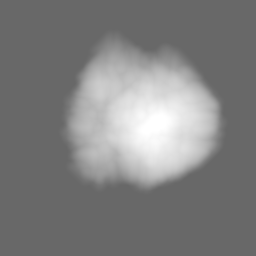
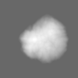
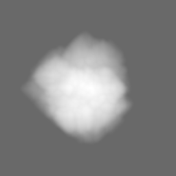
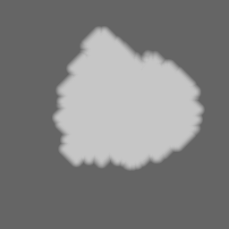
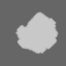
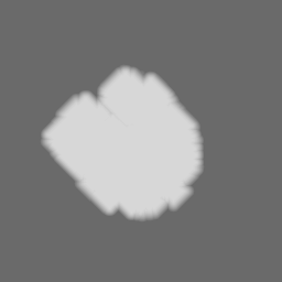
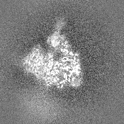
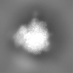
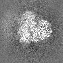
 マップデータ
 試料
試料 キーワード
キーワード
 データ登録者
 ドイツ, 2件
  引用
引用

 構造の表示
構造の表示
 ダウンロードとリンク
 EMDBマップデータ形式
 emd_18231.png
 http://ftp.pdbj.org/pub/emdb/structures/EMD-18231
 Z (Sec.)
 Y (Row.)
 X (Col.)


 試料の構成要素
 解析
解析 電子顕微鏡法
 FIELD EMISSION GUN