Movie
Movie Controller

[English] 日本語
 Yorodumi
- EMDB-17612: In-situ structure of the pentameric HEF trimers from influenza C ... -
+ Open data

- Basic information
| Entry |  | |||||||||
|---|---|---|---|---|---|---|---|---|---|---|
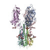
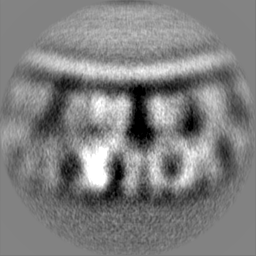
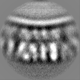
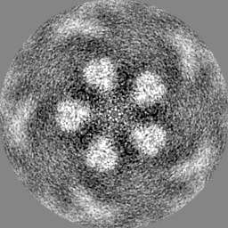
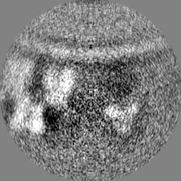
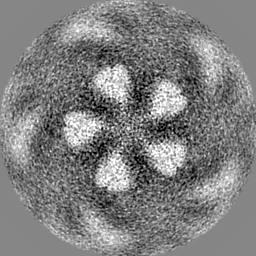
| Title | In-situ structure of the pentameric HEF trimers from influenza C viral particles | |||||||||
 Map data Map data | ||||||||||
 Sample Sample |
| |||||||||
 Keywords Keywords | Class 1 fusion protein / membrane fusion / viral assembly / pentameric / VIRUS | |||||||||
| Biological species |  Influenza C virus (C/Johannesburg/1/66) Influenza C virus (C/Johannesburg/1/66) | |||||||||
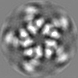
| Method | subtomogram averaging / cryo EM / Resolution: 12.2 Å | |||||||||
 Authors Authors | Liu ZB / Rosenthal PB | |||||||||
| Funding support |  United Kingdom, 1 items United Kingdom, 1 items
| |||||||||
 Citation Citation | Journal: To Be Published Title: Topological defects in the spherical virus crystal Authors: Liu ZB / Rosenthal PB | |||||||||
| History |
|
- Structure visualization
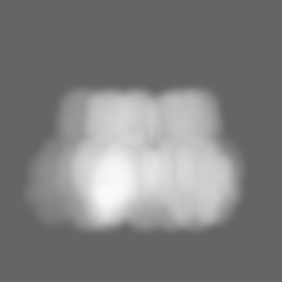
| Supplemental images |
|---|

- Downloads & links
-EMDB archive
| Map data | emd_17612.map.gz | 58.5 MB |  EMDB map data format EMDB map data format | |
|---|---|---|---|---|
| Header (meta data) | emd-17612-v30.xmlemd-17612.xml | 15.9 KB 15.9 KB | Display Display | EMDB header |
| FSC (resolution estimation) | emd_17612_fsc.xml | 9.2 KB | Display | FSC data file |
| Images | 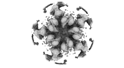 emd_17612.png emd_17612.png | 38.6 KB | ||
| Masks | emd_17612_msk_1.map | 64 MB | Mask map | |
| Filedesc metadata | emd-17612.cif.gz | 4.7 KB | ||
| Others | emd_17612_half_map_1.map.gzemd_17612_half_map_2.map.gz | 49.6 MB 49.6 MB | ||
| Archive directory |  http://ftp.pdbj.org/pub/emdb/structures/EMD-17612ftp://ftp.pdbj.org/pub/emdb/structures/EMD-17612 http://ftp.pdbj.org/pub/emdb/structures/EMD-17612ftp://ftp.pdbj.org/pub/emdb/structures/EMD-17612 | HTTPS FTP |
-Related structure data

-Links
| EMDB pages | EMDB (EBI/PDBe) / EMDataResource |
|---|
-Map
| File | Download / File: emd_17612.map.gz / Format: CCP4 / Size: 64 MB / Type: IMAGE STORED AS FLOATING POINT NUMBER (4 BYTES) | ||||||||||||||||||||||||||||||||||||
|---|---|---|---|---|---|---|---|---|---|---|---|---|---|---|---|---|---|---|---|---|---|---|---|---|---|---|---|---|---|---|---|---|---|---|---|---|---|
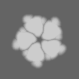
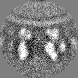
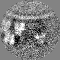
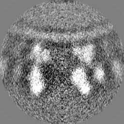
| Projections & slices | Image control
Images are generated by Spider. | ||||||||||||||||||||||||||||||||||||
| Voxel size | X=Y=Z: 1.38 Å | ||||||||||||||||||||||||||||||||||||
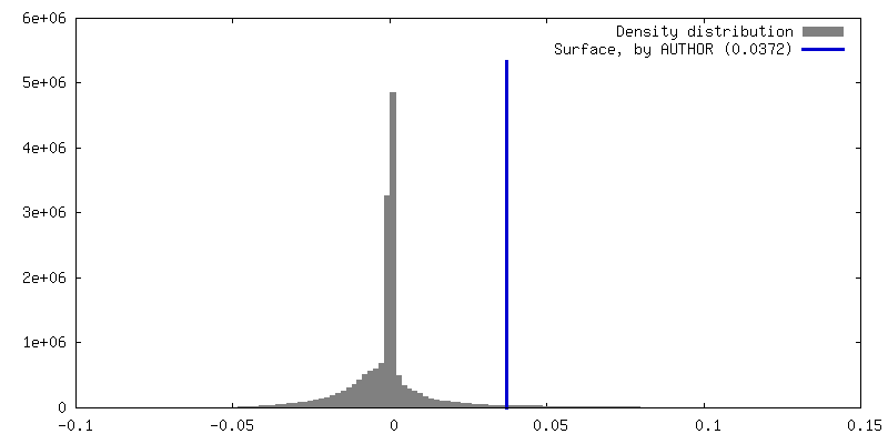
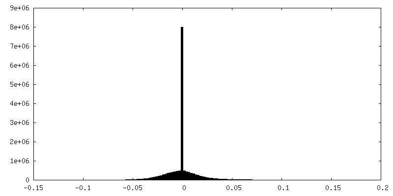
| Density |
| ||||||||||||||||||||||||||||||||||||
| Symmetry | Space group: 1 | ||||||||||||||||||||||||||||||||||||
| Details | EMDB XML:
|
 Z (Sec.)
 Y (Row.)
 X (Col.)


-Supplemental data
-Mask #1
| File | emd_17612_msk_1.map | ||||||||||||
|---|---|---|---|---|---|---|---|---|---|---|---|---|---|
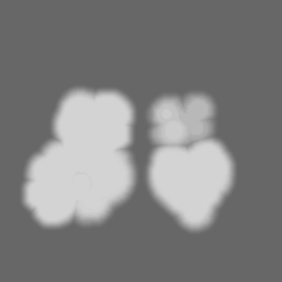
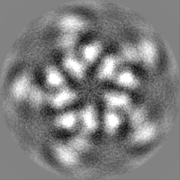
| Projections & Slices |
| ||||||||||||
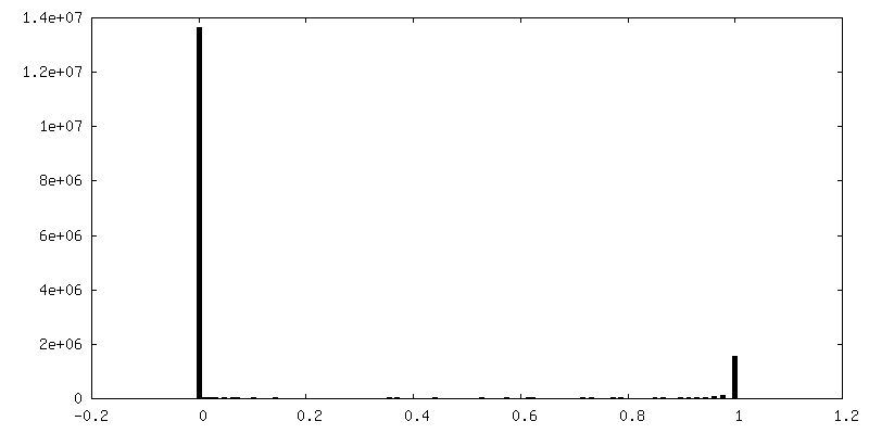
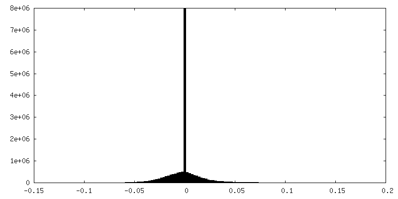
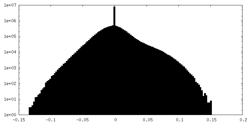
| Density Histograms |


-Half map: #1
| File | emd_17612_half_map_1.map | ||||||||||||
|---|---|---|---|---|---|---|---|---|---|---|---|---|---|
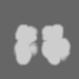
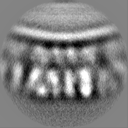
| Projections & Slices |
| ||||||||||||
| Density Histograms |


-Half map: #2
| File | emd_17612_half_map_2.map | ||||||||||||
|---|---|---|---|---|---|---|---|---|---|---|---|---|---|
| Projections & Slices |
| ||||||||||||
| Density Histograms |

- Sample components
-Entire : Influenza C virus (C/Johannesburg/1/66)
| Entire | Name: Influenza C virus (C/Johannesburg/1/66) |
|---|---|
| Components |
|
-Supramolecule #1: Influenza C virus (C/Johannesburg/1/66)
| Supramolecule | Name: Influenza C virus (C/Johannesburg/1/66) / type: virus / ID: 1 / Parent: 0 / Macromolecule list: #1-#2 / Details: Purified viruses / NCBI-ID: 100673 / Sci species name: Influenza C virus (C/Johannesburg/1/66) / Virus type: VIRION / Virus isolate: STRAIN / Virus enveloped: Yes / Virus empty: No |
|---|---|
| Molecular weight | Theoretical: 1.03 MDa |
-Experimental details
-Structure determination
| Method | cryo EM |
|---|---|
 Processing Processing | subtomogram averaging |
| Aggregation state | particle |
-Sample preparation
| Concentration | 2 mg/mL | ||||||||||||
|---|---|---|---|---|---|---|---|---|---|---|---|---|---|
| Buffer | pH: 7.4 Component:
| ||||||||||||
| Grid | Model: Quantifoil R2/2 / Material: COPPER / Support film - Material: CARBON / Support film - topology: HOLEY / Pretreatment - Type: GLOW DISCHARGE / Pretreatment - Time: 30 sec. / Pretreatment - Atmosphere: AMYLAMINE | ||||||||||||
| Vitrification | Cryogen name: ETHANE / Chamber humidity: 100 % / Chamber temperature: 277 K / Instrument: FEI VITROBOT MARK III | ||||||||||||
| Details | Heterogeneous mix of viral particles, viral like particles and cellular vesicles |
- Electron microscopy
| Microscope | FEI TITAN KRIOS |
|---|---|
| Specialist optics | Energy filter - Slit width: 20 eV |
| Image recording | Film or detector model: GATAN K2 SUMMIT (4k x 4k) / Detector mode: COUNTING / Digitization - Dimensions - Width: 3838 pixel / Digitization - Dimensions - Height: 3710 pixel / Number real images: 1 / Average exposure time: 1.1 sec. / Average electron dose: 2.1 e/Å2 |
| Electron beam | Acceleration voltage: 300 kV / Electron source:  FIELD EMISSION GUN FIELD EMISSION GUN |
| Electron optics | C2 aperture diameter: 100.0 µm / Illumination mode: FLOOD BEAM / Imaging mode: BRIGHT FIELD / Cs: 2.7 mm / Nominal defocus max: 4.5 µm / Nominal defocus min: 2.0 µm / Nominal magnification: 64000 |
| Sample stage | Specimen holder model: FEI TITAN KRIOS AUTOGRID HOLDER / Cooling holder cryogen: NITROGEN |
| Experimental equipment |  Model: Titan Krios / Image courtesy: FEI Company |
-Image processing
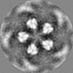
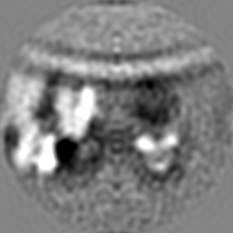
| Final reconstruction | Applied symmetry - Point group: C5 (5 fold cyclic) / Algorithm: BACK PROJECTION / Resolution.type: BY AUTHOR / Resolution: 12.2 Å / Resolution method: FSC 0.143 CUT-OFF / Software - Name: RELION (ver. 3.0.8) / Number subtomograms used: 3108 |
|---|---|
| Extraction | Number tomograms: 83 / Number images used: 83 / Software - Name: RELION (ver. 3.0.8) |
| Final angle assignment | Type: MAXIMUM LIKELIHOOD / Software - Name: RELION (ver. 3.0.8) |
| FSC plot (resolution estimation) | 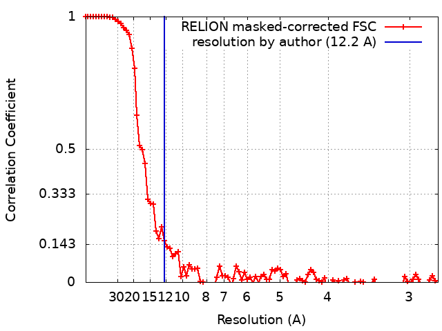 |
-Atomic model buiding 1
| Initial model | PDB ID: Chain - Source name: PDB / Chain - Initial model type: experimental model |
|---|