 ムービー
ムービー コントローラー


+ データを開く

- 基本情報
基本情報
| 登録情報 |  | |||||||||
|---|---|---|---|---|---|---|---|---|---|---|
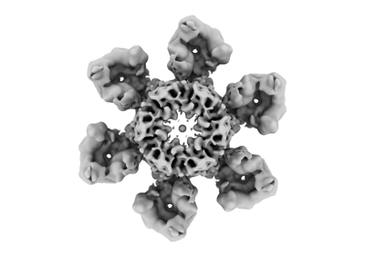
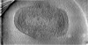
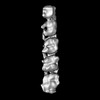
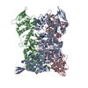
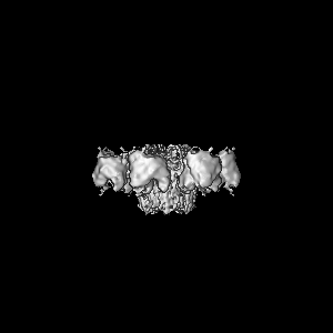
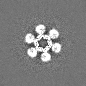
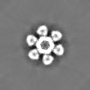
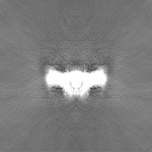
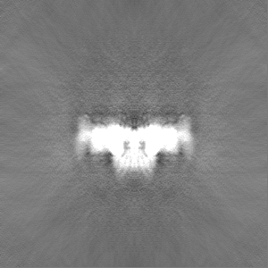
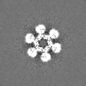
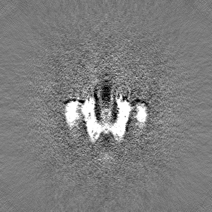
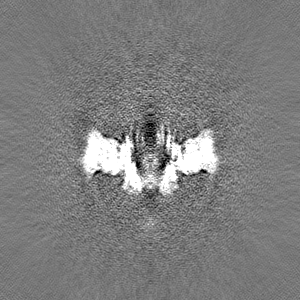
| タイトル | Vaccinia Virus flower-shaped pore-like structure | |||||||||
 マップデータ マップデータ | cryoSPARC 4.0 non-uniform 3D refine, locally filtered and sharpened within cryoSPARC using an FSC cutoff of 0.143. | |||||||||
 試料 試料 |
| |||||||||
 キーワード キーワード | Poxvirus / Vaccinia Virus / core / cryo-electron tomography / AlphaFold / VIRAL PROTEIN | |||||||||
| 生物種 |  Vaccinia virus Western Reserve (ウイルス) Vaccinia virus Western Reserve (ウイルス) | |||||||||
| 手法 | 単粒子再構成法 / クライオ電子顕微鏡法 / 解像度: 7.2 Å | |||||||||
 データ登録者 データ登録者 | Hansen J / Datler J / Thader A / Schloegl A / Hodirnau VV / Schur FKM | |||||||||
| 資金援助 | 1件
| |||||||||
 引用 引用 | ジャーナル: Nat Struct Mol Biol / 年: 2024 タイトル: Multi-modal cryo-EM reveals trimers of protein A10 to form the palisade layer in poxvirus cores. 著者: Julia Datler / Jesse M Hansen / Andreas Thader / Alois Schlögl / Lukas W Bauer / Victor-Valentin Hodirnau / Florian K M Schur /  要旨: Poxviruses are among the largest double-stranded DNA viruses, with members such as variola virus, monkeypox virus and the vaccination strain vaccinia virus (VACV). Knowledge about the structural ...Poxviruses are among the largest double-stranded DNA viruses, with members such as variola virus, monkeypox virus and the vaccination strain vaccinia virus (VACV). Knowledge about the structural proteins that form the viral core has remained sparse. While major core proteins have been annotated via indirect experimental evidence, their structures have remained elusive and they could not be assigned to individual core features. Hence, which proteins constitute which layers of the core, such as the palisade layer and the inner core wall, has remained enigmatic. Here we show, using a multi-modal cryo-electron microscopy (cryo-EM) approach in combination with AlphaFold molecular modeling, that trimers formed by the cleavage product of VACV protein A10 are the key component of the palisade layer. This allows us to place previously obtained descriptions of protein interactions within the core wall into perspective and to provide a detailed model of poxvirus core architecture. Importantly, we show that interactions within A10 trimers are likely generalizable over members of orthopox- and parapoxviruses. | |||||||||
| 履歴 |
|
- 構造の表示
構造の表示
| 添付画像 |
|---|
- ダウンロードとリンク
-EMDBアーカイブ
| マップデータ | emd_17412.map.gz | 2.2 MB |  EMDBマップデータ形式 EMDBマップデータ形式 | |
|---|---|---|---|---|
| ヘッダ (付随情報) | emd-17412-v30.xmlemd-17412.xml | 17.6 KB 17.6 KB | 表示 表示 | EMDBヘッダ |
| 画像 |  emd_17412.png emd_17412.png | 48.6 KB | ||
| Filedesc metadata | emd-17412.cif.gz | 4.8 KB | ||
| その他 | emd_17412_half_map_1.map.gzemd_17412_half_map_2.map.gz | 95.6 MB 95.6 MB | ||
| アーカイブディレクトリ |  http://ftp.pdbj.org/pub/emdb/structures/EMD-17412ftp://ftp.pdbj.org/pub/emdb/structures/EMD-17412 http://ftp.pdbj.org/pub/emdb/structures/EMD-17412ftp://ftp.pdbj.org/pub/emdb/structures/EMD-17412 | HTTPS FTP |
-検証レポート
| 文書・要旨 | emd_17412_validation.pdf.gz | 646.9 KB | 表示 | EMDB検証レポート |
|---|---|---|---|---|
| 文書・詳細版 | emd_17412_full_validation.pdf.gz | 646.4 KB | 表示 | |
| XML形式データ | emd_17412_validation.xml.gz | 13.5 KB | 表示 | |
| CIF形式データ | emd_17412_validation.cif.gz | 16 KB | 表示 | |
| アーカイブディレクトリ | https://ftp.pdbj.org/pub/emdb/validation_reports/EMD-17412ftp://ftp.pdbj.org/pub/emdb/validation_reports/EMD-17412 | HTTPS FTP |
-関連構造データ
-リンク
| EMDBのページ | EMDB (EBI/PDBe) / EMDataResource |
|---|
-マップ
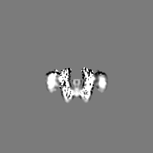
| ファイル | ダウンロード / ファイル: emd_17412.map.gz / 形式: CCP4 / 大きさ: 103 MB / タイプ: IMAGE STORED AS FLOATING POINT NUMBER (4 BYTES) | ||||||||||||||||||||||||||||||||||||
|---|---|---|---|---|---|---|---|---|---|---|---|---|---|---|---|---|---|---|---|---|---|---|---|---|---|---|---|---|---|---|---|---|---|---|---|---|---|
| 注釈 | cryoSPARC 4.0 non-uniform 3D refine, locally filtered and sharpened within cryoSPARC using an FSC cutoff of 0.143. | ||||||||||||||||||||||||||||||||||||
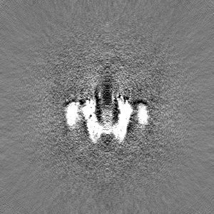
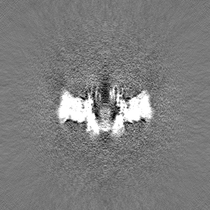
| 投影像・断面図 | 画像のコントロール
画像は Spider により作成 | ||||||||||||||||||||||||||||||||||||
| ボクセルのサイズ | X=Y=Z: 2.12 Å | ||||||||||||||||||||||||||||||||||||
| 密度 |
| ||||||||||||||||||||||||||||||||||||
| 対称性 | 空間群: 1 | ||||||||||||||||||||||||||||||||||||
| 詳細 | EMDB XML:
|
 Z (Sec.)
 Y (Row.)
 X (Col.)


-添付データ
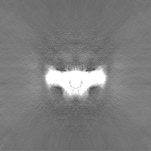
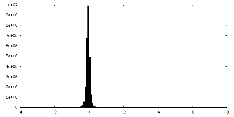
-ハーフマップ: cryoSPARC 4.0 non-uniform 3D refine half map A
| ファイル | emd_17412_half_map_1.map | ||||||||||||
|---|---|---|---|---|---|---|---|---|---|---|---|---|---|
| 注釈 | cryoSPARC 4.0 non-uniform 3D refine half map A | ||||||||||||
| 投影像・断面図 |
| ||||||||||||
| 密度ヒストグラム |

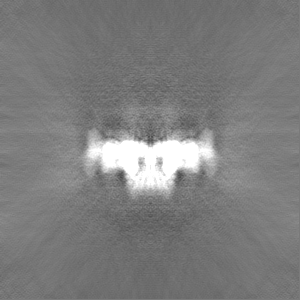


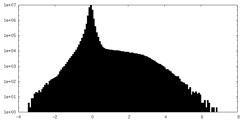
-ハーフマップ: cryoSPARC 4.0 non-uniform 3D refine half map B
| ファイル | emd_17412_half_map_2.map | ||||||||||||
|---|---|---|---|---|---|---|---|---|---|---|---|---|---|
| 注釈 | cryoSPARC 4.0 non-uniform 3D refine half map B | ||||||||||||
| 投影像・断面図 |
| ||||||||||||
| 密度ヒストグラム |
- 試料の構成要素
-全体 : Vaccinia virus Western Reserve
| 全体 | 名称: Vaccinia virus Western Reserve (ウイルス) |
|---|---|
| 要素 |
|
-超分子 #1: Vaccinia virus Western Reserve
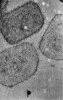
| 超分子 | 名称: Vaccinia virus Western Reserve / タイプ: virus / ID: 1 / 親要素: 0 / 含まれる分子: #1 詳細: Purified from HeLa cells infected with Vaccinia Virus Western Reserve. NCBI-ID: 696871 / 生物種: Vaccinia virus Western Reserve / ウイルスタイプ: VIRION / ウイルス・単離状態: STRAIN / ウイルス・エンベロープ: No / ウイルス・中空状態: No |
|---|
-実験情報
-構造解析
| 手法 | クライオ電子顕微鏡法 |
|---|---|
 解析 解析 | 単粒子再構成法 |
| 試料の集合状態 | particle |
-試料調製
| 緩衝液 | pH: 9 構成要素:
詳細: diluted 1:1 with 0.25% Trypsin (final concentration 0.125%) and 1:1 4% paraformaldehyde (final concentration 2%) in 1 mM Tris-HCl. | |||||||||
|---|---|---|---|---|---|---|---|---|---|---|
| グリッド | モデル: Quantifoil R2/2 / 材質: COPPER / メッシュ: 200 / 支持フィルム - 材質: CARBON / 支持フィルム - トポロジー: HOLEY / 前処理 - タイプ: GLOW DISCHARGE / 前処理 - 時間: 180 sec. / 前処理 - 雰囲気: AIR | |||||||||
| 凍結 | 凍結剤: ETHANE / チャンバー内湿度: 80 % / チャンバー内温度: 277 K / 装置: LEICA EM GP / 詳細: Leica GP2. | |||||||||
| 詳細 | Intact viral cores with some detached soluble monodispersed particles. |
- 電子顕微鏡法
| 顕微鏡 | FEI TITAN KRIOS |
|---|---|
| 特殊光学系 | エネルギーフィルター - 名称: GIF Bioquantum / エネルギーフィルター - スリット幅: 20 eV |
| 撮影 | フィルム・検出器のモデル: GATAN K3 BIOQUANTUM (6k x 4k) デジタル化 - サイズ - 横: 5760 pixel / デジタル化 - サイズ - 縦: 4092 pixel / 実像数: 9264 / 平均電子線量: 53.04 e/Å2 / 詳細: 34 frames total. |
| 電子線 | 加速電圧: 300 kV / 電子線源:  FIELD EMISSION GUN FIELD EMISSION GUN |
| 電子光学系 | C2レンズ絞り径: 50.0 µm / 照射モード: FLOOD BEAM / 撮影モード: BRIGHT FIELD / Cs: 2.7 mm / 最大 デフォーカス(公称値): 3.0 µm / 最小 デフォーカス(公称値): 1.25 µm / 倍率(公称値): 81000 |
| 試料ステージ | 試料ホルダーモデル: FEI TITAN KRIOS AUTOGRID HOLDER ホルダー冷却材: NITROGEN |
| 実験機器 |  モデル: Titan Krios / 画像提供: FEI Company |
