 ムービー
ムービー コントローラー


+ データを開く

- 基本情報
基本情報
| 登録情報 |  | |||||||||||||||
|---|---|---|---|---|---|---|---|---|---|---|---|---|---|---|---|---|
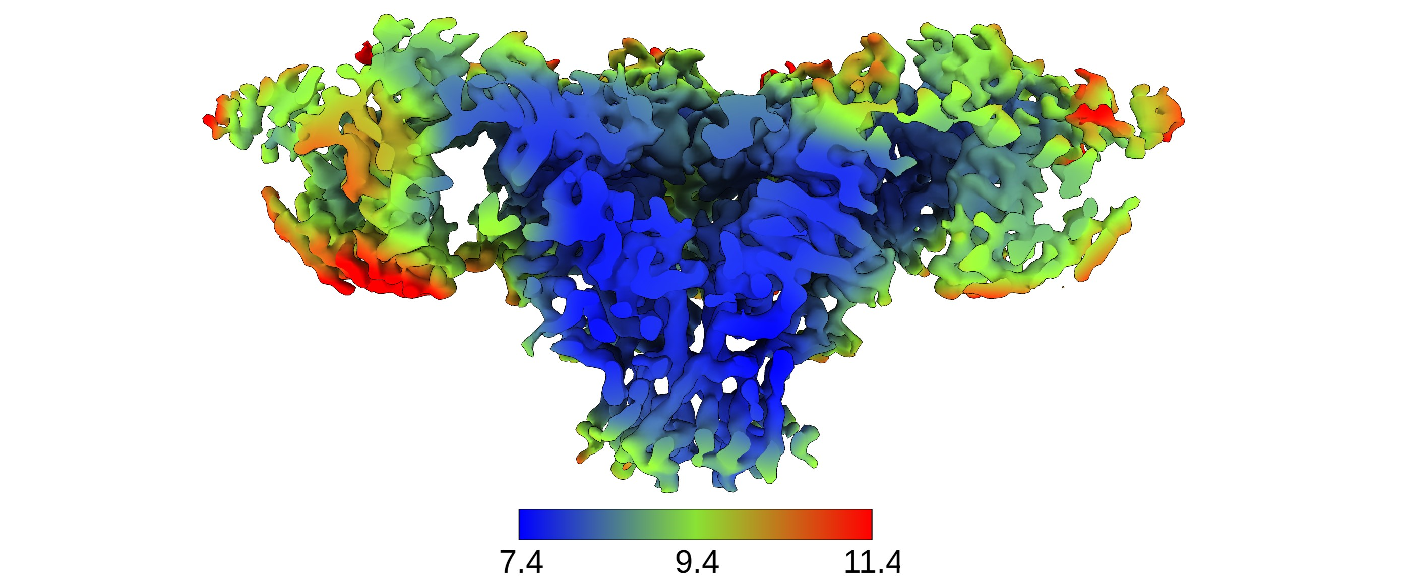
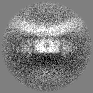
| タイトル | Structure of RyR1 obtained from SR vesicles tilt series (EMPIAR-10452), reprocessed with TomoBEAR | |||||||||||||||
 マップデータ マップデータ | the nominal apix is 1.8, the calibrated value was found to be 1.7 on that microscope with used mag | |||||||||||||||
 試料 試料 |
| |||||||||||||||
 キーワード キーワード | SR / RyR1 / Ryanodine receptor type 1 / skeletal muscle / sarcoplasmic reticulum / cryo-ET / tomography / StA / subtomogram averaging / tilt series / subnanometer resolution / native membrane / MEMBRANE PROTEIN | |||||||||||||||
| 生物種 |  | |||||||||||||||
| 手法 | サブトモグラム平均法 / クライオ電子顕微鏡法 / 解像度: 8.9 Å | |||||||||||||||
 データ登録者 データ登録者 | Balyschew N / Yushkevich A / Mikirtumov V / Sanchez RM / Sprink T / Kudryashev M | |||||||||||||||
| 資金援助 |  ドイツ, 4件 ドイツ, 4件
| |||||||||||||||
 引用 引用 | ジャーナル: Nat Commun / 年: 2023 タイトル: Streamlined structure determination by cryo-electron tomography and subtomogram averaging using TomoBEAR. 著者: Nikita Balyschew / Artsemi Yushkevich / Vasilii Mikirtumov / Ricardo M Sanchez / Thiemo Sprink / Mikhail Kudryashev / 要旨: Structures of macromolecules in their native state provide unique unambiguous insights into their functions. Cryo-electron tomography combined with subtomogram averaging demonstrated the power to ...Structures of macromolecules in their native state provide unique unambiguous insights into their functions. Cryo-electron tomography combined with subtomogram averaging demonstrated the power to solve such structures in situ at resolutions in the range of 3 Angstrom for some macromolecules. In order to be applicable to the structural determination of the majority of macromolecules observable in cells in limited amounts, processing of tomographic data has to be performed in a high-throughput manner. Here we present TomoBEAR-a modular configurable workflow engine for streamlined processing of cryo-electron tomographic data for subtomogram averaging. TomoBEAR combines commonly used cryo-EM packages with reasonable presets to provide a transparent ("white box") approach for data management and processing. We demonstrate applications of TomoBEAR to two data sets of purified macromolecular targets, to an ion channel RyR1 in a membrane, and the tomograms of plasma FIB-milled lamellae and demonstrate the ability to produce high-resolution structures. TomoBEAR speeds up data processing, minimizes human interventions, and will help accelerate the adoption of in situ structural biology by cryo-ET. The source code and the documentation are freely available. #1: ジャーナル: bioRxiv / 年: 2023タイトル: Streamlined Structure Determination by Cryo-Electron Tomography and Subtomogram Averaging using TomoBEAR 著者: Balyschew N / Yushkevich A / Mikirtumov V / Sanchez RM / Sprink T / Kudryashev M | |||||||||||||||
| 履歴 |
|
- 構造の表示
構造の表示
| 添付画像 |
|---|
- ダウンロードとリンク
-EMDBアーカイブ
| マップデータ | emd_17272.map.gz | 69.7 MB |  EMDBマップデータ形式 EMDBマップデータ形式 | |
|---|---|---|---|---|
| ヘッダ (付随情報) | emd-17272-v30.xmlemd-17272.xml | 16.2 KB 16.2 KB | 表示 表示 | EMDBヘッダ |
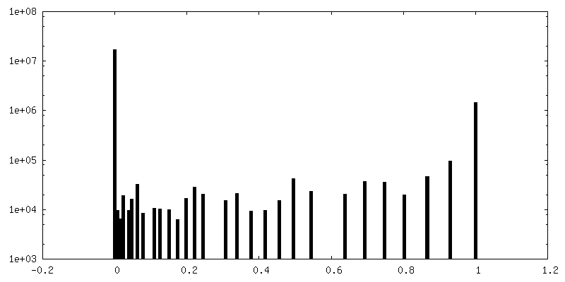
| FSC (解像度算出) | emd_17272_fsc.xml | 11.5 KB | 表示 | FSCデータファイル |
| 画像 |  emd_17272.png emd_17272.png | 2 MB | ||
| マスクデータ | emd_17272_msk_1.map | 125 MB | マスクマップ | |
| Filedesc metadata | emd-17272.cif.gz | 4.5 KB | ||
| その他 | emd_17272_half_map_1.map.gzemd_17272_half_map_2.map.gz | 61.5 MB 61.5 MB | ||
| アーカイブディレクトリ |  http://ftp.pdbj.org/pub/emdb/structures/EMD-17272ftp://ftp.pdbj.org/pub/emdb/structures/EMD-17272 http://ftp.pdbj.org/pub/emdb/structures/EMD-17272ftp://ftp.pdbj.org/pub/emdb/structures/EMD-17272 | HTTPS FTP |
-検証レポート
| 文書・要旨 | emd_17272_validation.pdf.gz | 1 MB | 表示 | EMDB検証レポート |
|---|---|---|---|---|
| 文書・詳細版 | emd_17272_full_validation.pdf.gz | 1 MB | 表示 | |
| XML形式データ | emd_17272_validation.xml.gz | 19.7 KB | 表示 | |
| CIF形式データ | emd_17272_validation.cif.gz | 25.1 KB | 表示 | |
| アーカイブディレクトリ | https://ftp.pdbj.org/pub/emdb/validation_reports/EMD-17272ftp://ftp.pdbj.org/pub/emdb/validation_reports/EMD-17272 | HTTPS FTP |
-関連構造データ

-リンク
| EMDBのページ | EMDB (EBI/PDBe) / EMDataResource |
|---|
-マップ
| ファイル | ダウンロード / ファイル: emd_17272.map.gz / 形式: CCP4 / 大きさ: 125 MB / タイプ: IMAGE STORED AS FLOATING POINT NUMBER (4 BYTES) | ||||||||||||||||||||
|---|---|---|---|---|---|---|---|---|---|---|---|---|---|---|---|---|---|---|---|---|---|
| 注釈 | the nominal apix is 1.8, the calibrated value was found to be 1.7 on that microscope with used mag | ||||||||||||||||||||
| ボクセルのサイズ | X=Y=Z: 1.7 Å | ||||||||||||||||||||
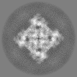
| 密度 |
| ||||||||||||||||||||
| 対称性 | 空間群: 1 | ||||||||||||||||||||
| 詳細 | EMDB XML:
|
-添付データ
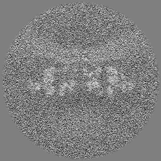
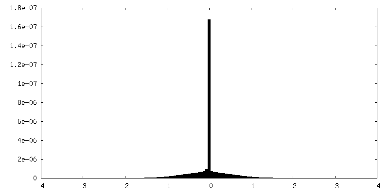
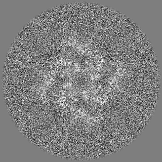
-マスク #1
| ファイル | emd_17272_msk_1.map | ||||||||||||
|---|---|---|---|---|---|---|---|---|---|---|---|---|---|
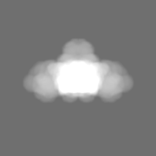
| 投影像・断面図 |
| ||||||||||||
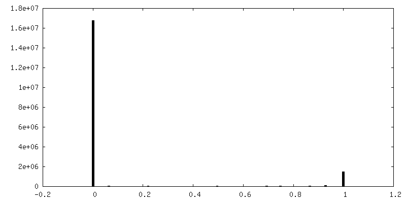
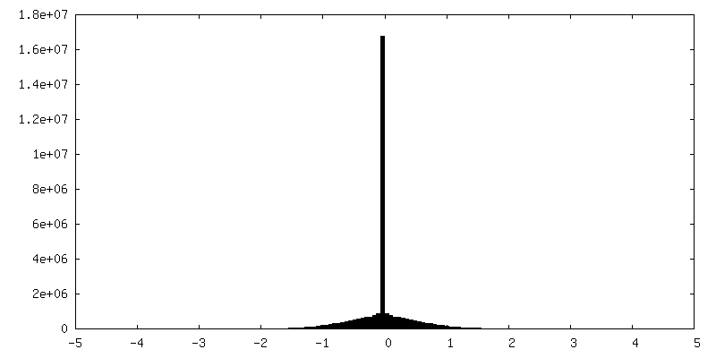
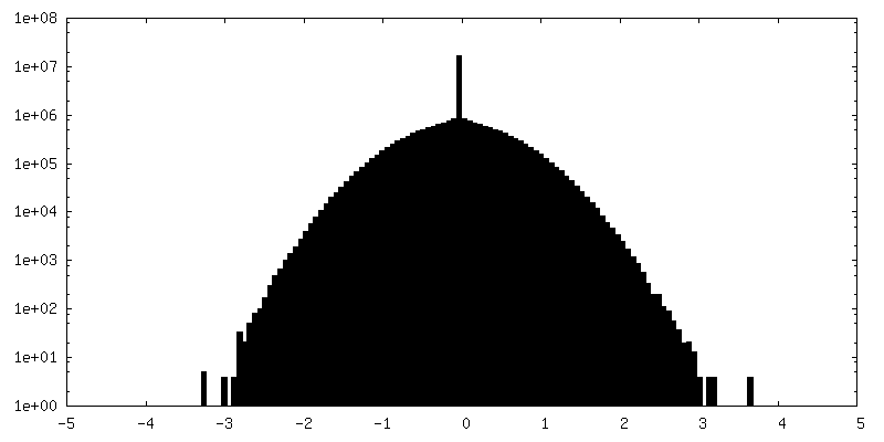
| 密度ヒストグラム |
 Z
Z Y
Y X
X


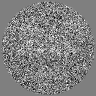
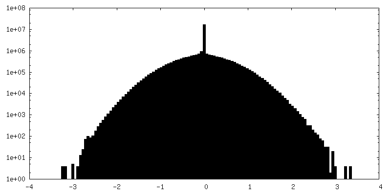
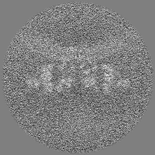
-ハーフマップ: #1
| ファイル | emd_17272_half_map_1.map | ||||||||||||
|---|---|---|---|---|---|---|---|---|---|---|---|---|---|
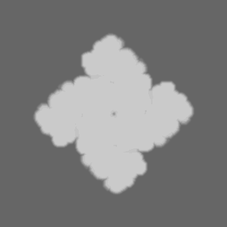
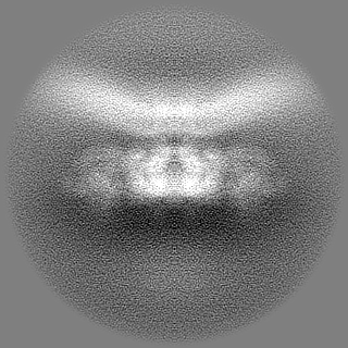
| 投影像・断面図 |
| ||||||||||||
| 密度ヒストグラム |

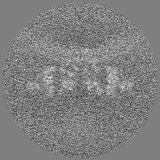
-ハーフマップ: #2
| ファイル | emd_17272_half_map_2.map | ||||||||||||
|---|---|---|---|---|---|---|---|---|---|---|---|---|---|
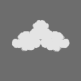
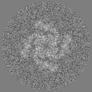
| 投影像・断面図 |
| ||||||||||||
| 密度ヒストグラム |


- 試料の構成要素
-全体 : Sarcoplasmic reticulum
| 全体 | 名称: Sarcoplasmic reticulum |
|---|---|
| 要素 |
|
-超分子 #1: Sarcoplasmic reticulum
| 超分子 | 名称: Sarcoplasmic reticulum / タイプ: organelle_or_cellular_component / ID: 1 / 親要素: 0 |
|---|---|
| 由来(天然) | 生物種: |
-実験情報
-構造解析
| 手法 | クライオ電子顕微鏡法 |
|---|---|
 解析 解析 | サブトモグラム平均法 |
| 試料の集合状態 | tissue |
-試料調製
| 緩衝液 | pH: 7.1 |
|---|---|
| 凍結 | 凍結剤: ETHANE |
- 電子顕微鏡法
| 顕微鏡 | FEI TITAN KRIOS |
|---|---|
| 撮影 | フィルム・検出器のモデル: GATAN K2 SUMMIT (4k x 4k) 検出モード: COUNTING / 平均電子線量: 2.0 e/Å2 |
| 電子線 | 加速電圧: 300 kV / 電子線源:  FIELD EMISSION GUN FIELD EMISSION GUN |
| 電子光学系 | 最大 デフォーカス(補正後): 4.5 µm / 最小 デフォーカス(補正後): 3.5 µm / 倍率(補正後): 81000 / 照射モード: FLOOD BEAM / 撮影モード: BRIGHT FIELD / Cs: 2.7 mm / 最大 デフォーカス(公称値): 4.5 µm / 最小 デフォーカス(公称値): 3.5 µm / 倍率(公称値): 81000 |
| 試料ステージ | 試料ホルダーモデル: FEI TITAN KRIOS AUTOGRID HOLDER ホルダー冷却材: NITROGEN |
| 実験機器 |  モデル: Titan Krios / 画像提供: FEI Company |
-画像解析
| 最終 再構成 | 使用したクラス数: 1 / 想定した対称性 - 点群: C4 (4回回転対称) / アルゴリズム: FOURIER SPACE / 解像度のタイプ: BY AUTHOR / 解像度: 8.9 Å / 解像度の算出法: FSC 0.143 CUT-OFF / ソフトウェア - 名称: RELION / 使用したサブトモグラム数: 3169 |
|---|---|
| 抽出 | トモグラム数: 52 / 使用した粒子像数: 5200 / 参照モデル: emd-10840 / 手法: template matching / ソフトウェア - 名称: Dynamo / 詳細: Dynamo GPU template matching in TomoBEAR |
| 最終 角度割当 | タイプ: MAXIMUM LIKELIHOOD / ソフトウェア - 名称: RELION (ver. relion 4 beta) |
| FSC曲線 (解像度の算出) | 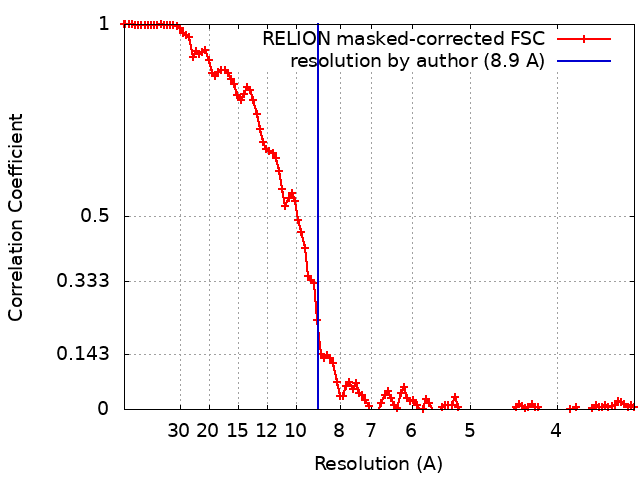 |
