 ムービー
ムービー コントローラー


+ データを開く

- 基本情報
基本情報
| 登録情報 |  | |||||||||
|---|---|---|---|---|---|---|---|---|---|---|
| タイトル | C. elegans L1 larva 80S ribosome class 3 | |||||||||
 マップデータ マップデータ | Main map | |||||||||
 試料 試料 |
| |||||||||
 キーワード キーワード | Caenorhabditis / elegans / ribosome / 80S / L1 / larva / C. elegans | |||||||||
| 生物種 |  | |||||||||
| 手法 | サブトモグラム平均法 / クライオ電子顕微鏡法 / 解像度: 15.0 Å | |||||||||
 データ登録者 データ登録者 | Schioetz OH / Kaiser CJO / Klumpe S / Beck F / Plitzko JM | |||||||||
| 資金援助 |  ドイツ, 1件 ドイツ, 1件
| |||||||||
 引用 引用 | ジャーナル: Nat Methods / 年: 2024 タイトル: Serial Lift-Out: sampling the molecular anatomy of whole organisms. 著者: Oda Helene Schiøtz / Christoph J O Kaiser / Sven Klumpe / Dustin R Morado / Matthias Poege / Jonathan Schneider / Florian Beck / David P Klebl / Christopher Thompson / Jürgen M Plitzko /   要旨: Cryo-focused ion beam milling of frozen-hydrated cells and subsequent cryo-electron tomography (cryo-ET) has enabled the structural elucidation of macromolecular complexes directly inside cells. ...Cryo-focused ion beam milling of frozen-hydrated cells and subsequent cryo-electron tomography (cryo-ET) has enabled the structural elucidation of macromolecular complexes directly inside cells. Application of the technique to multicellular organisms and tissues, however, is still limited by sample preparation. While high-pressure freezing enables the vitrification of thicker samples, it prolongs subsequent preparation due to increased thinning times and the need for extraction procedures. Additionally, thinning removes large portions of the specimen, restricting the imageable volume to the thickness of the final lamella, typically <300 nm. Here we introduce Serial Lift-Out, an enhanced lift-out technique that increases throughput and obtainable contextual information by preparing multiple sections from single transfers. We apply Serial Lift-Out to Caenorhabditis elegans L1 larvae, yielding a cryo-ET dataset sampling the worm's anterior-posterior axis, and resolve its ribosome structure to 7 Å and a subregion of the 11-protofilament microtubule to 13 Å, illustrating how Serial Lift-Out enables the study of multicellular molecular anatomy. | |||||||||
| 履歴 |
|
- 構造の表示
構造の表示
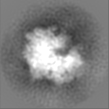
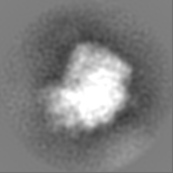
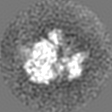
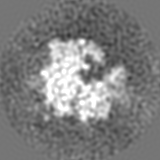
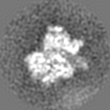
| 添付画像 |
|---|

- ダウンロードとリンク
-EMDBアーカイブ
| マップデータ | emd_17244.map.gz | 11.9 MB |  EMDBマップデータ形式 EMDBマップデータ形式 | |
|---|---|---|---|---|
| ヘッダ (付随情報) | emd-17244-v30.xmlemd-17244.xml | 16.4 KB 16.4 KB | 表示 表示 | EMDBヘッダ |
| FSC (解像度算出) | emd_17244_fsc.xml | 5.8 KB | 表示 | FSCデータファイル |
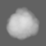
| 画像 | 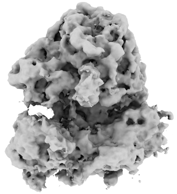 emd_17244.png emd_17244.png | 90.6 KB | ||
| マスクデータ | emd_17244_msk_1.map | 15.6 MB | マスクマップ | |
| Filedesc metadata | emd-17244.cif.gz | 4.7 KB | ||
| その他 | emd_17244_additional_1.map.gzemd_17244_half_map_1.map.gzemd_17244_half_map_2.map.gz | 14.2 MB 11.9 MB 11.9 MB | ||
| アーカイブディレクトリ |  http://ftp.pdbj.org/pub/emdb/structures/EMD-17244ftp://ftp.pdbj.org/pub/emdb/structures/EMD-17244 http://ftp.pdbj.org/pub/emdb/structures/EMD-17244ftp://ftp.pdbj.org/pub/emdb/structures/EMD-17244 | HTTPS FTP |
-関連構造データ


-リンク
| EMDBのページ | EMDB (EBI/PDBe) / EMDataResource |
|---|
-マップ
| ファイル | ダウンロード / ファイル: emd_17244.map.gz / 形式: CCP4 / 大きさ: 15.6 MB / タイプ: IMAGE STORED AS FLOATING POINT NUMBER (4 BYTES) | ||||||||||||||||||||||||||||||||||||
|---|---|---|---|---|---|---|---|---|---|---|---|---|---|---|---|---|---|---|---|---|---|---|---|---|---|---|---|---|---|---|---|---|---|---|---|---|---|
| 注釈 | Main map | ||||||||||||||||||||||||||||||||||||
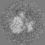
| 投影像・断面図 | 画像のコントロール
画像は Spider により作成 | ||||||||||||||||||||||||||||||||||||
| ボクセルのサイズ | X=Y=Z: 2.98 Å | ||||||||||||||||||||||||||||||||||||
| 密度 |
| ||||||||||||||||||||||||||||||||||||
| 対称性 | 空間群: 1 | ||||||||||||||||||||||||||||||||||||
| 詳細 | EMDB XML:
|
 Z (Sec.)
 Y (Row.)
 X (Col.)


-添付データ
-マスク #1
| ファイル | emd_17244_msk_1.map | ||||||||||||
|---|---|---|---|---|---|---|---|---|---|---|---|---|---|
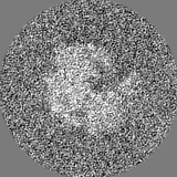
| 投影像・断面図 |
| ||||||||||||
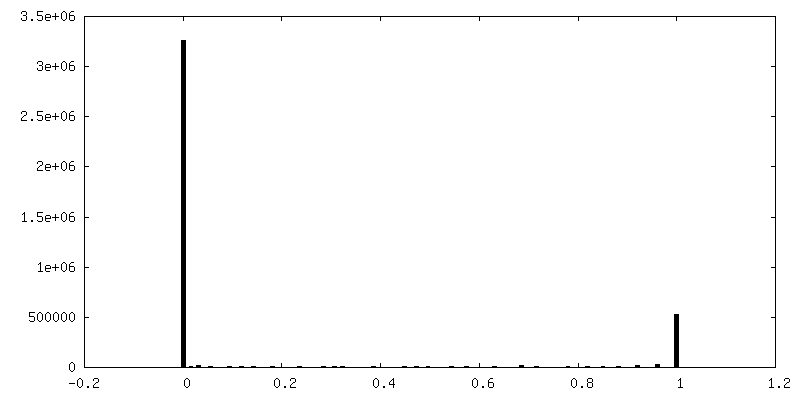
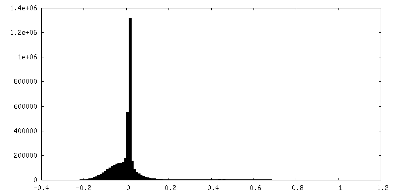
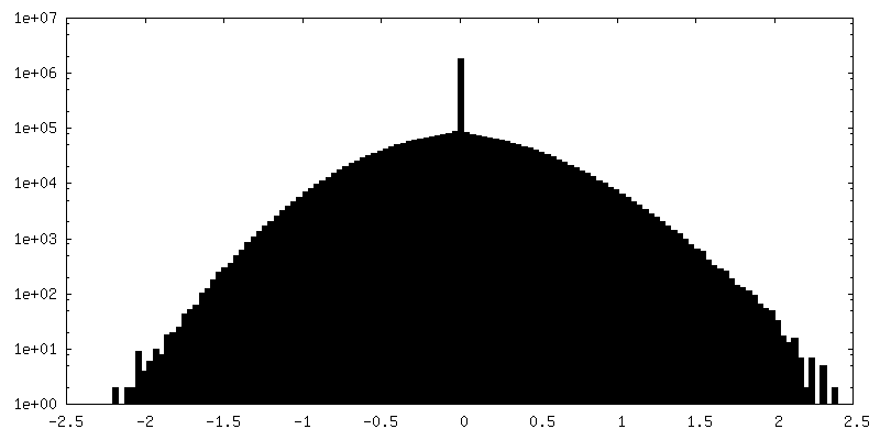
| 密度ヒストグラム |


-追加マップ: postprocessed
| ファイル | emd_17244_additional_1.map | ||||||||||||
|---|---|---|---|---|---|---|---|---|---|---|---|---|---|
| 注釈 | postprocessed | ||||||||||||
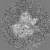
| 投影像・断面図 |
| ||||||||||||
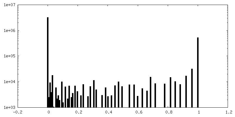
| 密度ヒストグラム |


-ハーフマップ: Half map 1
| ファイル | emd_17244_half_map_1.map | ||||||||||||
|---|---|---|---|---|---|---|---|---|---|---|---|---|---|
| 注釈 | Half map 1 | ||||||||||||
| 投影像・断面図 |
| ||||||||||||
| 密度ヒストグラム |


- 試料の構成要素
-全体 : C. elegans L1 larva, CGC strain NK2476
| 全体 | 名称: C. elegans L1 larva, CGC strain NK2476 |
|---|---|
| 要素 |
|
-超分子 #1: C. elegans L1 larva, CGC strain NK2476
| 超分子 | 名称: C. elegans L1 larva, CGC strain NK2476 / タイプ: complex / ID: 1 / 親要素: 0 |
|---|---|
| 由来(天然) | 生物種: |
-実験情報
-構造解析
| 手法 | クライオ電子顕微鏡法 |
|---|---|
 解析 解析 | サブトモグラム平均法 |
| 試料の集合状態 | tissue |
-試料調製
| 緩衝液 | pH: 7.5 / 詳細: M9 buffer + 20% Ficoll 400 |
|---|---|
| グリッド | モデル: Homemade / 支持フィルム - 材質: FORMVAR / 支持フィルム - トポロジー: CONTINUOUS / 支持フィルム - Film thickness: 100 |
| 凍結 | 凍結剤: NITROGEN / 詳細: High pressure freezing, Leica EM ICE. |
| 詳細 | Developmentally arrested L1 larvae |
- 電子顕微鏡法
| 顕微鏡 | FEI TITAN KRIOS |
|---|---|
| 特殊光学系 | エネルギーフィルター - 名称: TFS Selectris X / エネルギーフィルター - スリット幅: 10 eV |
| 撮影 | フィルム・検出器のモデル: FEI FALCON IV (4k x 4k) デジタル化 - サイズ - 横: 4096 pixel / デジタル化 - サイズ - 縦: 4096 pixel / 実像数: 1 / 平均露光時間: 0.68 sec. / 平均電子線量: 3.2 e/Å2 |
| 電子線 | 加速電圧: 300 kV / 電子線源:  FIELD EMISSION GUN FIELD EMISSION GUN |
| 電子光学系 | C2レンズ絞り径: 50.0 µm / 照射モード: FLOOD BEAM / 撮影モード: BRIGHT FIELD / Cs: 2.7 mm / 最大 デフォーカス(公称値): 4.5 µm / 最小 デフォーカス(公称値): 1.5 µm / 倍率(公称値): 64000 |
| 試料ステージ | 試料ホルダーモデル: FEI TITAN KRIOS AUTOGRID HOLDER ホルダー冷却材: NITROGEN |
| 実験機器 |  モデル: Titan Krios / 画像提供: FEI Company |
-画像解析
| 最終 再構成 | 想定した対称性 - 点群: C1 (非対称) / 解像度のタイプ: BY AUTHOR / 解像度: 15.0 Å / 解像度の算出法: FSC 0.143 CUT-OFF / ソフトウェア - 名称: RELION (ver. 3.0) / 使用したサブトモグラム数: 877 |
|---|---|
| 抽出 | トモグラム数: 200 / 使用した粒子像数: 65451 |
| 最終 角度割当 | タイプ: MAXIMUM LIKELIHOOD |
| FSC曲線 (解像度の算出) | 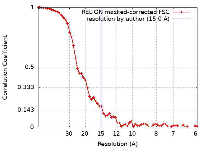 |
