- EMDB-1712: Three-dimensional CryoEM Structure of a Membrane-Anchored AAA Protease -
+
データを開く
IDまたはキーワード:
読み込み中...
-
基本情報
登録情報
データベース: EMDB / ID: EMD-1712
タイトル
Three-dimensional CryoEM Structure of a Membrane-Anchored AAA Protease
マップデータ
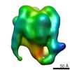
Isosurface representation of m-AAA protease. The density of transmembrane and intermembrane domains is weaker than those of AAA and protease domains. In the publication, the map was split into two parts and contoured separately, 91 kDa and 440 kDa.
試料
試料: m-AAA Protease
タンパク質・ペプチド: Yta10
タンパク質・ペプチド: Yta12
キーワード
AAA protein / protease / ATPase / membrane protein / mitocondria
ジャーナル: J Biol Chem / 年: 2011 タイトル: Electron cryomicroscopy structure of a membrane-anchored mitochondrial AAA protease. 著者: Sukyeong Lee / Steffen Augustin / Takashi Tatsuta / Florian Gerdes / Thomas Langer / Francis T F Tsai / 要旨: FtsH-related AAA proteases are conserved membrane-anchored, ATP-dependent molecular machines, which mediate the processing and turnover of soluble and membrane-embedded proteins in eubacteria, ...FtsH-related AAA proteases are conserved membrane-anchored, ATP-dependent molecular machines, which mediate the processing and turnover of soluble and membrane-embedded proteins in eubacteria, mitochondria, and chloroplasts. Homo- and hetero-oligomeric proteolytic complexes exist, which are composed of homologous subunits harboring an ATPase domain of the AAA family and an H41 metallopeptidase domain. Mutations in subunits of mitochondrial m-AAA proteases have been associated with different neurodegenerative disorders in human, raising questions on the functional differences between homo- and hetero-oligomeric AAA proteases. Here, we have analyzed the hetero-oligomeric yeast m-AAA protease composed of homologous Yta10 and Yta12 subunits. We combined genetic and structural approaches to define the molecular determinants for oligomer assembly and to assess functional similarities between Yta10 and Yta12. We demonstrate that replacement of only two amino acid residues within the metallopeptidase domain of Yta12 allows its assembly into homo-oligomeric complexes. To provide a molecular explanation, we determined the 12 Å resolution structure of the intact yeast m-AAA protease with its transmembrane domains by electron cryomicroscopy (cryo-EM) and atomic structure fitting. The full-length m-AAA protease has a bipartite structure and is a hexamer in solution. We found that residues in Yta12, which facilitate homo-oligomerization when mutated, are located at the interface between neighboring protomers in the hexamer ring. Notably, the transmembrane and intermembrane space domains are separated from the main body, creating a passage on the matrix side, which is wide enough to accommodate unfolded but not folded polypeptides. These results suggest a mechanism regarding how proteins are recognized and degraded by m-AAA proteases.
ダウンロード / ファイル: emd_1712.map.gz / 形式: CCP4 / 大きさ: 7.8 MB / タイプ: IMAGE STORED AS FLOATING POINT NUMBER (4 BYTES)
注釈
Isosurface representation of m-AAA protease. The density of transmembrane and intermembrane domains is weaker than those of AAA and protease domains. In the publication, the map was split into two parts and contoured separately, 91 kDa and 440 kDa.
 ムービー
ムービー コントローラー

 万見
万見 データを開く
 基本情報
基本情報 マップデータ
 試料
試料 キーワード
キーワード
 データ登録者
 引用
引用
 構造の表示
構造の表示 ムービービューア
 ダウンロードとリンク
 em1712.png
 http://ftp.pdbj.org/pub/emdb/structures/EMD-1712


 Z (Sec.)
 X (Row.)
 Y (Col.)


 試料の構成要素
 解析
解析 電子顕微鏡法
 FIELD EMISSION GUN