 Movie
Movie Controller

[English] 日本語
 Yorodumi
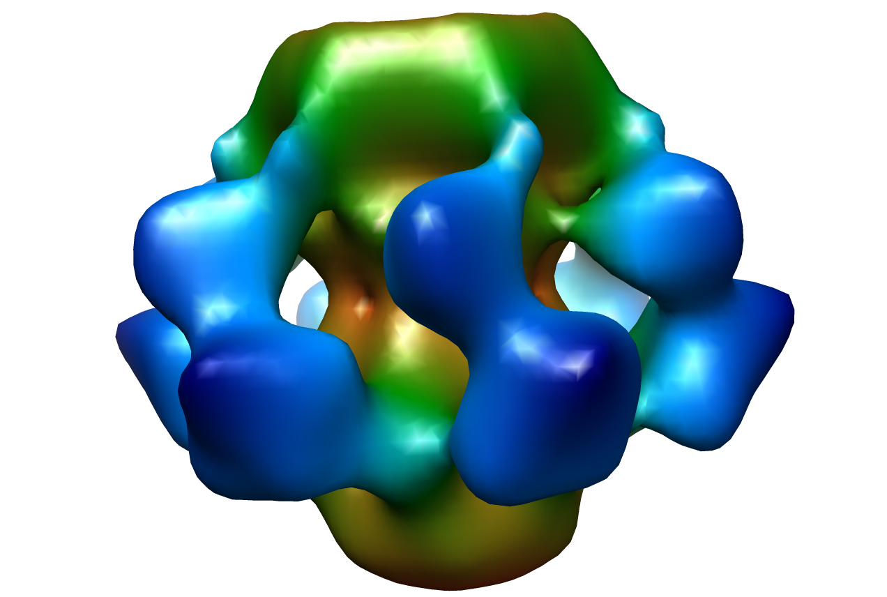
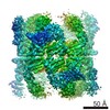
- EMDB-1706: Cryo-EM map of Lactococcal phage p2 baseplate consisting of ORF 1... -
+ Open data

- Basic information
| Entry | Database: EMDB / ID: EMD-1706 | |||||||||
|---|---|---|---|---|---|---|---|---|---|---|
| Title | Cryo-EM map of Lactococcal phage p2 baseplate consisting of ORF 15, 16 and 18. | |||||||||
 Map data Map data | Cryo-EM map of p2 phage baseplate | |||||||||
 Sample Sample |
| |||||||||
 Keywords Keywords | Baseplate / phage / P2 / Siphoviridae / orf 15 / orf 16 / orf 18 / RBP / cryo-EM | |||||||||
| Biological species |  Lactococcus lactis phage p2 (virus) Lactococcus lactis phage p2 (virus) | |||||||||
| Method | single particle reconstruction / cryo EM / Resolution: 26.0 Å | |||||||||
 Authors Authors | Sciara G / Bebeacua C / Bron P / Tremblay D / Ortiz-Lombardia M / Lichiere J / Van Heel M / Campanacci V / Moineau S / Cambillau C | |||||||||
 Citation Citation | Journal: Proc Natl Acad Sci U S A / Year: 2010 Title: Structure of lactococcal phage p2 baseplate and its mechanism of activation. Authors: Giuliano Sciara / Cecilia Bebeacua / Patrick Bron / Denise Tremblay / Miguel Ortiz-Lombardia / Julie Lichière / Marin van Heel / Valérie Campanacci / Sylvain Moineau / Christian Cambillau /  Abstract: Siphoviridae is the most abundant viral family on earth which infects bacteria as well as archaea. All known siphophages infecting gram+ Lactococcus lactis possess a baseplate at the tip of their ...Siphoviridae is the most abundant viral family on earth which infects bacteria as well as archaea. All known siphophages infecting gram+ Lactococcus lactis possess a baseplate at the tip of their tail involved in host recognition and attachment. Here, we report analysis of the p2 phage baseplate structure by X-ray crystallography and electron microscopy and propose a mechanism for the baseplate activation during attachment to the host cell. This approximately 1 MDa, Escherichia coli-expressed baseplate is composed of three protein species, including six trimers of the receptor-binding protein (RBP). RBPs host-recognition domains point upwards, towards the capsid, in agreement with the electron-microscopy map of the free virion. In the presence of Ca(2+), a cation mandatory for infection, the RBPs rotated 200 degrees downwards, presenting their binding sites to the host, and a channel opens at the bottom of the baseplate for DNA passage. These conformational changes reveal a novel siphophage activation and host-recognition mechanism leading ultimately to DNA ejection. | |||||||||
| History |
|
- Structure visualization
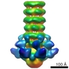
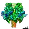
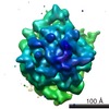
| Movie |
 Movie viewer Movie viewer |
|---|---|
| Structure viewer | EM map: SurfViewMolmilJmol/JSmol |
| Supplemental images |
- Downloads & links
-EMDB archive
| Map data | emd_1706.map.gz | 625.4 KB | EMDB map data format | |
|---|---|---|---|---|
| Header (meta data) | emd-1706-v30.xmlemd-1706.xml | 10.1 KB 10.1 KB | Display Display | EMDB header |
| Images |  1706.png 1706.png | 360.1 KB | ||
| Archive directory |  http://ftp.pdbj.org/pub/emdb/structures/EMD-1706ftp://ftp.pdbj.org/pub/emdb/structures/EMD-1706 http://ftp.pdbj.org/pub/emdb/structures/EMD-1706ftp://ftp.pdbj.org/pub/emdb/structures/EMD-1706 | HTTPS FTP |
-Related structure data


-Links
| EMDB pages | EMDB (EBI/PDBe) / EMDataResource |
|---|
-Map
| File | Download / File: emd_1706.map.gz / Format: CCP4 / Size: 8.2 MB / Type: IMAGE STORED AS FLOATING POINT NUMBER (4 BYTES) | ||||||||||||||||||||||||||||||||||||||||||||||||||||||||||||||||||||
|---|---|---|---|---|---|---|---|---|---|---|---|---|---|---|---|---|---|---|---|---|---|---|---|---|---|---|---|---|---|---|---|---|---|---|---|---|---|---|---|---|---|---|---|---|---|---|---|---|---|---|---|---|---|---|---|---|---|---|---|---|---|---|---|---|---|---|---|---|---|
| Annotation | Cryo-EM map of p2 phage baseplate | ||||||||||||||||||||||||||||||||||||||||||||||||||||||||||||||||||||
| Projections & slices | Image control
Images are generated by Spider. | ||||||||||||||||||||||||||||||||||||||||||||||||||||||||||||||||||||
| Voxel size | X=Y=Z: 4.37 Å | ||||||||||||||||||||||||||||||||||||||||||||||||||||||||||||||||||||
| Density |
| ||||||||||||||||||||||||||||||||||||||||||||||||||||||||||||||||||||
| Symmetry | Space group: 1 | ||||||||||||||||||||||||||||||||||||||||||||||||||||||||||||||||||||
| Details | EMDB XML:
CCP4 map header:
| ||||||||||||||||||||||||||||||||||||||||||||||||||||||||||||||||||||
 Z (Sec.)
 Y (Row.)
 X (Col.)


-Supplemental data
- Sample components
-Entire : Over-expressed Lactococcal phage p2 baseplate
| Entire | Name: Over-expressed Lactococcal phage p2 baseplate |
|---|---|
| Components |
|
-Supramolecule #1000: Over-expressed Lactococcal phage p2 baseplate
| Supramolecule | Name: Over-expressed Lactococcal phage p2 baseplate / type: sample / ID: 1000 Oligomeric state: One homotrimer of ORF16 binds to two hexamers of ORF15 and binds to six homotrimers of ORF18 Number unique components: 1 |
|---|---|
| Molecular weight | Experimental: 1 MDa |
-Macromolecule #1: p2 Lactoccocal phage baseplate
| Macromolecule | Name: p2 Lactoccocal phage baseplate / type: protein_or_peptide / ID: 1 / Name.synonym: p2 baseplate / Details: Complexes was cross-linked by glutaraldehyde / Recombinant expression: Yes |
|---|---|
| Source (natural) | Organism: Lactococcus lactis phage p2 (virus) |
| Molecular weight | Experimental: 1 MDa |
| Recombinant expression | Organism:  |
-Experimental details
-Structure determination
| Method | cryo EM |
|---|---|
 Processing Processing | single particle reconstruction |
| Aggregation state | particle |
-Sample preparation
| Concentration | 0.07 mg/mL |
|---|---|
| Buffer | pH: 7.5 / Details: 10mM HEPES, 150mM NaCl, 0.02% Glutaraldehyde |
| Grid | Details: R2.2 Quantifoil |
| Vitrification | Cryogen name: ETHANE / Chamber humidity: 92 % / Chamber temperature: 103.15 K / Instrument: GATAN CRYOPLUNGE 3 / Details: Vitrification instrument: CP3 Gatan / Method: Blot for 1 second before plunging |
- Electron microscopy
| Microscope | JEOL 2200FS |
|---|---|
| Temperature | Average: 98.15 K |
| Alignment procedure | Legacy - Astigmatism: Objective lens astigmatism was corrected at 200,000 times magnification |
| Specialist optics | Energy filter - Name: Omega / Energy filter - Lower energy threshold: 0.0 eV / Energy filter - Upper energy threshold: 20.0 eV |
| Image recording | Category: FILM / Film or detector model: KODAK SO-163 FILM / Digitization - Scanner: NIKON SUPER COOLSCAN 9000 / Digitization - Sampling interval: 10 µm / Number real images: 46 / Average electron dose: 18 e/Å2 / Bits/pixel: 8 |
| Electron beam | Acceleration voltage: 200 kV / Electron source:  FIELD EMISSION GUN FIELD EMISSION GUN |
| Electron optics | Calibrated magnification: 45710 / Illumination mode: FLOOD BEAM / Imaging mode: BRIGHT FIELD / Cs: 2.0 mm / Nominal defocus max: 2.6 µm / Nominal defocus min: 1.2 µm / Nominal magnification: 50000 |
| Sample stage | Specimen holder: Eucentric / Specimen holder model: GATAN LIQUID NITROGEN |
-Image processing
| CTF correction | Details: Each particle |
|---|---|
| Final reconstruction | Applied symmetry - Point group: C6 (6 fold cyclic) / Resolution.type: BY AUTHOR / Resolution: 26.0 Å / Resolution method: FSC 0.5 CUT-OFF / Software - Name: IMAGIC V |
| Final angle assignment | Details: beta 0 degrees, gamma 90 |
| Final two d classification | Number classes: 263 |
