 Movie
Movie Controller

[English] 日本語
 Yorodumi
- EMDB-1700: Negative stain EM structure of human native COP9 Signalosome (CSN) -
+ Open data

- Basic information
| Entry | Database: EMDB / ID: EMD-1700 | |||||||||
|---|---|---|---|---|---|---|---|---|---|---|
| Title | Negative stain EM structure of human native COP9 Signalosome (CSN) | |||||||||
 Map data Map data | Apo CSN map | |||||||||
 Sample Sample |
| |||||||||
| Biological species |  Homo sapiens (human) Homo sapiens (human) | |||||||||
| Method | single particle reconstruction / negative staining / Resolution: 25.0 Å | |||||||||
 Authors Authors | Enchev R / Schreiber A / Beuron F / Morris E | |||||||||
 Citation Citation | Journal: Structure / Year: 2010 Title: Structural insights into the COP9 signalosome and its common architecture with the 26S proteasome lid and eIF3. Authors: Radoslav I Enchev / Anne Schreiber / Fabienne Beuron / Edward P Morris /  Abstract: The evolutionary conserved COP9 signalosome (CSN), a large multisubunit complex, plays a central role in regulating ubiquitination and cell signaling. Here we report recombinant insect cell ...The evolutionary conserved COP9 signalosome (CSN), a large multisubunit complex, plays a central role in regulating ubiquitination and cell signaling. Here we report recombinant insect cell expression and two-step purification of human CSN and demonstrate its functional assembly. We further obtain a three-dimensional structure of both native and recombinant CSN using electron microscopy and single particle analysis. Antibody labeling of CSN5 and segmentation of the structure suggest a likely subunit distribution and the architecture of its helical repeat subunits is revealed. We compare the structure of CSN with its homologous complexes, the 26S proteasome lid and eIF3, and propose a conserved architecture implying similar assembly pathways and/or conserved substrate interaction modes. | |||||||||
| History |
|
- Structure visualization
| Movie |
 Movie viewer Movie viewer |
|---|---|
| Structure viewer | EM map: SurfViewMolmilJmol/JSmol |
| Supplemental images |

- Downloads & links
-EMDB archive
| Map data | emd_1700.map.gz | 6.9 MB | EMDB map data format | |
|---|---|---|---|---|
| Header (meta data) | emd-1700-v30.xmlemd-1700.xml | 8.3 KB 8.3 KB | Display Display | EMDB header |
| Images | 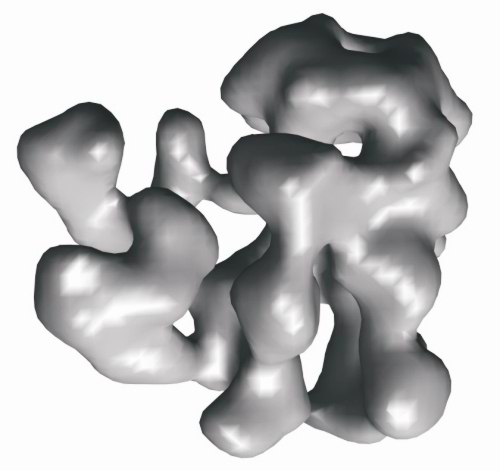 1700.jpg 1700.jpg | 54.9 KB | ||
| Archive directory |  http://ftp.pdbj.org/pub/emdb/structures/EMD-1700ftp://ftp.pdbj.org/pub/emdb/structures/EMD-1700 http://ftp.pdbj.org/pub/emdb/structures/EMD-1700ftp://ftp.pdbj.org/pub/emdb/structures/EMD-1700 | HTTPS FTP |
-Related structure data
| Similar structure data |
|---|


-Links
| EMDB pages | EMDB (EBI/PDBe) / EMDataResource |
|---|
-Map
| File | Download / File: emd_1700.map.gz / Format: CCP4 / Size: 7.8 MB / Type: IMAGE STORED AS FLOATING POINT NUMBER (4 BYTES) | ||||||||||||||||||||||||||||||||||||||||||||||||||||||||||||||||||||
|---|---|---|---|---|---|---|---|---|---|---|---|---|---|---|---|---|---|---|---|---|---|---|---|---|---|---|---|---|---|---|---|---|---|---|---|---|---|---|---|---|---|---|---|---|---|---|---|---|---|---|---|---|---|---|---|---|---|---|---|---|---|---|---|---|---|---|---|---|---|
| Annotation | Apo CSN map | ||||||||||||||||||||||||||||||||||||||||||||||||||||||||||||||||||||
| Projections & slices | Image control
Images are generated by Spider. | ||||||||||||||||||||||||||||||||||||||||||||||||||||||||||||||||||||
| Voxel size | X=Y=Z: 3.47 Å | ||||||||||||||||||||||||||||||||||||||||||||||||||||||||||||||||||||
| Density |
| ||||||||||||||||||||||||||||||||||||||||||||||||||||||||||||||||||||
| Symmetry | Space group: 1 | ||||||||||||||||||||||||||||||||||||||||||||||||||||||||||||||||||||
| Details | EMDB XML:
CCP4 map header:
| ||||||||||||||||||||||||||||||||||||||||||||||||||||||||||||||||||||
 Z (Sec.)
 Y (Row.)
 X (Col.)


-Supplemental data
- Sample components
-Entire : Human erythrocyte COP9 Signalosome
| Entire | Name: Human erythrocyte COP9 Signalosome |
|---|---|
| Components |
|
-Supramolecule #1000: Human erythrocyte COP9 Signalosome
| Supramolecule | Name: Human erythrocyte COP9 Signalosome / type: sample / ID: 1000 / Oligomeric state: Heterooctamer / Number unique components: 1 |
|---|---|
| Molecular weight | Experimental: 321 KDa / Theoretical: 321 KDa / Method: Native mass spectrometry |
-Macromolecule #1: Cop9 Signalosome
| Macromolecule | Name: Cop9 Signalosome / type: protein_or_peptide / ID: 1 / Name.synonym: CSN / Number of copies: 1 / Oligomeric state: Octamer / Recombinant expression: No |
|---|---|
| Source (natural) | Organism: Homo sapiens (human) / synonym: Human / Cell: Erythrocytes / Location in cell: Nucleus |
-Experimental details
-Structure determination
| Method | negative staining |
|---|---|
 Processing Processing | single particle reconstruction |
| Aggregation state | particle |
-Sample preparation
| Concentration | 0.1 mg/mL |
|---|---|
| Buffer | pH: 7.8 Details: 15 mM HEPES pH 7.8, 200 mM NaCl, 1% glycerol and 2 mM DTT |
| Staining | Type: NEGATIVE Details: Sample was applied to glow-discharged quantifoil grids coated with thin carbon, washed with buffer and stained with 2% uranyl acetate |
| Grid | Details: Quantifoil 400 mesh R2/2 |
| Vitrification | Cryogen name: NONE / Instrument: OTHER |
- Electron microscopy
| Microscope | FEI TECNAI F20 |
|---|---|
| Image recording | Category: CCD / Film or detector model: GENERIC TVIPS (4k x 4k) / Digitization - Sampling interval: 3.47 µm / Average electron dose: 100 e/Å2 Details: Initial selection was made manually from more than 100 2048x2048 CCD frames |
| Tilt angle min | 0 |
| Tilt angle max | 0 |
| Electron beam | Acceleration voltage: 200 kV / Electron source:  FIELD EMISSION GUN FIELD EMISSION GUN |
| Electron optics | Illumination mode: FLOOD BEAM / Imaging mode: BRIGHT FIELD / Nominal defocus max: 1.5 µm / Nominal defocus min: 1.0 µm / Nominal magnification: 50000 |
| Sample stage | Specimen holder: Eucentric / Specimen holder model: SIDE ENTRY, EUCENTRIC |
| Experimental equipment |  Model: Tecnai F20 / Image courtesy: FEI Company |
-Image processing
| Final reconstruction | Applied symmetry - Point group: C1 (asymmetric) / Algorithm: OTHER / Resolution.type: BY AUTHOR / Resolution: 25.0 Å / Resolution method: FSC 0.5 CUT-OFF / Software - Name: IMAGIC, SPIDER, EMAN / Number images used: 7877 |
|---|
