- EMDB-1699: Structure of Lactococcal Phage p2 Baseplate and its Mechanism of ... -
+
データを開く
IDまたはキーワード:
読み込み中...
-
基本情報
登録情報
データベース: EMDB / ID: EMD-1699
タイトル
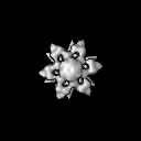
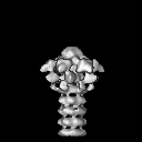
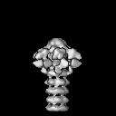
Structure of Lactococcal Phage p2 Baseplate and its Mechanism of Activation
マップデータ
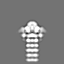
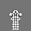
This is the ccp4 file of the EM 3D reconstruction of the baseplate of the wild-type p2 bacteriophage. The map is associated to the following PDB entries: PDB: 2WZP: BP closed form PDB: 2X53: BP Activated form C2 PDB: 2X54 + 2X5A: BP Activated form P2
試料
試料: P2 baseplate wild-type
タンパク質・ペプチド: P2 baseplate
キーワード
p2 / baseplate / phage / EM
機能・相同性
機能・相同性情報
symbiont genome ejection through host cell envelope, long flexible tail mechanism / virus tail, baseplate / virus tail / entry receptor-mediated virion attachment to host cell / cell adhesion / symbiont entry into host cell / virion attachment to host cell 類似検索 - 分子機能
Phage tail base-plate attachment protein ORF16 / Baseplate protein Gp15-like / Baseplate protein Gp16, domain D4 / Baseplate protein Gp16, domain 1 and 2 / : / : / : / : / Distal tail protein, N-terminal domain / Phage tail base-plate attachment protein N-terminal barrel domain ...Phage tail base-plate attachment protein ORF16 / Baseplate protein Gp15-like / Baseplate protein Gp16, domain D4 / Baseplate protein Gp16, domain 1 and 2 / : / : / : / : / Distal tail protein, N-terminal domain / Phage tail base-plate attachment protein N-terminal barrel domain / Phage tail base-plate attachment protein C-terminal insertion domain / Phage tail base-plate attachment protein N0 domain / Phage tail base-plate attachment protein C-terminal barrel domain / Dit, C-terminal domain / : / Lactococcus phage p2, Receptor binding protein, neck domain / : / Lactophage receptor-binding protein N-terminal shoulder domain / Receptor-binding protein of phage tail base-plate Siphoviridae, head / Lactophage receptor-binding protein C-terminal head domain / Adenovirus pIV-like, attachment domain 類似検索 - ドメイン・相同性
Baseplate protein gp16 / Distal tail protein / Receptor binding protein 類似検索 - 構成要素
ジャーナル: Proc Natl Acad Sci U S A / 年: 2010 タイトル: Structure of lactococcal phage p2 baseplate and its mechanism of activation. 著者: Giuliano Sciara / Cecilia Bebeacua / Patrick Bron / Denise Tremblay / Miguel Ortiz-Lombardia / Julie Lichière / Marin van Heel / Valérie Campanacci / Sylvain Moineau / Christian Cambillau / 要旨: Siphoviridae is the most abundant viral family on earth which infects bacteria as well as archaea. All known siphophages infecting gram+ Lactococcus lactis possess a baseplate at the tip of their ...Siphoviridae is the most abundant viral family on earth which infects bacteria as well as archaea. All known siphophages infecting gram+ Lactococcus lactis possess a baseplate at the tip of their tail involved in host recognition and attachment. Here, we report analysis of the p2 phage baseplate structure by X-ray crystallography and electron microscopy and propose a mechanism for the baseplate activation during attachment to the host cell. This approximately 1 MDa, Escherichia coli-expressed baseplate is composed of three protein species, including six trimers of the receptor-binding protein (RBP). RBPs host-recognition domains point upwards, towards the capsid, in agreement with the electron-microscopy map of the free virion. In the presence of Ca(2+), a cation mandatory for infection, the RBPs rotated 200 degrees downwards, presenting their binding sites to the host, and a channel opens at the bottom of the baseplate for DNA passage. These conformational changes reveal a novel siphophage activation and host-recognition mechanism leading ultimately to DNA ejection.
ダウンロード / ファイル: emd_1699.map.gz / 形式: CCP4 / 大きさ: 7.8 MB / タイプ: IMAGE STORED AS FLOATING POINT NUMBER (4 BYTES)
注釈
This is the ccp4 file of the EM 3D reconstruction of the baseplate of the wild-type p2 bacteriophage. The map is associated to the following PDB entries: PDB: 2WZP: BP closed form PDB: 2X53: BP Activated form C2 PDB: 2X54 + 2X5A: BP Activated form P2
タイプ: NEGATIVE 詳細: Sample was incubated on glow-discharged grid for approximately one minute. 2% uranyl acetate was applied onto the sample and left for about 30 seconds.
凍結
凍結剤: NONE / 装置: OTHER
-
電子顕微鏡法
顕微鏡
FEI/PHILIPS CM200FEG/UT
アライメント法
Legacy - 非点収差: corrected at 200,000 times magnification
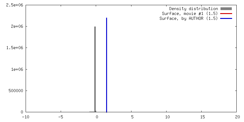
想定した対称性 - 点群: C6 (6回回転対称) / アルゴリズム: OTHER / 解像度のタイプ: BY AUTHOR / 解像度: 22.0 Å / 解像度の算出法: OTHER / ソフトウェア - 名称: IMAGIC-5 詳細: Initial map calculated with class averages. Final map calculated after projection matching refinement. 使用した粒子像数: 9486
 ムービー
ムービー コントローラー


 データを開く
 基本情報
基本情報 マップデータ
 試料
試料 キーワード
キーワード 機能・相同性情報
 Lactococcus phage p2 (ウイルス)
 データ登録者
 引用
引用
 構造の表示
構造の表示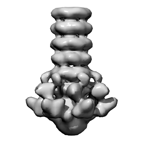
 ダウンロードとリンク
 3d_p2baseplate.png
 http://ftp.pdbj.org/pub/emdb/structures/EMD-1699


 Z (Sec.)
 Y (Row.)
 X (Col.)


 試料の構成要素
 解析
解析 電子顕微鏡法
 FIELD EMISSION GUN