[English] 日本語
 Yorodumi
- EMDB-1697: Outer dynein arms with a microtubule doublet in the presence of A... -
+ Open data
- Basic information
| Entry | Database: EMDB / ID: EMD-1697 | |||||||||
|---|---|---|---|---|---|---|---|---|---|---|
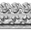
| Title | Outer dynein arms with a microtubule doublet in the presence of ADP.Vanadate | |||||||||
 Map data Map data | This is an image of outer dynein arms with a microtubule doublet in the presence of ADP.Vanadate | |||||||||
 Sample Sample |
| |||||||||
 Keywords Keywords | Dynein / flagella / ATP / tomography / motility / tubulin / microtubule | |||||||||
| Biological species |  | |||||||||
| Method | subtomogram averaging / cryo EM / Resolution: 39.0 Å | |||||||||
 Authors Authors | Movassagh T / Bui KH / Sakakibara H / Oiwa K / Ishikawa T | |||||||||
 Citation Citation |  Journal: Nat Struct Mol Biol / Year: 2010 Journal: Nat Struct Mol Biol / Year: 2010Title: Nucleotide-induced global conformational changes of flagellar dynein arms revealed by in situ analysis. Authors: Tandis Movassagh / Khanh Huy Bui / Hitoshi Sakakibara / Kazuhiro Oiwa / Takashi Ishikawa /  Abstract: Outer and inner dynein arms generate force for the flagellar/ciliary bending motion. Although nucleotide-induced structural change of dynein heavy chains (the ATP-driven motor) was proven in vitro, ...Outer and inner dynein arms generate force for the flagellar/ciliary bending motion. Although nucleotide-induced structural change of dynein heavy chains (the ATP-driven motor) was proven in vitro, our lack of knowledge in situ has precluded an understanding of the bending mechanism. Here we reveal nucleotide-induced global structural changes of the outer and inner dynein arms of Chlamydomonas reinhardtii flagella in situ using electron cryotomography. The ATPase domains of the dynein heavy chains move toward the distal end, and the N-terminal tail bends sharply during product release. This motion could drive the adjacent microtubule to cause a sliding motion. In contrast to in vitro results, in the presence of nucleotides, outer dynein arms coexist as clusters of apo or nucleotide-bound forms in situ. This implies a cooperative switching, which may be related to the mechanism of bending. | |||||||||
| History |
|
- Structure visualization
| Movie |
 Movie viewer Movie viewer |
|---|---|
| Structure viewer | EM map:  SurfView SurfView Molmil Molmil Jmol/JSmol Jmol/JSmol |
| Supplemental images |
- Downloads & links
-EMDB archive
| Map data |  emd_1697.map.gz emd_1697.map.gz | 2.7 MB |  EMDB map data format EMDB map data format | |
|---|---|---|---|---|
| Header (meta data) |  emd-1697-v30.xml emd-1697-v30.xml emd-1697.xml emd-1697.xml | 9.2 KB 9.2 KB | Display Display |  EMDB header EMDB header |
| Images |  1697ADPVi.jpg 1697ADPVi.jpg | 74.6 KB | ||
| Archive directory |  http://ftp.pdbj.org/pub/emdb/structures/EMD-1697 http://ftp.pdbj.org/pub/emdb/structures/EMD-1697 ftp://ftp.pdbj.org/pub/emdb/structures/EMD-1697 ftp://ftp.pdbj.org/pub/emdb/structures/EMD-1697 | HTTPS FTP |
-Related structure data
- Links
Links
| EMDB pages |  EMDB (EBI/PDBe) / EMDB (EBI/PDBe) /  EMDataResource EMDataResource |
|---|
- Map
Map
| File |  Download / File: emd_1697.map.gz / Format: CCP4 / Size: 3 MB / Type: IMAGE STORED AS FLOATING POINT NUMBER (4 BYTES) Download / File: emd_1697.map.gz / Format: CCP4 / Size: 3 MB / Type: IMAGE STORED AS FLOATING POINT NUMBER (4 BYTES) | ||||||||||||||||||||||||||||||||||||||||||||||||||||||||||||||||||||
|---|---|---|---|---|---|---|---|---|---|---|---|---|---|---|---|---|---|---|---|---|---|---|---|---|---|---|---|---|---|---|---|---|---|---|---|---|---|---|---|---|---|---|---|---|---|---|---|---|---|---|---|---|---|---|---|---|---|---|---|---|---|---|---|---|---|---|---|---|---|
| Annotation | This is an image of outer dynein arms with a microtubule doublet in the presence of ADP.Vanadate | ||||||||||||||||||||||||||||||||||||||||||||||||||||||||||||||||||||
| Projections & slices | Image control
Images are generated by Spider. generated in cubic-lattice coordinate | ||||||||||||||||||||||||||||||||||||||||||||||||||||||||||||||||||||
| Voxel size | X=Y=Z: 6.85 Å | ||||||||||||||||||||||||||||||||||||||||||||||||||||||||||||||||||||
| Density |
| ||||||||||||||||||||||||||||||||||||||||||||||||||||||||||||||||||||
| Symmetry | Space group: 1 | ||||||||||||||||||||||||||||||||||||||||||||||||||||||||||||||||||||
| Details | EMDB XML:
CCP4 map header:
| ||||||||||||||||||||||||||||||||||||||||||||||||||||||||||||||||||||
-Supplemental data
- Sample components
-Entire : Outer dynein arm, microtubule doublet
| Entire | Name: Outer dynein arm, microtubule doublet |
|---|---|
| Components |
|
-Supramolecule #1000: Outer dynein arm, microtubule doublet
| Supramolecule | Name: Outer dynein arm, microtubule doublet / type: sample / ID: 1000 / Details: The sample was flagella in situ / Number unique components: 1 |
|---|
-Supramolecule #1: Flagella
| Supramolecule | Name: Flagella / type: organelle_or_cellular_component / ID: 1 / Name.synonym: Outer dynein arm and Microtubule doublet / Recombinant expression: No / Database: NCBI |
|---|---|
| Source (natural) | Organism:  |
-Experimental details
-Structure determination
| Method | cryo EM |
|---|---|
 Processing Processing | subtomogram averaging |
| Aggregation state | particle |
- Sample preparation
| Buffer | pH: 7.4 Details: 30 mM HEPES (pH 7.4), 5 mM MgSO4, 1 mM DTT, 1 mM EGTA, 50 mM Potassium Acetate, 0.5% (w/v) Polyethylene Glycol (MW 20,000), 0.1 mM ATP with 0.05 mM sodium Orthovanadate |
|---|---|
| Grid | Details: 300 mesh lacey carbon grid |
| Vitrification | Cryogen name: ETHANE / Chamber humidity: 95 % / Instrument: OTHER / Details: Vitrification instrument: Vitrobot / Method: Blot for 3 seconds before plunging |
- Electron microscopy
| Microscope | FEI TECNAI 20 |
|---|---|
| Temperature | Average: 100 K |
| Specialist optics | Energy filter - Name: GIF / Energy filter - Lower energy threshold: 10.0 eV / Energy filter - Upper energy threshold: 25.0 eV |
| Image recording | Category: CCD / Film or detector model: GENERIC GATAN / Digitization - Sampling interval: 6.85 µm / Number real images: 1200 / Average electron dose: 30 e/Å2 / Bits/pixel: 16 |
| Electron beam | Acceleration voltage: 200 kV / Electron source:  FIELD EMISSION GUN FIELD EMISSION GUN |
| Electron optics | Illumination mode: FLOOD BEAM / Imaging mode: BRIGHT FIELD |
| Sample stage | Specimen holder: Eucentric Gatan cryo / Specimen holder model: GATAN LIQUID NITROGEN / Tilt series - Axis1 - Min angle: -60 ° / Tilt series - Axis1 - Max angle: 60 ° |
- Image processing
| Details | Average number of projections used in the 3D reconstructions: 6097. Average number of class averages: 2. |
|---|---|
| Final reconstruction | Algorithm: OTHER / Resolution.type: BY AUTHOR / Resolution: 39.0 Å / Resolution method: FSC 0.5 CUT-OFF / Software - Name:  IMOD IMOD |
 Movie
Movie Controller


 Z (Sec.)
 Y (Row.)
 X (Col.)


