H2020 Marie Curie Actions of the European Commission
846688
European Union
Citation
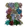
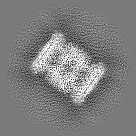
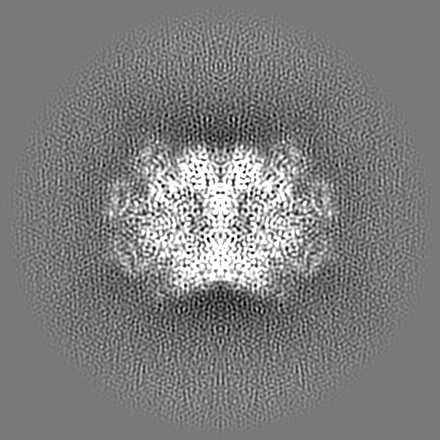
Journal: Nat Commun / Year: 2024 Title: Structural elucidation of recombinant Trichomonas vaginalis 20S proteasome bound to covalent inhibitors. Authors: Jan Silhan / Pavla Fajtova / Jitka Bartosova / Brianna M Hurysz / Jehad Almaliti / Yukiko Miyamoto / Lars Eckmann / William H Gerwick / Anthony J O'Donoghue / Evzen Boura / Abstract: The proteasome is a proteolytic enzyme complex essential for protein homeostasis in mammalian cells and protozoan parasites like Trichomonas vaginalis (Tv), the cause of the most common, non-viral ...The proteasome is a proteolytic enzyme complex essential for protein homeostasis in mammalian cells and protozoan parasites like Trichomonas vaginalis (Tv), the cause of the most common, non-viral sexually transmitted disease. Tv and other protozoan 20S proteasomes have been validated as druggable targets for antimicrobials. However, low yields and purity of the native proteasome have hindered studies of the Tv 20S proteasome (Tv20S). We address this challenge by creating a recombinant protozoan proteasome by expressing all seven α and seven β subunits of Tv20S alongside the Ump-1 chaperone in insect cells. The recombinant Tv20S displays biochemical equivalence to its native counterpart, confirmed by various assays. Notably, the marizomib (MZB) inhibits all catalytic subunits of Tv20S, while the peptide inhibitor carmaphycin-17 (CP-17) specifically targets β2 and β5. Cryo-electron microscopy (cryo-EM) unveils the structures of Tv20S bound to MZB and CP-17 at 2.8 Å. These findings explain MZB's low specificity for Tv20S compared to the human proteasome and demonstrate CP-17's higher specificity. Overall, these data provide a structure-based strategy for the development of specific Tv20S inhibitors to treat trichomoniasis.
Model: Quantifoil R2/1 / Material: COPPER / Mesh: 300 / Support film - Material: CARBON / Support film - topology: HOLEY ARRAY / Pretreatment - Type: GLOW DISCHARGE / Pretreatment - Time: 30 sec. / Pretreatment - Atmosphere: AIR / Pretreatment - Pressure: 0.015 kPa / Details: 15 mA
Vitrification
Cryogen name: ETHANE / Chamber humidity: 100 % / Chamber temperature: 277 K / Instrument: FEI VITROBOT MARK IV
-
Electron microscopy
Microscope
FEI TITAN KRIOS
Specialist optics
Energy filter - Name: TFS Selectris
Image recording
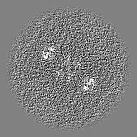
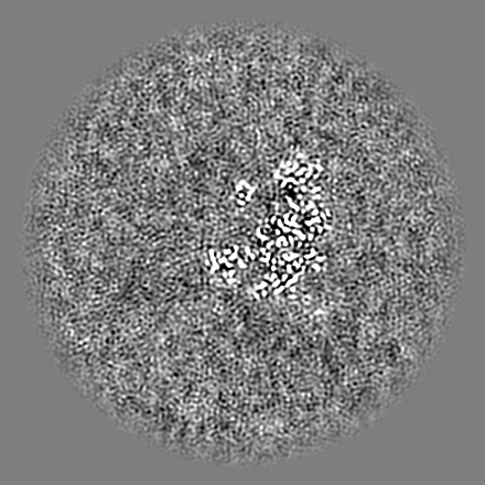
Film or detector model: FEI FALCON IV (4k x 4k) / Digitization - Dimensions - Width: 4096 pixel / Digitization - Dimensions - Height: 4096 pixel / Number grids imaged: 1 / Number real images: 7983 / Average electron dose: 50.0 e/Å2
Electron beam
Acceleration voltage: 300 kV / Electron source: FIELD EMISSION GUN
Electron optics
Illumination mode: OTHER / Imaging mode: BRIGHT FIELD / Nominal defocus max: 3.6 µm / Nominal defocus min: 0.8 µm
Type of model: INSILICO MODEL In silico model: a startup model was generated using AlphaFold predicted monomers that were superimposed onto 20S PDB:7ZYJ Details: 7ZYJ LEISHMANIA TARENTOLAE PROTEASOME 20S SUBUNIT IN COMPLEX WITH COMPOUND was used as a template for the superimposition of all 14 AlphaFold-generated models.
Final reconstruction
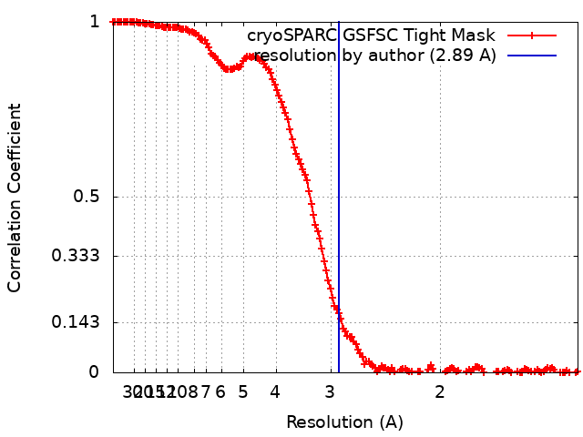
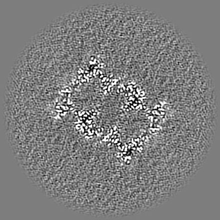
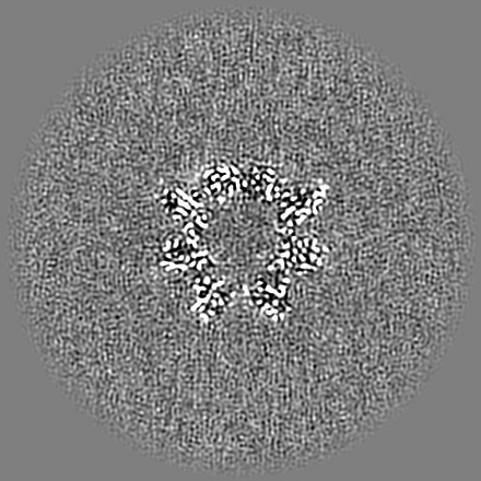
Applied symmetry - Point group: C2 (2 fold cyclic) / Resolution.type: BY AUTHOR / Resolution: 2.89 Å / Resolution method: FSC 0.143 CUT-OFF / Details: Standard Homo Refine job in Cryosparc v4 / Number images used: 14257
Initial angle assignment
Type: RANDOM ASSIGNMENT
Final angle assignment
Type: OTHER
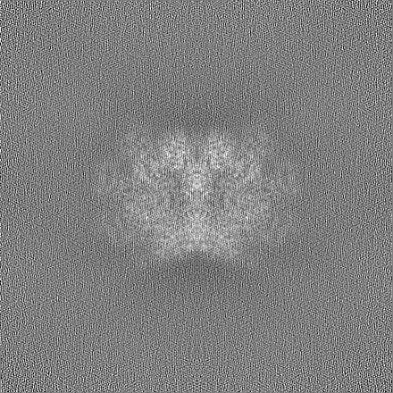
FSC plot (resolution estimation)
+
About Yorodumi
-
News
-
Feb 9, 2022. New format data for meta-information of EMDB entries
New format data for meta-information of EMDB entries
Version 3 of the EMDB header file is now the official format.
The previous official version 1.9 will be removed from the archive.
In the structure databanks used in Yorodumi, some data are registered as the other names, "COVID-19 virus" and "2019-nCoV". Here are the details of the virus and the list of structure data.
Jan 31, 2019. EMDB accession codes are about to change! (news from PDBe EMDB page)
EMDB accession codes are about to change! (news from PDBe EMDB page)
The allocation of 4 digits for EMDB accession codes will soon come to an end. Whilst these codes will remain in use, new EMDB accession codes will include an additional digit and will expand incrementally as the available range of codes is exhausted. The current 4-digit format prefixed with “EMD-” (i.e. EMD-XXXX) will advance to a 5-digit format (i.e. EMD-XXXXX), and so on. It is currently estimated that the 4-digit codes will be depleted around Spring 2019, at which point the 5-digit format will come into force.
The EM Navigator/Yorodumi systems omit the EMD- prefix.
Related info.:Q: What is EMD? / ID/Accession-code notation in Yorodumi/EM Navigator
Yorodumi is a browser for structure data from EMDB, PDB, SASBDB, etc.
This page is also the successor to EM Navigator detail page, and also detail information page/front-end page for Omokage search.
The word "yorodu" (or yorozu) is an old Japanese word meaning "ten thousand". "mi" (miru) is to see.
Related info.:EMDB / PDB / SASBDB / Comparison of 3 databanks / Yorodumi Search / Aug 31, 2016. New EM Navigator & Yorodumi / Yorodumi Papers / Jmol/JSmol / Function and homology information / Changes in new EM Navigator and Yorodumi
 Movie
Movie Controller

 Yorodumi
 Open data
 Basic information
 Map data
 Sample
 Keywords
 Function and homology information
 Trichomonas vaginalis G3 (eukaryote)
 Authors
 Citation

 Structure visualization
 Downloads & links
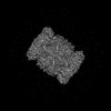
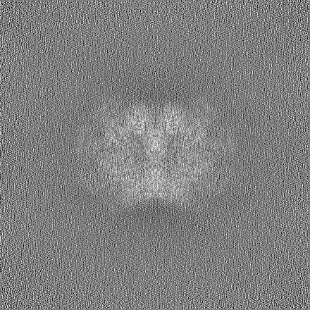
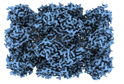 emd_16901.png
 http://ftp.pdbj.org/pub/emdb/structures/EMD-16901


 Z (Sec.)
 Y (Row.)
 X (Col.)


 Sample components
 Spodoptera frugiperda (fall armyworm)
 Processing
 Electron microscopy
 FIELD EMISSION GUN