 ムービー
ムービー コントローラー


+ データを開く

- 基本情報
基本情報
| 登録情報 |  | |||||||||
|---|---|---|---|---|---|---|---|---|---|---|
| タイトル | Subtomogram averaging structure of cofilactin filament inside microtubule lumen of Drosophila S2 cell protrusion. | |||||||||
 マップデータ マップデータ | Masked map at 16.5 A resolution. Sharpened with a B-factor of -500 A2. | |||||||||
 試料 試料 |
| |||||||||
 キーワード キーワード | Cytoskeleton / Filament / Actin / Cofilactin / CONTRACTILE PROTEIN | |||||||||
| 機能・相同性 |  機能・相同性情報 機能・相同性情報establishment of imaginal disc-derived wing hair orientation / establishment of ommatidial planar polarity / imaginal disc-derived leg segmentation / Gap junction degradation / Formation of annular gap junctions / EPHB-mediated forward signaling / EPH-ephrin mediated repulsion of cells / Cell-extracellular matrix interactions / RHOBTB2 GTPase cycle / RHOF GTPase cycle ...establishment of imaginal disc-derived wing hair orientation / establishment of ommatidial planar polarity / imaginal disc-derived leg segmentation / Gap junction degradation / Formation of annular gap junctions / EPHB-mediated forward signaling / EPH-ephrin mediated repulsion of cells / Cell-extracellular matrix interactions / RHOBTB2 GTPase cycle / RHOF GTPase cycle / VEGFA-VEGFR2 Pathway / ovarian fusome organization / Platelet degranulation / MAP2K and MAPK activation / RHO GTPases Activate WASPs and WAVEs / Regulation of actin dynamics for phagocytic cup formation / compound eye morphogenesis / DNA Damage Recognition in GG-NER / rhabdomere development / meiotic cytokinesis / mushroom body development / actomyosin contractile ring assembly / Clathrin-mediated endocytosis / border follicle cell migration / actin filament fragmentation / compound eye development / sperm individualization / centrosome separation / UCH proteinases / establishment of planar polarity / epithelial structure maintenance / maintenance of protein location in cell / brahma complex / actin filament severing / actin filament depolymerization / tube formation / regulation of lamellipodium assembly / Ino80 complex / lamellipodium assembly / female gonad development / mitotic cytokinesis / actin filament polymerization / axonogenesis / actin filament organization / positive regulation of protein secretion / 加水分解酵素; 酸無水物に作用; 酸無水物に作用・細胞または細胞小器官の運動に関与 / nuclear matrix / actin filament binding / actin cytoskeleton / actin binding / hydrolase activity / chromatin remodeling / nucleoplasm / ATP binding / cytoplasm / cytosol 類似検索 - 分子機能 | |||||||||
| 生物種 |  | |||||||||
| 手法 | サブトモグラム平均法 / クライオ電子顕微鏡法 / 解像度: 16.5 Å | |||||||||
 データ登録者 データ登録者 | Ventura Santos C / Carter AP | |||||||||
| 資金援助 |  英国, 2件 英国, 2件
| |||||||||
 引用 引用 | ジャーナル: bioRxiv / 年: 2023 タイトル: CryoET shows cofilactin filaments inside the microtubule lumen. 著者: Camilla Ventura Santos / Stephen L Rogers / Andrew P Carter / 要旨: Cytoplasmic microtubules are tubular polymers that can harbor small proteins or filaments inside their lumen. The identity of these objects and what causes their accumulation has not been ...Cytoplasmic microtubules are tubular polymers that can harbor small proteins or filaments inside their lumen. The identity of these objects and what causes their accumulation has not been conclusively established. Here, we used cryogenic electron tomography (cryoET) of S2 cell protrusions and found filaments inside the microtubule lumen, which resemble those reported recently in human HAP1 cells. The frequency of these filaments increased upon inhibition of the sarco/endoplasmic reticulum Ca ATPase (SERCA) with the small-molecule drug thapsigargin. Subtomogram averaging showed that the luminal filaments adopt a helical structure reminiscent of cofilin-bound actin (cofilactin). Consistent with this, cofilin was activated in cells under the same conditions that increased luminal filament occurrence. Furthermore, RNAi knock-down of cofilin reduced the frequency of luminal filaments with cofilactin morphology. These results suggest that cofilin activation stimulates its accumulation on actin filaments inside the microtubule lumen. | |||||||||
| 履歴 |
|
- 構造の表示
構造の表示
| 添付画像 |
|---|

- ダウンロードとリンク
-EMDBアーカイブ
| マップデータ | emd_16877.map.gz | 5.1 MB | EMDBマップデータ形式 | |
|---|---|---|---|---|
| ヘッダ (付随情報) | emd-16877-v30.xmlemd-16877.xml | 19.2 KB 19.2 KB | 表示 表示 | EMDBヘッダ |
| FSC (解像度算出) | emd_16877_fsc.xml | 9.2 KB | 表示 | FSCデータファイル |
| 画像 | 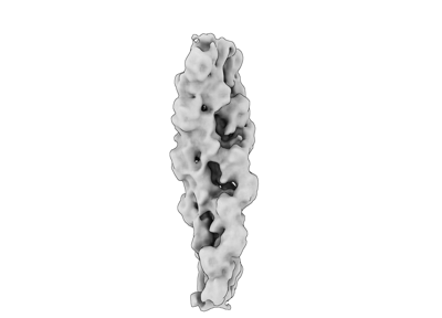 emd_16877.png emd_16877.png | 23.4 KB | ||
| マスクデータ | emd_16877_msk_1.map | 64 MB | マスクマップ | |
| Filedesc metadata | emd-16877.cif.gz | 6.2 KB | ||
| その他 | emd_16877_additional_1.map.gzemd_16877_half_map_1.map.gzemd_16877_half_map_2.map.gz | 59.6 MB 49.6 MB 49.6 MB | ||
| アーカイブディレクトリ |  http://ftp.pdbj.org/pub/emdb/structures/EMD-16877ftp://ftp.pdbj.org/pub/emdb/structures/EMD-16877 http://ftp.pdbj.org/pub/emdb/structures/EMD-16877ftp://ftp.pdbj.org/pub/emdb/structures/EMD-16877 | HTTPS FTP |
-検証レポート
| 文書・要旨 | emd_16877_validation.pdf.gz | 877 KB | 表示 | EMDB検証レポート |
|---|---|---|---|---|
| 文書・詳細版 | emd_16877_full_validation.pdf.gz | 876.6 KB | 表示 | |
| XML形式データ | emd_16877_validation.xml.gz | 16 KB | 表示 | |
| CIF形式データ | emd_16877_validation.cif.gz | 21.2 KB | 表示 | |
| アーカイブディレクトリ | https://ftp.pdbj.org/pub/emdb/validation_reports/EMD-16877ftp://ftp.pdbj.org/pub/emdb/validation_reports/EMD-16877 | HTTPS FTP |
-関連構造データ
-リンク
| EMDBのページ | EMDB (EBI/PDBe) / EMDataResource |
|---|---|
| 「今月の分子」の関連する項目 |


-マップ
| ファイル | ダウンロード / ファイル: emd_16877.map.gz / 形式: CCP4 / 大きさ: 64 MB / タイプ: IMAGE STORED AS FLOATING POINT NUMBER (4 BYTES) | ||||||||||||||||||||||||||||||||||||
|---|---|---|---|---|---|---|---|---|---|---|---|---|---|---|---|---|---|---|---|---|---|---|---|---|---|---|---|---|---|---|---|---|---|---|---|---|---|
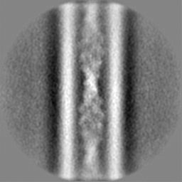
| 注釈 | Masked map at 16.5 A resolution. Sharpened with a B-factor of -500 A2. | ||||||||||||||||||||||||||||||||||||
| 投影像・断面図 | 画像のコントロール
画像は Spider により作成 | ||||||||||||||||||||||||||||||||||||
| ボクセルのサイズ | X=Y=Z: 2.83 Å | ||||||||||||||||||||||||||||||||||||
| 密度 |
| ||||||||||||||||||||||||||||||||||||
| 対称性 | 空間群: 1 | ||||||||||||||||||||||||||||||||||||
| 詳細 | EMDB XML:
|
 Z (Sec.)
 Y (Row.)
 X (Col.)


-添付データ
-マスク #1
| ファイル | emd_16877_msk_1.map | ||||||||||||
|---|---|---|---|---|---|---|---|---|---|---|---|---|---|
| 投影像・断面図 |
| ||||||||||||
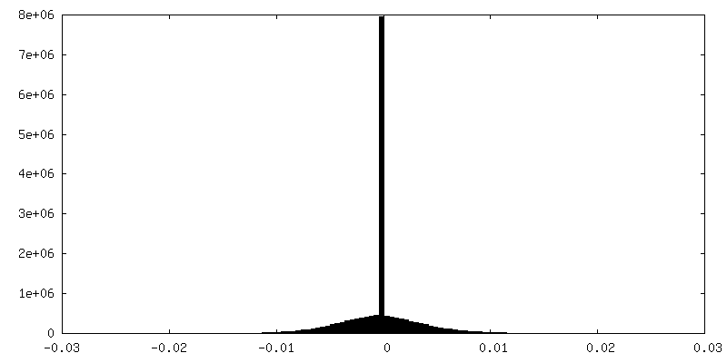
| 密度ヒストグラム |


-追加マップ: Unmasked map at 16.5 A resolution. Sharpened with...
| ファイル | emd_16877_additional_1.map | ||||||||||||
|---|---|---|---|---|---|---|---|---|---|---|---|---|---|
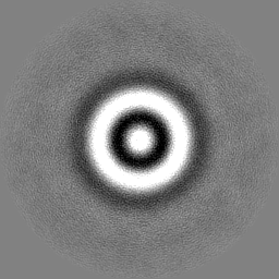
| 注釈 | Unmasked map at 16.5 A resolution. Sharpened with a B-factor of -500 A2. | ||||||||||||
| 投影像・断面図 |
| ||||||||||||
| 密度ヒストグラム |


-ハーフマップ: Half map 2.
| ファイル | emd_16877_half_map_1.map | ||||||||||||
|---|---|---|---|---|---|---|---|---|---|---|---|---|---|
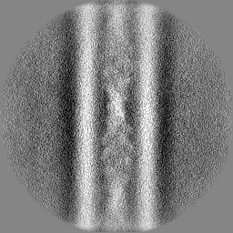
| 注釈 | Half map 2. | ||||||||||||
| 投影像・断面図 |
| ||||||||||||
| 密度ヒストグラム |


-ハーフマップ: Half map 1.
| ファイル | emd_16877_half_map_2.map | ||||||||||||
|---|---|---|---|---|---|---|---|---|---|---|---|---|---|
| 注釈 | Half map 1. | ||||||||||||
| 投影像・断面図 |
| ||||||||||||
| 密度ヒストグラム |


- 試料の構成要素
-全体 : Cofilactin filament inside the microtubule lumen of induced Droso...
| 全体 | 名称: Cofilactin filament inside the microtubule lumen of induced Drosophila S2 cell protrusion |
|---|---|
| 要素 |
|
-超分子 #1: Cofilactin filament inside the microtubule lumen of induced Droso...
| 超分子 | 名称: Cofilactin filament inside the microtubule lumen of induced Drosophila S2 cell protrusion タイプ: cell / ID: 1 / 親要素: 0 / 含まれる分子: all |
|---|---|
| 由来(天然) | 生物種: |
-分子 #1: Actin-5C
| 分子 | 名称: Actin-5C / タイプ: protein_or_peptide / ID: 1 / コピー数: 8 / 光学異性体: LEVO EC番号: 加水分解酵素; 酸無水物に作用; 酸無水物に作用・細胞または細胞小器官の運動に関与 |
|---|---|
| 由来(天然) | 生物種: |
| 分子量 | 理論値: 41.16098 KDa |
| 配列 | 文字列: AALVVDNGSG MCKAGFAGDD APRAVFPSIV GRPRHQGVMV GMGQKDSYVG DEAQSKRGIL TLKYPIEHGI VTNWDDMEKI WHHTFYNEL RVAPEEHPVL LTEAPLNPKA NREKMTQIMF ETFNTPAMYV AIQAVLSLYA SGRTTGIVLD SGDGVSHTVP I YEGYALPH ...文字列: AALVVDNGSG MCKAGFAGDD APRAVFPSIV GRPRHQGVMV GMGQKDSYVG DEAQSKRGIL TLKYPIEHGI VTNWDDMEKI WHHTFYNEL RVAPEEHPVL LTEAPLNPKA NREKMTQIMF ETFNTPAMYV AIQAVLSLYA SGRTTGIVLD SGDGVSHTVP I YEGYALPH AILRLDLAGR DLTDYLMKIL TERGYSFTTT AEREIVRDIK EKLCYVALDF EQEMATAASS SSLEKSYELP DG QVITIGN ERFRCPEALF QPSFLGMEAC GIHETTYNSI MKCDVDIRKD LYANTVLSGG TTMYPGIADR MQKEITALAP STM KIKIIA PPERKYSVWI GGSILASLST FQQMWISKQE YDESGPSIVH RKCF UniProtKB: Actin-5C |
-分子 #2: Cofilin/actin-depolymerizing factor homolog
| 分子 | 名称: Cofilin/actin-depolymerizing factor homolog / タイプ: protein_or_peptide / ID: 2 / コピー数: 6 / 光学異性体: LEVO |
|---|---|
| 由来(天然) | 生物種: |
| 分子量 | 理論値: 17.180529 KDa |
| 配列 | 文字列: MASGVTVSDV CKTTYEEIKK DKKHRYVIFY IRDEKQIDVE TVADRNAEYD QFLEDIQKCG PGECRYGLFD FEYMHQCQGT SESSKKQKL FLMSWCPDTA KVKKKMLYSS SFDALKKSLV GVQKYIQATD LSEASREAVE EKLRATDRQ UniProtKB: Cofilin/actin-depolymerizing factor homolog |
-実験情報
-構造解析
| 手法 | クライオ電子顕微鏡法 |
|---|---|
 解析 解析 | サブトモグラム平均法 |
| 試料の集合状態 | filament |
-試料調製
| 緩衝液 | pH: 7 |
|---|---|
| グリッド | モデル: Quantifoil R3.5/1 / 材質: GOLD / メッシュ: 200 / 前処理 - タイプ: GLOW DISCHARGE / 前処理 - 時間: 30 sec. 詳細: Quantifoil R3.5/1 Au200 grids were glow discharged for 30s at 20 - 30 mA and subsequently coated with 0.25 ug/mL Concanavalin Aater for 1 - 16 h at 37 degrees Celcius. |
| 凍結 | 凍結剤: ETHANE / チャンバー内湿度: 95 % / チャンバー内温度: 198.15 K / 装置: FEI VITROBOT MARK III |
- 電子顕微鏡法
| 顕微鏡 | FEI TITAN KRIOS |
|---|---|
| 特殊光学系 | エネルギーフィルター - 名称: GIF Bioquantum / エネルギーフィルター - スリット幅: 20 eV |
| 撮影 | フィルム・検出器のモデル: GATAN K2 SUMMIT (4k x 4k) 検出モード: COUNTING / 平均電子線量: 3.0 e/Å2 詳細: Data was collected on Gatan K2 summit (2.952 A/pixel) and Gatan K3 summit (2.659 A/pixel) with 3 degree increments (3 e/A2 dose per tilt). The total dose was between 118 and 122 e/A2. |
| 電子線 | 加速電圧: 300 kV / 電子線源:  FIELD EMISSION GUN FIELD EMISSION GUN |
| 電子光学系 | 照射モード: FLOOD BEAM / 撮影モード: BRIGHT FIELD / 最大 デフォーカス(公称値): 6.0 µm / 最小 デフォーカス(公称値): 2.5 µm |
| 実験機器 |  モデル: Titan Krios / 画像提供: FEI Company |
+画像解析

-原子モデル構築 1
| 詳細 | Drosophila cofilin and actin structures were predicted as a complex with Alphafold 2-Multimer. Actin from the predicted cofilin-actin complex was iteratively aligned with 8 actin subunits in the chicken cofilactin PDB model 5yU8. Two cofilin moieties at the pointed end were removed. Side chains were truncated. The model was not refined. |
|---|---|
| 得られたモデル |  PDB-8oh4: |
