 Movie
Movie Controller

[English] 日本語
 Yorodumi
- EMDB-16731: Alpha-ketoglutarate dehydrogenase complex core (E2, dihydrolipoyl... -
+ Open data

- Basic information
| Entry |  | |||||||||
|---|---|---|---|---|---|---|---|---|---|---|
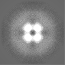
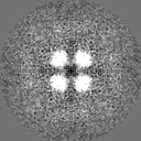
| Title | Alpha-ketoglutarate dehydrogenase complex core (E2, dihydrolipoyl transsuccinylase), Branched-chain alpha-ketoacid dehydrogenase complex core (E2, dihydrolipoyl transacylase), indistinguishable | |||||||||
 Map data Map data | ||||||||||
 Sample Sample |
| |||||||||
 Keywords Keywords | transferase / cube / mitochondrion | |||||||||
| Biological species |  | |||||||||
| Method | subtomogram averaging / cryo EM / Resolution: 32.0 Å | |||||||||
 Authors Authors | Plokhikh KS / Chesnokov YM | |||||||||
| Funding support | 1 items
| |||||||||
 Citation Citation | Journal: FEBS J / Year: 2024 Title: Association of 2-oxoacid dehydrogenase complexes with respirasomes in mitochondria. Authors: Konstantin S Plokhikh / Semen V Nesterov / Yuriy M Chesnokov / Anton G Rogov / Roman A Kamyshinsky / Aleksandr L Vasiliev / Lev S Yaguzhinsky / Raif G Vasilov /  Abstract: In the present study, cryo-electron tomography was used to investigate the localization of 2-oxoacid dehydrogenase complexes (OADCs) in cardiac mitochondria and mitochondrial inner membrane samples. ...In the present study, cryo-electron tomography was used to investigate the localization of 2-oxoacid dehydrogenase complexes (OADCs) in cardiac mitochondria and mitochondrial inner membrane samples. Two classes of ordered OADC inner cores with different symmetries were distinguished and their quaternary structures modeled. One class corresponds to pyruvate dehydrogenase complexes and the other to dehydrogenase complexes of α-ketoglutarate and branched-chain α-ketoacids. OADCs were shown to be localized in close proximity to membrane-embedded respirasomes, as observed both in densely packed lamellar cristae of cardiac mitochondria and in ruptured mitochondrial samples where the dense packing is absent. This suggests the specificity of the OADC-respirasome interaction, which allows localized NADH/NAD exchange between OADCs and complex I of the respiratory chain. The importance of this local coupling is based on OADCs being the link between respiration, glycolysis and amino acid metabolism. The coupling of these basic metabolic processes can vary in different tissues and conditions and may be involved in the development of various pathologies. The present study shows that this important and previously missing parameter of mitochondrial complex coupling can be successfully assessed using cryo-electron tomography. | |||||||||
| History |
|
- Structure visualization
| Supplemental images |
|---|

- Downloads & links
-EMDB archive
| Map data | emd_16731.map.gz | 367 KB |  EMDB map data format EMDB map data format | |
|---|---|---|---|---|
| Header (meta data) | emd-16731-v30.xmlemd-16731.xml | 14.4 KB 14.4 KB | Display Display | EMDB header |
| FSC (resolution estimation) | emd_16731_fsc.xml | 6 KB | Display | FSC data file |
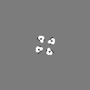
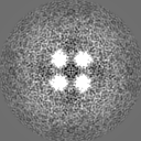
| Images | 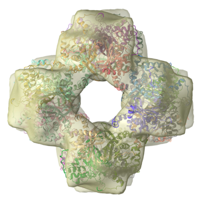 emd_16731.png emd_16731.png | 166.4 KB | ||
| Masks | emd_16731_msk_1.map | 8 MB | Mask map | |
| Filedesc metadata | emd-16731.cif.gz | 4.2 KB | ||
| Others | emd_16731_half_map_1.map.gzemd_16731_half_map_2.map.gz | 4.6 MB 4.6 MB | ||
| Archive directory |  http://ftp.pdbj.org/pub/emdb/structures/EMD-16731ftp://ftp.pdbj.org/pub/emdb/structures/EMD-16731 http://ftp.pdbj.org/pub/emdb/structures/EMD-16731ftp://ftp.pdbj.org/pub/emdb/structures/EMD-16731 | HTTPS FTP |
-Related structure data
-Links
| EMDB pages | EMDB (EBI/PDBe) / EMDataResource |
|---|
-Map
| File | Download / File: emd_16731.map.gz / Format: CCP4 / Size: 8 MB / Type: IMAGE STORED AS FLOATING POINT NUMBER (4 BYTES) | ||||||||||||||||||||||||||||||||||||
|---|---|---|---|---|---|---|---|---|---|---|---|---|---|---|---|---|---|---|---|---|---|---|---|---|---|---|---|---|---|---|---|---|---|---|---|---|---|
| Projections & slices | Image control
Images are generated by Spider. | ||||||||||||||||||||||||||||||||||||
| Voxel size | X=Y=Z: 3.7 Å | ||||||||||||||||||||||||||||||||||||
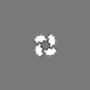
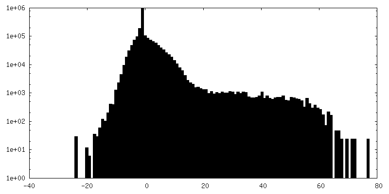
| Density |
| ||||||||||||||||||||||||||||||||||||
| Symmetry | Space group: 1 | ||||||||||||||||||||||||||||||||||||
| Details | EMDB XML:
|
 Z (Sec.)
 Y (Row.)
 X (Col.)


-Supplemental data
-Mask #1
| File | emd_16731_msk_1.map | ||||||||||||
|---|---|---|---|---|---|---|---|---|---|---|---|---|---|
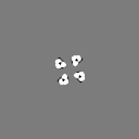
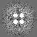
| Projections & Slices |
| ||||||||||||
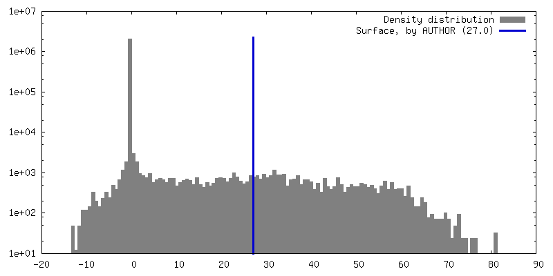
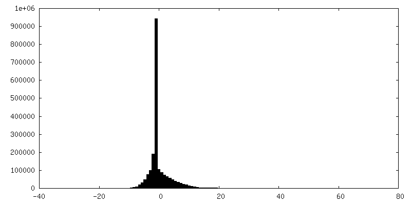
| Density Histograms |


-Half map: #1
| File | emd_16731_half_map_1.map | ||||||||||||
|---|---|---|---|---|---|---|---|---|---|---|---|---|---|
| Projections & Slices |
| ||||||||||||
| Density Histograms |


-Half map: #2
| File | emd_16731_half_map_2.map | ||||||||||||
|---|---|---|---|---|---|---|---|---|---|---|---|---|---|
| Projections & Slices |
| ||||||||||||
| Density Histograms |


- Sample components
-Entire : ruptured mitochondrial membrane samples
| Entire | Name: ruptured mitochondrial membrane samples |
|---|---|
| Components |
|
-Supramolecule #1: ruptured mitochondrial membrane samples
| Supramolecule | Name: ruptured mitochondrial membrane samples / type: organelle_or_cellular_component / ID: 1 / Parent: 0 / Macromolecule list: #1 |
|---|---|
| Source (natural) | Organism: |
-Experimental details
-Structure determination
| Method | cryo EM |
|---|---|
 Processing Processing | subtomogram averaging |
| Aggregation state | cell |
-Sample preparation
| Buffer | pH: 7.4 / Component - Concentration: 10.0 mM / Component - Name: Hepes/KOH |
|---|---|
| Grid | Model: EMS Lacey Carbon / Material: COPPER / Mesh: 300 / Support film - Material: CARBON / Support film - topology: LACEY / Pretreatment - Type: GLOW DISCHARGE / Pretreatment - Time: 30 sec. |
| Vitrification | Cryogen name: ETHANE / Chamber humidity: 100 % / Chamber temperature: 277 K / Instrument: FEI VITROBOT MARK IV |
- Electron microscopy
| Microscope | FEI TITAN KRIOS |
|---|---|
| Image recording | Film or detector model: FEI FALCON II (4k x 4k) / Average electron dose: 1.7 e/Å2 |
| Electron beam | Acceleration voltage: 300 kV / Electron source:  FIELD EMISSION GUN FIELD EMISSION GUN |
| Electron optics | C2 aperture diameter: 100.0 µm / Illumination mode: FLOOD BEAM / Imaging mode: BRIGHT FIELD / Nominal defocus max: 8.0 µm / Nominal defocus min: 6.0 µm / Nominal magnification: 18000 |
| Sample stage | Specimen holder model: FEI TITAN KRIOS AUTOGRID HOLDER / Cooling holder cryogen: NITROGEN |
| Experimental equipment |  Model: Titan Krios / Image courtesy: FEI Company |

