 ムービー
ムービー コントローラー


+ データを開く

- 基本情報
基本情報
| 登録情報 |  | |||||||||
|---|---|---|---|---|---|---|---|---|---|---|
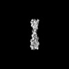
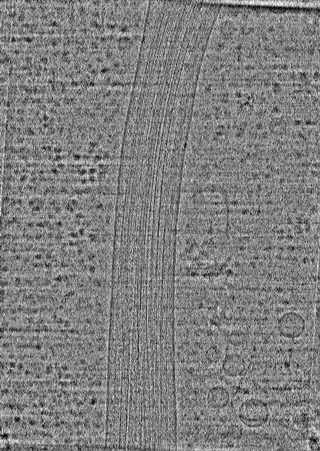
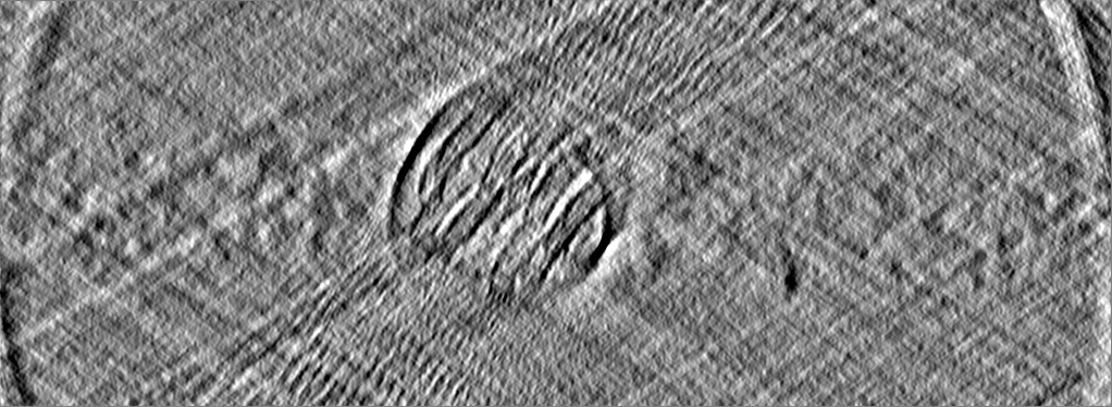
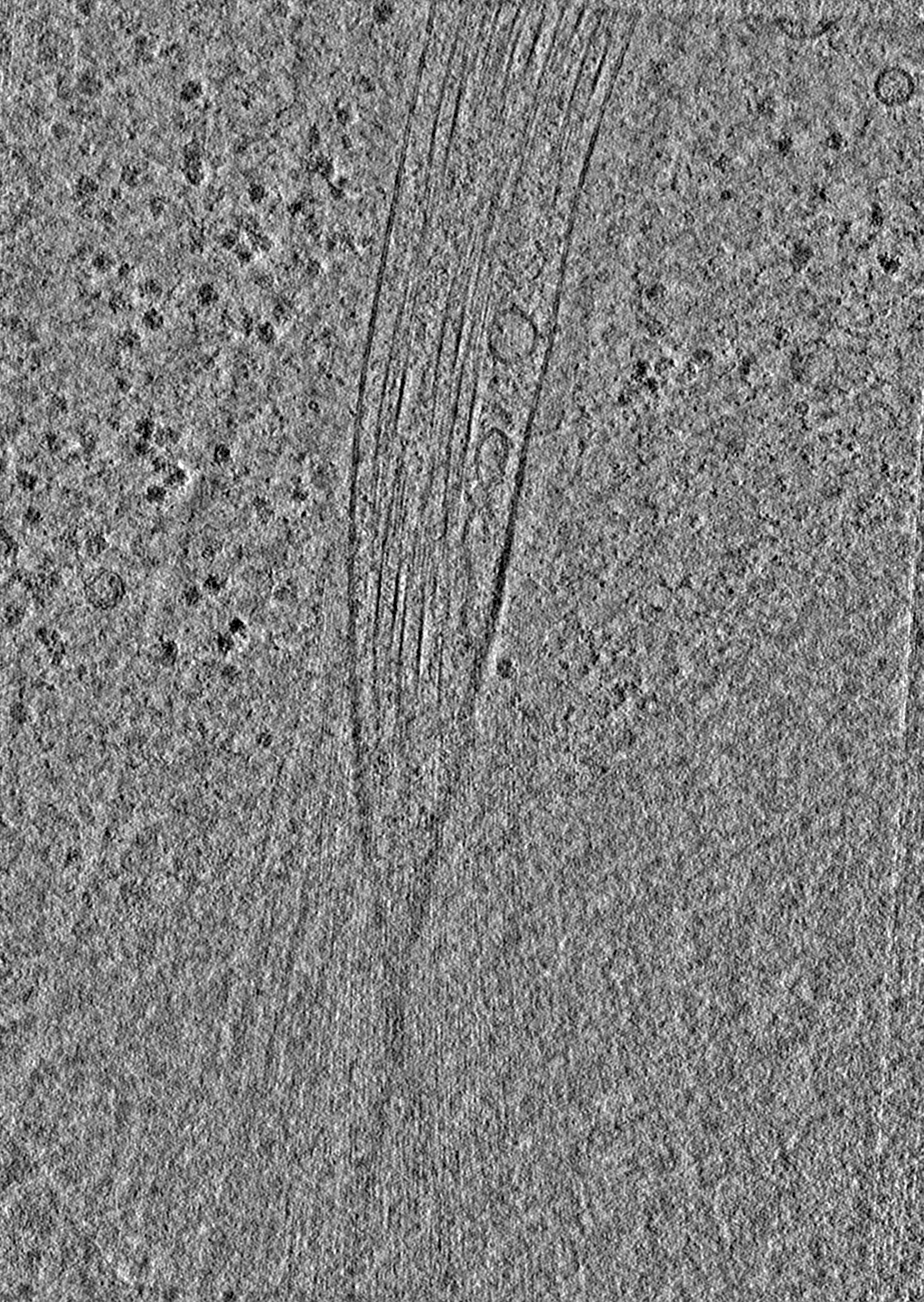
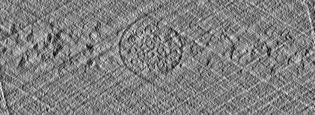
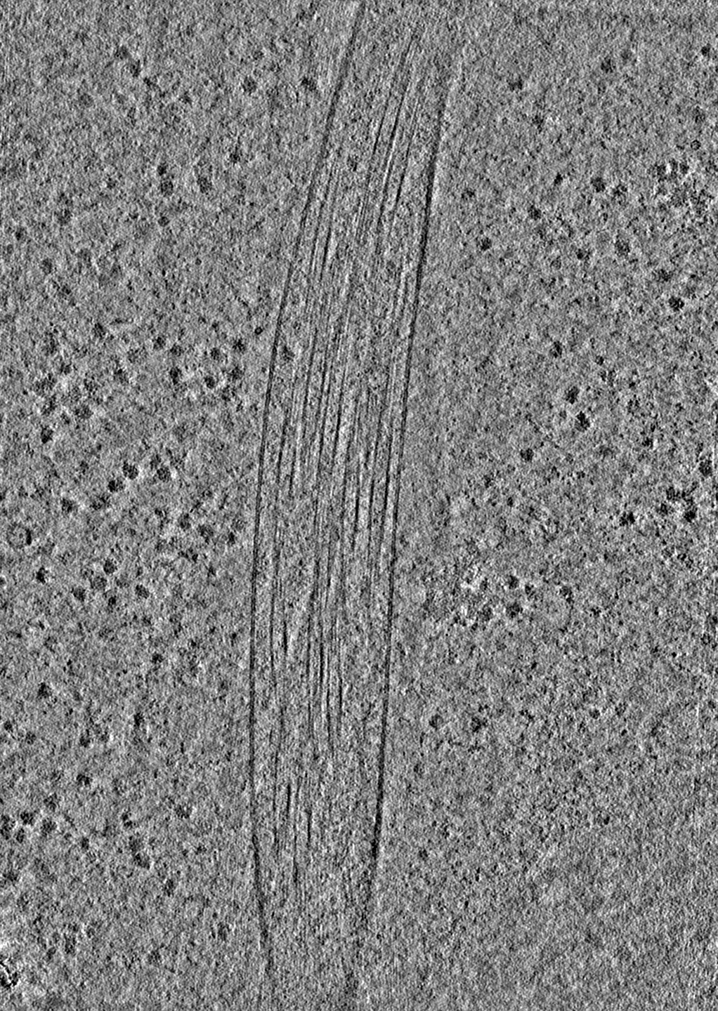
| タイトル | Tomogram of an induced protrusion of a Drosophila S2 cell with filaments inside the microtubule lumen. | |||||||||
 マップデータ マップデータ | Deconvolved tomogram (binned by four) of an induced protrusion from Drosophila S2 cells with multiple filaments inside the microtubule lumen. From Dataset 7. | |||||||||
 試料 試料 |
| |||||||||
| 生物種 |  | |||||||||
| 手法 | 電子線トモグラフィー法 / クライオ電子顕微鏡法 | |||||||||
 データ登録者 データ登録者 | Ventura Santos C / Carter AP | |||||||||
| 資金援助 |  英国, 2件 英国, 2件
| |||||||||
 引用 引用 | ジャーナル: bioRxiv / 年: 2023 タイトル: CryoET shows cofilactin filaments inside the microtubule lumen. 著者: Camilla Ventura Santos / Stephen L Rogers / Andrew P Carter / 要旨: Cytoplasmic microtubules are tubular polymers that can harbor small proteins or filaments inside their lumen. The identity of these objects and what causes their accumulation has not been ...Cytoplasmic microtubules are tubular polymers that can harbor small proteins or filaments inside their lumen. The identity of these objects and what causes their accumulation has not been conclusively established. Here, we used cryogenic electron tomography (cryoET) of S2 cell protrusions and found filaments inside the microtubule lumen, which resemble those reported recently in human HAP1 cells. The frequency of these filaments increased upon inhibition of the sarco/endoplasmic reticulum Ca ATPase (SERCA) with the small-molecule drug thapsigargin. Subtomogram averaging showed that the luminal filaments adopt a helical structure reminiscent of cofilin-bound actin (cofilactin). Consistent with this, cofilin was activated in cells under the same conditions that increased luminal filament occurrence. Furthermore, RNAi knock-down of cofilin reduced the frequency of luminal filaments with cofilactin morphology. These results suggest that cofilin activation stimulates its accumulation on actin filaments inside the microtubule lumen. | |||||||||
| 履歴 |
|
- 構造の表示
構造の表示
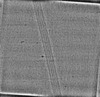
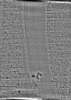
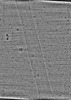
| 添付画像 |
|---|

- ダウンロードとリンク
-EMDBアーカイブ
| マップデータ | emd_16720.map.gz | 1.9 GB |  EMDBマップデータ形式 EMDBマップデータ形式 | |
|---|---|---|---|---|
| ヘッダ (付随情報) | emd-16720-v30.xmlemd-16720.xml | 9.9 KB 9.9 KB | 表示 表示 | EMDBヘッダ |
| 画像 | 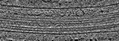 emd_16720.png emd_16720.png | 93.6 KB | ||
| アーカイブディレクトリ |  http://ftp.pdbj.org/pub/emdb/structures/EMD-16720ftp://ftp.pdbj.org/pub/emdb/structures/EMD-16720 http://ftp.pdbj.org/pub/emdb/structures/EMD-16720ftp://ftp.pdbj.org/pub/emdb/structures/EMD-16720 | HTTPS FTP |
-検証レポート
| 文書・要旨 | emd_16720_validation.pdf.gz | 531.8 KB | 表示 | EMDB検証レポート |
|---|---|---|---|---|
| 文書・詳細版 | emd_16720_full_validation.pdf.gz | 531.3 KB | 表示 | |
| XML形式データ | emd_16720_validation.xml.gz | 4.6 KB | 表示 | |
| CIF形式データ | emd_16720_validation.cif.gz | 5 KB | 表示 | |
| アーカイブディレクトリ | https://ftp.pdbj.org/pub/emdb/validation_reports/EMD-16720ftp://ftp.pdbj.org/pub/emdb/validation_reports/EMD-16720 | HTTPS FTP |
-関連構造データ

-リンク
| EMDBのページ | EMDB (EBI/PDBe) / EMDataResource |
|---|
-マップ
| ファイル | ダウンロード / ファイル: emd_16720.map.gz / 形式: CCP4 / 大きさ: 2.1 GB / タイプ: IMAGE STORED AS FLOATING POINT NUMBER (4 BYTES) | ||||||||||||||||||||||||||||||||
|---|---|---|---|---|---|---|---|---|---|---|---|---|---|---|---|---|---|---|---|---|---|---|---|---|---|---|---|---|---|---|---|---|---|
| 注釈 | Deconvolved tomogram (binned by four) of an induced protrusion from Drosophila S2 cells with multiple filaments inside the microtubule lumen. From Dataset 7. | ||||||||||||||||||||||||||||||||
| 投影像・断面図 | 画像のコントロール
画像は Spider により作成 これらの図は立方格子座標系で作成されたものです | ||||||||||||||||||||||||||||||||
| ボクセルのサイズ | X=Y=Z: 10.64 Å | ||||||||||||||||||||||||||||||||
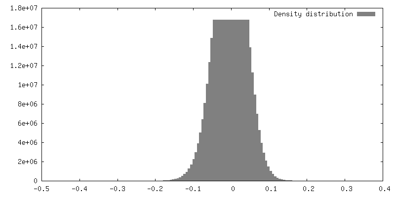
| 密度 |
| ||||||||||||||||||||||||||||||||
| 対称性 | 空間群: 1 | ||||||||||||||||||||||||||||||||
| 詳細 | EMDB XML:
|
 Z (Sec.)
 Y (Row.)
 X (Col.)


-添付データ
- 試料の構成要素
-全体 : Tomogram of an induced protrusion of a Drosophila S2 cell with fi...
| 全体 | 名称: Tomogram of an induced protrusion of a Drosophila S2 cell with filaments inside the microtubule lumen |
|---|---|
| 要素 |
|
-超分子 #1: Tomogram of an induced protrusion of a Drosophila S2 cell with fi...
| 超分子 | 名称: Tomogram of an induced protrusion of a Drosophila S2 cell with filaments inside the microtubule lumen タイプ: cell / ID: 1 / 親要素: 0 詳細: Cells were treated with 2.5 uM Cytochalasin D for 4 hours and with 2 uM thapsigargin for 5 hours before vitrification. Example tomogram from dataset 7. |
|---|---|
| 由来(天然) | 生物種: 株: S2 cells |
-実験情報
-構造解析
| 手法 | クライオ電子顕微鏡法 |
|---|---|
 解析 解析 | 電子線トモグラフィー法 |
| 試料の集合状態 | cell |
-試料調製
| 緩衝液 | pH: 7 |
|---|---|
| グリッド | モデル: Quantifoil / 材質: GOLD / メッシュ: 200 詳細: Grids were glow discharged for 30s at 20mA and then coated with 0.25 ug/mL Concanavalin A for at least 2 hours at 37 degrees. |
| 凍結 | 凍結剤: ETHANE / チャンバー内湿度: 95 % / チャンバー内温度: 298.15 K |
| 詳細 | Cells were treated with 2.5 uM Cytochalasin D for 4 hours and with 2 uM thapsigargin for 5 hours before vitrification. |
| 切片作成 | その他: NO SECTIONING |
| 位置合わせマーカー | Manufacturer: BBI Solutions / 直径: 10 nm |
- 電子顕微鏡法
| 顕微鏡 | FEI TITAN KRIOS |
|---|---|
| 特殊光学系 | エネルギーフィルター - 名称: GIF Bioquantum / エネルギーフィルター - スリット幅: 20 eV |
| 撮影 | フィルム・検出器のモデル: GATAN K3 BIOQUANTUM (6k x 4k) 平均電子線量: 3.0 e/Å2 詳細: Data was collected at a pixel size of 2.659 A/pixel with a total dose of 121.54 A/e^2. |
| 電子線 | 加速電圧: 300 kV / 電子線源:  FIELD EMISSION GUN FIELD EMISSION GUN |
| 電子光学系 | 照射モード: FLOOD BEAM / 撮影モード: BRIGHT FIELD / 最大 デフォーカス(公称値): 6.0 µm / 最小 デフォーカス(公称値): 2.5 µm |
| 実験機器 |  モデル: Titan Krios / 画像提供: FEI Company |
-画像解析
| 最終 再構成 | 使用した粒子像数: 41 |
|---|
