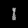
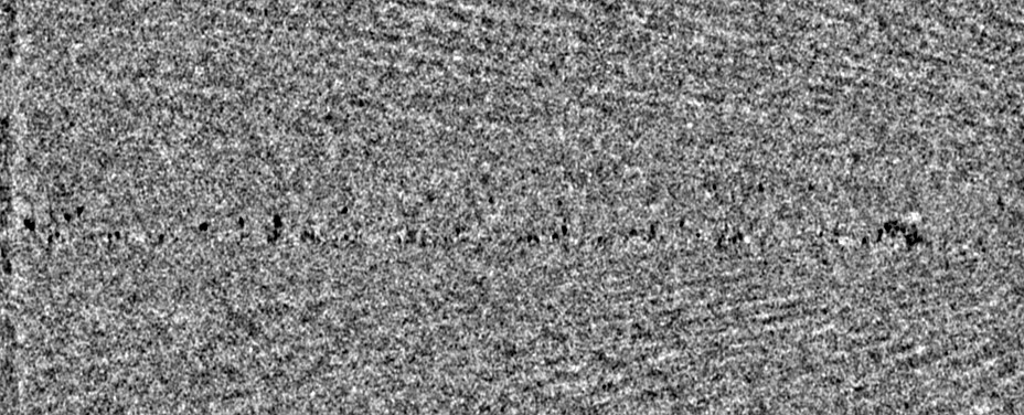
- EMDB-16695: Tomogram of an induced protrusion of a Drosophila S2 alpha-tubuli... -
+
データを開く
IDまたはキーワード:
読み込み中...
-
基本情報
登録情報
データベース: EMDB / ID: EMD-16695
タイトル
Tomogram of an induced protrusion of a Drosophila S2 alpha-tubulin acetyltransferase knock-out (dTAT KO) cell with a filament inside the microtubule lumen
マップデータ
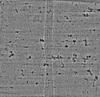
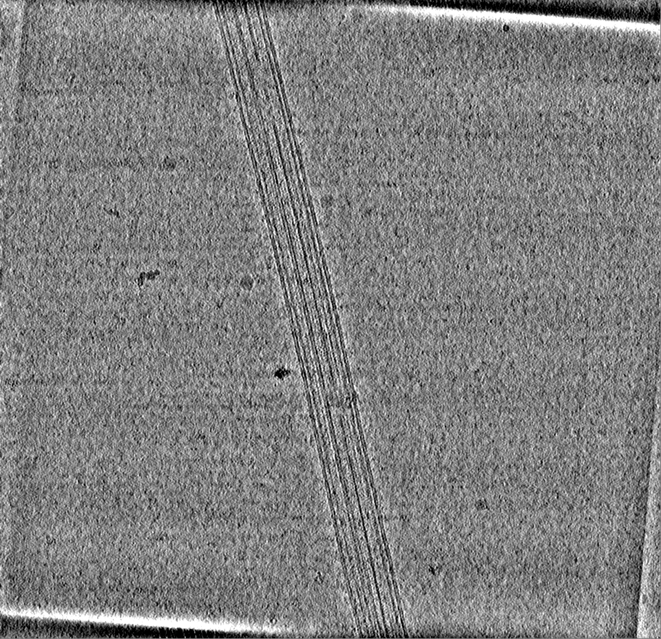
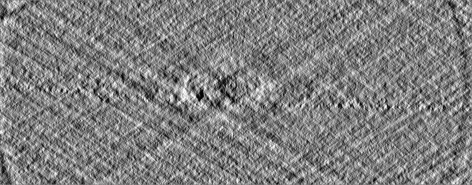
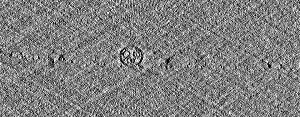
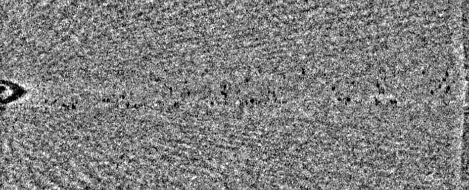
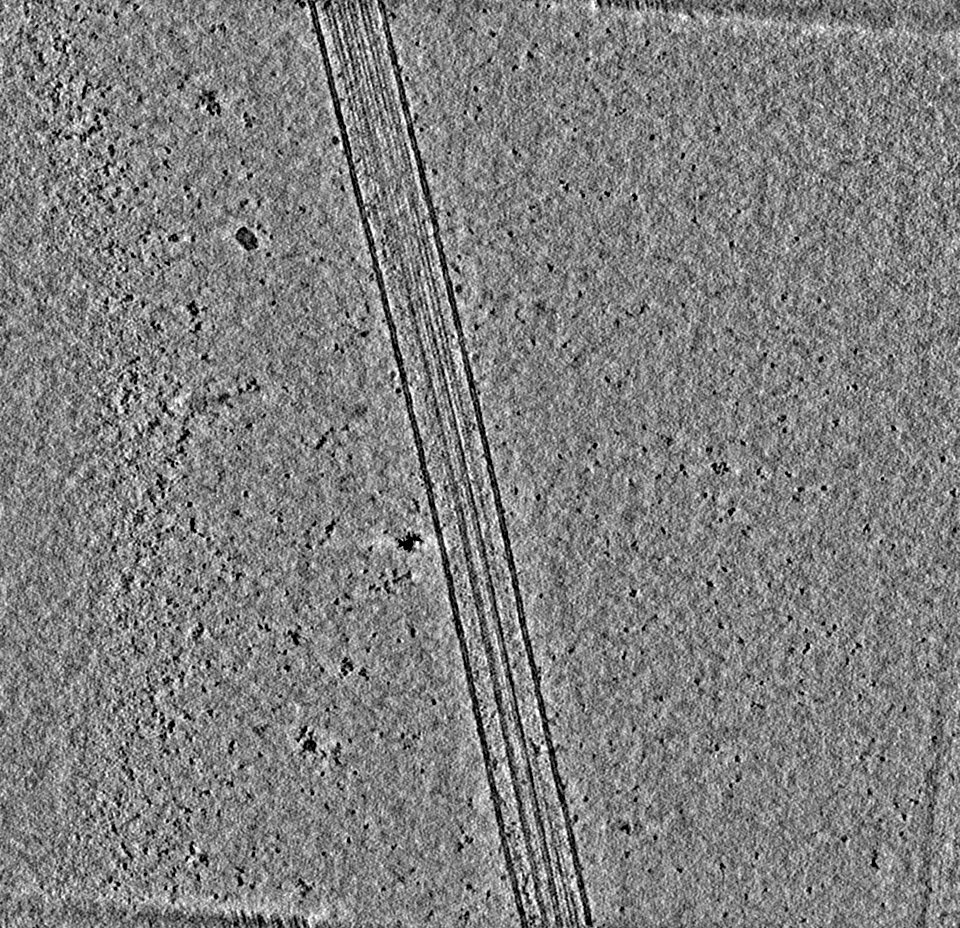
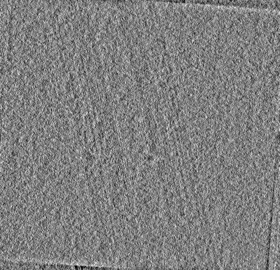
Deconvolved tomogram (binned by four) of an induced protrusion from Drosophila S2 alpha-tubulin acetyltransferase knock-out (dTAT KO) cells. From Dataset 8.
試料
細胞: Tomogram of an induced protrusion of a Drosophila S2 alpha-tublin acetyltransferase knock-out cell.
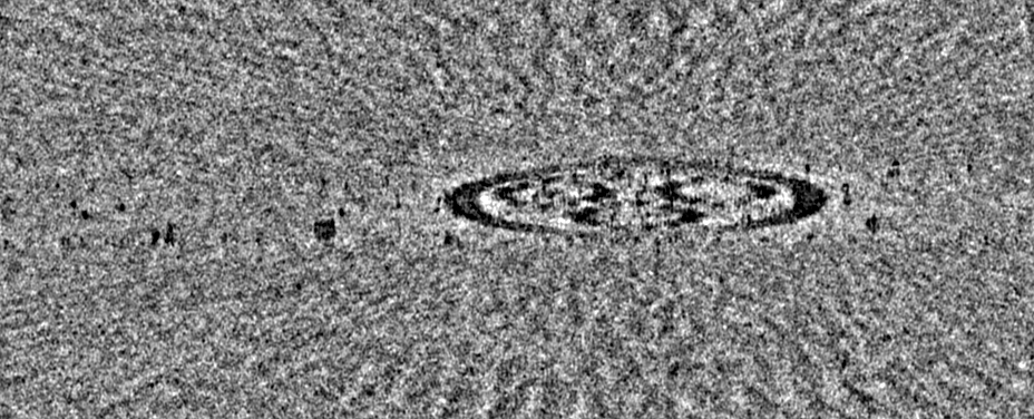
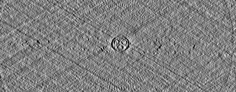
ジャーナル: bioRxiv / 年: 2023 タイトル: CryoET shows cofilactin filaments inside the microtubule lumen. 著者: Camilla Ventura Santos / Stephen L Rogers / Andrew P Carter / 要旨: Cytoplasmic microtubules are tubular polymers that can harbor small proteins or filaments inside their lumen. The identity of these objects and what causes their accumulation has not been ...Cytoplasmic microtubules are tubular polymers that can harbor small proteins or filaments inside their lumen. The identity of these objects and what causes their accumulation has not been conclusively established. Here, we used cryogenic electron tomography (cryoET) of S2 cell protrusions and found filaments inside the microtubule lumen, which resemble those reported recently in human HAP1 cells. The frequency of these filaments increased upon inhibition of the sarco/endoplasmic reticulum Ca ATPase (SERCA) with the small-molecule drug thapsigargin. Subtomogram averaging showed that the luminal filaments adopt a helical structure reminiscent of cofilin-bound actin (cofilactin). Consistent with this, cofilin was activated in cells under the same conditions that increased luminal filament occurrence. Furthermore, RNAi knock-down of cofilin reduced the frequency of luminal filaments with cofilactin morphology. These results suggest that cofilin activation stimulates its accumulation on actin filaments inside the microtubule lumen.
ダウンロード / ファイル: emd_16695.map.gz / 形式: CCP4 / 大きさ: 1.2 GB / タイプ: IMAGE STORED AS FLOATING POINT NUMBER (4 BYTES)
注釈
Deconvolved tomogram (binned by four) of an induced protrusion from Drosophila S2 alpha-tubulin acetyltransferase knock-out (dTAT KO) cells. From Dataset 8.
A: 11335.68 Å / B: 10957.823 Å / C: 4439.8076 Å α=β=γ: 90.0 °
-
添付データ
-
試料の構成要素
-
全体 : Tomogram of an induced protrusion of a Drosophila S2 alpha-tublin...
全体
名称: Tomogram of an induced protrusion of a Drosophila S2 alpha-tublin acetyltransferase knock-out cell.
要素
細胞: Tomogram of an induced protrusion of a Drosophila S2 alpha-tublin acetyltransferase knock-out cell.
-
超分子 #1: Tomogram of an induced protrusion of a Drosophila S2 alpha-tublin...
超分子
名称: Tomogram of an induced protrusion of a Drosophila S2 alpha-tublin acetyltransferase knock-out cell. タイプ: cell / ID: 1 / 親要素: 0 詳細: Protrusion formation was induced by treatment with 5 uM Cytochalasin D for 4h. Example tomogram from Dataset 8.
フィルム・検出器のモデル: GATAN K2 SUMMIT (4k x 4k) 検出モード: COUNTING / 平均電子線量: 3.0 e/Å2 詳細: Data was collected on Gatan K2 summit (2.952 A/pixel) with a total dose of 122.3 e/A2.
 ムービー
ムービー コントローラー


 データを開く
 基本情報
基本情報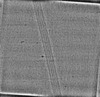
 マップデータ
 試料
試料
 データ登録者
 英国, 2件
  引用
引用 構造の表示
構造の表示
 ダウンロードとリンク
 EMDBマップデータ形式
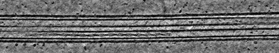 emd_16695.png
 http://ftp.pdbj.org/pub/emdb/structures/EMD-16695
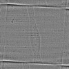

 Z (Sec.)
 Y (Row.)
 X (Col.)


 試料の構成要素
 解析
解析 電子顕微鏡法
 FIELD EMISSION GUN
