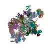
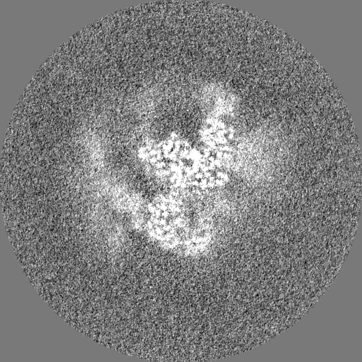
ジャーナル: Nature / 年: 2023 タイトル: Structural basis of catalytic activation in human splicing. 著者: Jana Schmitzová / Constantin Cretu / Christian Dienemann / Henning Urlaub / Vladimir Pena / 要旨: Pre-mRNA splicing follows a pathway driven by ATP-dependent RNA helicases. A crucial event of the splicing pathway is the catalytic activation, which takes place at the transition between the ...Pre-mRNA splicing follows a pathway driven by ATP-dependent RNA helicases. A crucial event of the splicing pathway is the catalytic activation, which takes place at the transition between the activated B and the branching-competent B spliceosomes. Catalytic activation occurs through an ATP-dependent remodelling mediated by the helicase PRP2 (also known as DHX16). However, because PRP2 is observed only at the periphery of spliceosomes, its function has remained elusive. Here we show that catalytic activation occurs in two ATP-dependent stages driven by two helicases: PRP2 and Aquarius. The role of Aquarius in splicing has been enigmatic. Here the inactivation of Aquarius leads to the stalling of a spliceosome intermediate-the B complex-found halfway through the catalytic activation process. The cryogenic electron microscopy structure of B reveals how PRP2 and Aquarius remodel B and B, respectively. Notably, PRP2 translocates along the intron while it strips away the RES complex, opens the SF3B1 clamp and unfastens the branch helix. Translocation terminates six nucleotides downstream of the branch site through an assembly of PPIL4, SKIP and the amino-terminal domain of PRP2. Finally, Aquarius enables the dissociation of PRP2, plus the SF3A and SF3B complexes, which promotes the relocation of the branch duplex for catalysis. This work elucidates catalytic activation in human splicing, reveals how a DEAH helicase operates and provides a paradigm for how helicases can coordinate their activities.
超分子 #1: Late human activated spliceosome arrested with a dominant-negativ...
超分子
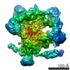
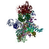
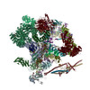
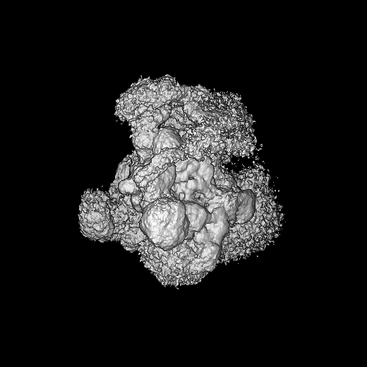
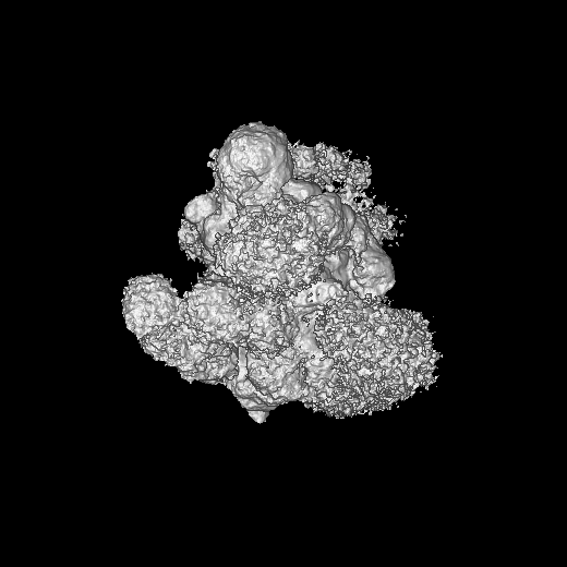
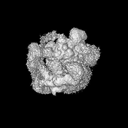
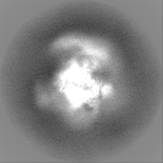
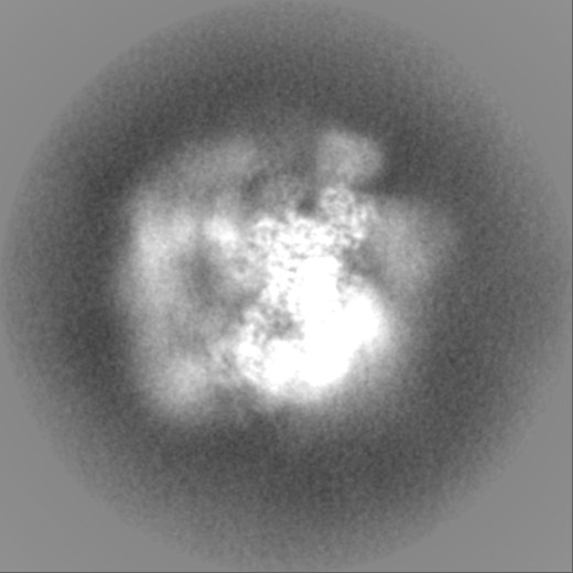
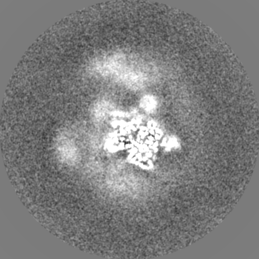
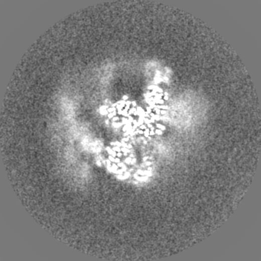
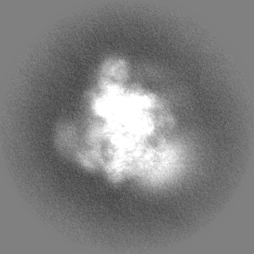
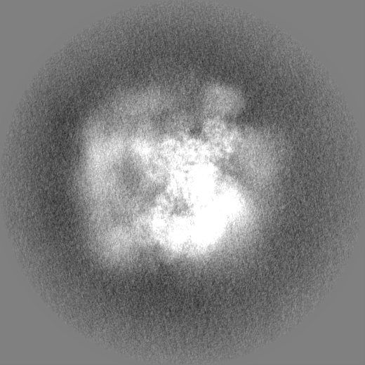
名称: Late human activated spliceosome arrested with a dominant-negative mutant of the splicing helicase Aquarius (Aquarius K829A) タイプ: complex / ID: 1 / 親要素: 0 / 含まれる分子: #1-#49 詳細: The spliceosome complex was assembled in vitro in the HeLa nuclear extract on a model pre-mRNA substrate (MINX) tagged with three MS2 aptamer sequences for affinity purification.
由来(天然)
生物種: Homo sapiens (ヒト)
+
分子 #1: Small nuclear ribonucleoprotein E
分子
名称: Small nuclear ribonucleoprotein E / タイプ: protein_or_peptide / ID: 1 / コピー数: 2 / 光学異性体: LEVO
凍結剤: ETHANE / チャンバー内湿度: 100 % / チャンバー内温度: 277.15 K / 装置: FEI VITROBOT MARK IV 詳細: Volumes of 4 ul of the concentrated sample were applied to one side of glow-discharged UltrAuFoil 200 2/2 grids (Quantifoil) in a Vitrobot Mark IV (FEI), operated at 4 degrees Celsius and ...詳細: Volumes of 4 ul of the concentrated sample were applied to one side of glow-discharged UltrAuFoil 200 2/2 grids (Quantifoil) in a Vitrobot Mark IV (FEI), operated at 4 degrees Celsius and 100% humidity. The grids were blotted for 2s with blotting force 5 and immediately frozen by plunging into liquid ethane..
詳細
The Baqr spliceosome was purified by affinity selection and gradient ultracentrifugation and crosslinked with 0.1% (v/v) glutaraldehyde in batch for cryo-EM grid preparation.
-
電子顕微鏡法
顕微鏡
FEI TITAN KRIOS
特殊光学系
エネルギーフィルター - 名称: GIF Quantum LS / エネルギーフィルター - スリット幅: 30 eV
撮影
フィルム・検出器のモデル: GATAN K2 SUMMIT (4k x 4k) 検出モード: COUNTING / 実像数: 10013 / 平均露光時間: 9.0 sec. / 平均電子線量: 45.47 e/Å2 詳細: Automated data acquisition for dataset 1 (untilted, 5229 micrographs) and dataset 2 (tilted, 25 degrees, 4784 micrographs) was performed with FEI EPU software package at a nominal ...詳細: Automated data acquisition for dataset 1 (untilted, 5229 micrographs) and dataset 2 (tilted, 25 degrees, 4784 micrographs) was performed with FEI EPU software package at a nominal magnification of 130,000 (1.05 A per pixel). Micrographs for these two datasets, dose fractionated over 40 frames, were collected at a dose rate of 5.04 or 5.06 e/A2/s-1 over 9 s, resulting in a total dose of 45.38 and 45.55 e/A2, respectively.
 ムービー
ムービー コントローラー


 データを開く
 基本情報
基本情報
 マップデータ
 試料
試料 キーワード
キーワード 機能・相同性情報
 Homo sapiens (ヒト) /
  unidentified adenovirus (ウイルス)
 データ登録者
 ドイツ, 1件
  引用
引用
 構造の表示
構造の表示
 ダウンロードとリンク
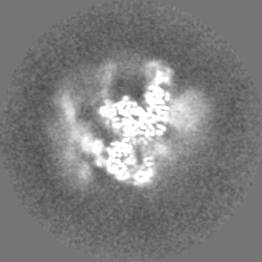
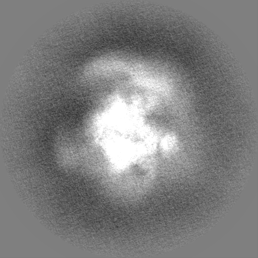
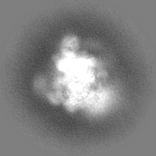
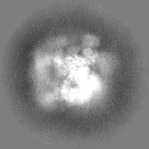
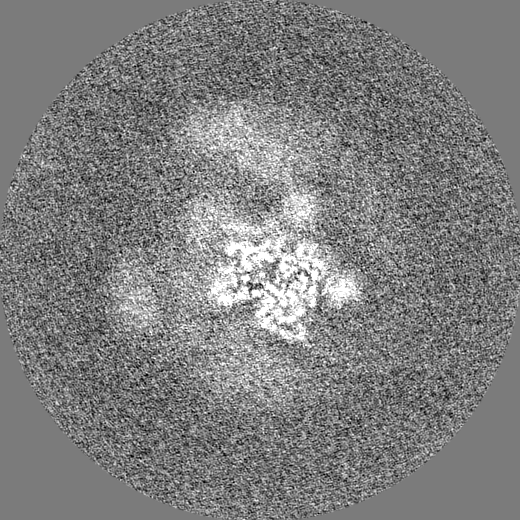
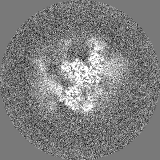
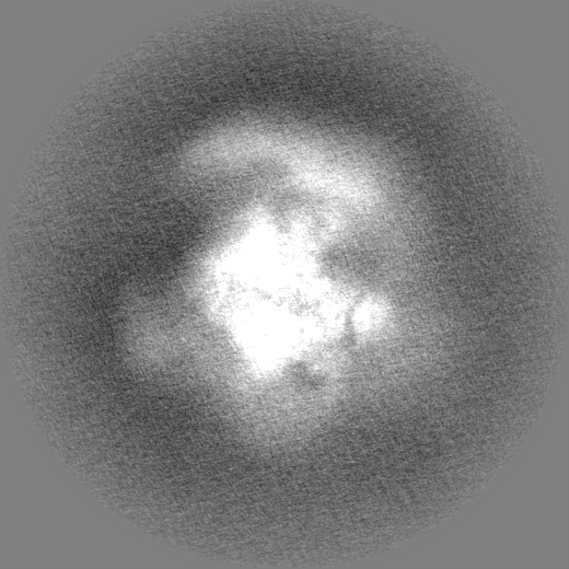
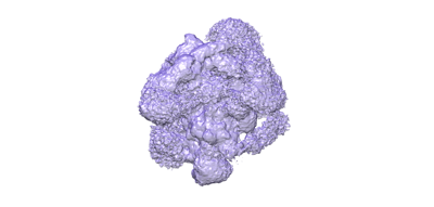 emd_16658.png
 http://ftp.pdbj.org/pub/emdb/structures/EMD-16658


 Z (Sec.)
 Y (Row.)
 X (Col.)


 試料の構成要素

 解析
解析 電子顕微鏡法
 FIELD EMISSION GUN