Movie
Movie Controller

[English] 日本語
 Yorodumi
- EMDB-16560: Structure of the yeast delta mtg1 mitochondrial ribosome assembly... -
+ Open data

- Basic information
| Entry |  | |||||||||||||||
|---|---|---|---|---|---|---|---|---|---|---|---|---|---|---|---|---|
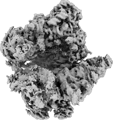
| Title | Structure of the yeast delta mtg1 mitochondrial ribosome assembly intermediate - State 2 | |||||||||||||||
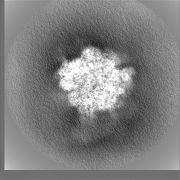
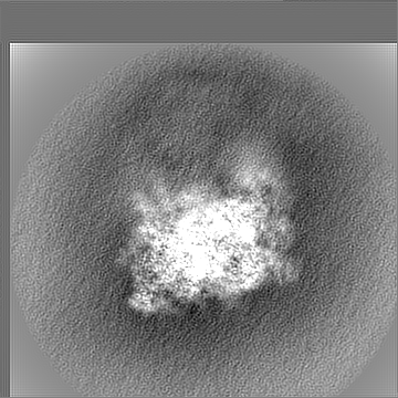
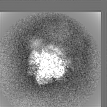
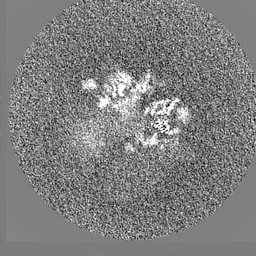
 Map data Map data | State 2, mt-monosome, consensus | |||||||||||||||
 Sample Sample |
| |||||||||||||||
 Keywords Keywords | Yeast mitochondrial ribosome / monosome / RIBOSOME | |||||||||||||||
| Biological species |  | |||||||||||||||
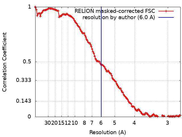
| Method | single particle reconstruction / cryo EM / Resolution: 6.0 Å | |||||||||||||||
 Authors Authors | Conrad J / Rathore S / Barrientos A | |||||||||||||||
| Funding support |  United States, United States,  Sweden, Sweden,  Germany, 4 items Germany, 4 items
| |||||||||||||||
 Citation Citation | Journal: To Be Published Title: The late stages of yeast mitoribosome large subunit biogenesis Authors: Rathore S / Conrad J / De Silva D / Ferrari A / Bouquio D / Kim H-J / Urlaub H / Ott M / Barrientos A | |||||||||||||||
| History |
|
- Structure visualization
| Supplemental images |
|---|
- Downloads & links
-EMDB archive
| Map data | emd_16560.map.gz | 163.9 MB |  EMDB map data format EMDB map data format | |
|---|---|---|---|---|
| Header (meta data) | emd-16560-v30.xmlemd-16560.xml | 21.9 KB 21.9 KB | Display Display | EMDB header |
| FSC (resolution estimation) | emd_16560_fsc.xml | 12.8 KB | Display | FSC data file |
| Images |  emd_16560.png emd_16560.png | 123.5 KB | ||
| Filedesc metadata | emd-16560.cif.gz | 4.5 KB | ||
| Others | emd_16560_additional_1.map.gzemd_16560_additional_2.map.gzemd_16560_additional_3.map.gzemd_16560_additional_4.map.gzemd_16560_half_map_1.map.gzemd_16560_half_map_2.map.gz | 162.6 MB 162.5 MB 156.9 MB 157.6 MB 140.4 MB 139 MB | ||
| Archive directory |  http://ftp.pdbj.org/pub/emdb/structures/EMD-16560ftp://ftp.pdbj.org/pub/emdb/structures/EMD-16560 http://ftp.pdbj.org/pub/emdb/structures/EMD-16560ftp://ftp.pdbj.org/pub/emdb/structures/EMD-16560 | HTTPS FTP |
-Validation report
| Summary document | emd_16560_validation.pdf.gz | 847.1 KB | Display | EMDB validaton report |
|---|---|---|---|---|
| Full document | emd_16560_full_validation.pdf.gz | 846.7 KB | Display | |
| Data in XML | emd_16560_validation.xml.gz | 20.3 KB | Display | |
| Data in CIF | emd_16560_validation.cif.gz | 26.7 KB | Display | |
| Arichive directory | https://ftp.pdbj.org/pub/emdb/validation_reports/EMD-16560ftp://ftp.pdbj.org/pub/emdb/validation_reports/EMD-16560 | HTTPS FTP |
-Related structure data


-Links
| EMDB pages | EMDB (EBI/PDBe) / EMDataResource |
|---|
-Map
| File | Download / File: emd_16560.map.gz / Format: CCP4 / Size: 178 MB / Type: IMAGE STORED AS FLOATING POINT NUMBER (4 BYTES) | ||||||||||||||||||||||||||||||||||||
|---|---|---|---|---|---|---|---|---|---|---|---|---|---|---|---|---|---|---|---|---|---|---|---|---|---|---|---|---|---|---|---|---|---|---|---|---|---|
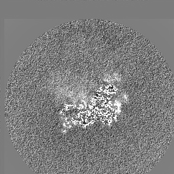
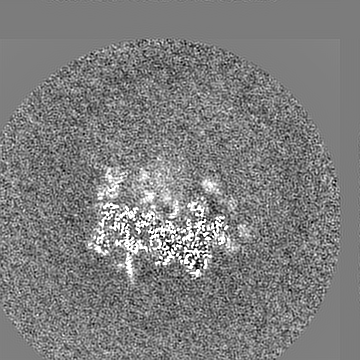
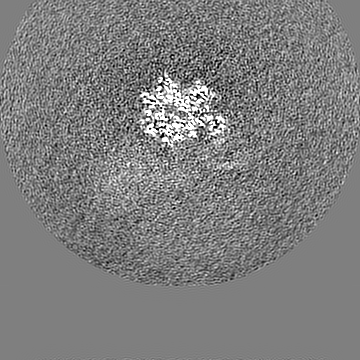
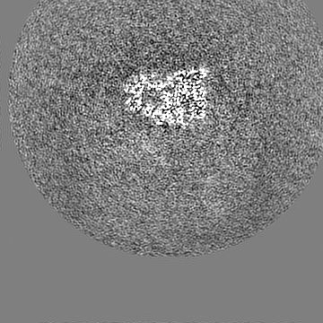
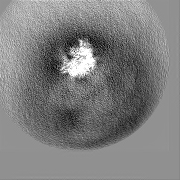
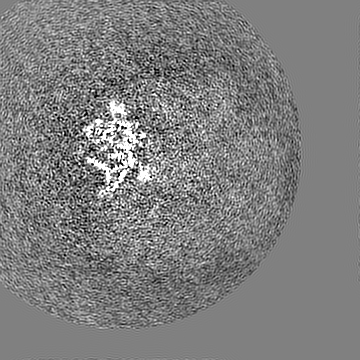
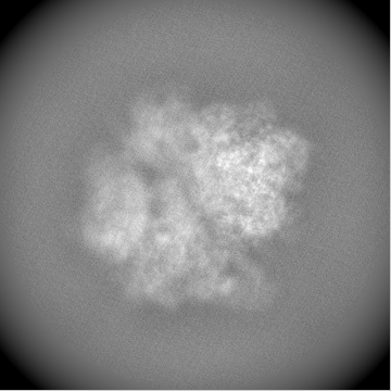
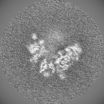
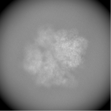
| Annotation | State 2, mt-monosome, consensus | ||||||||||||||||||||||||||||||||||||
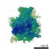
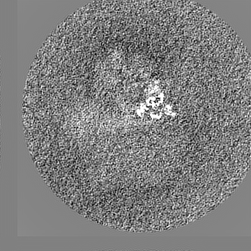
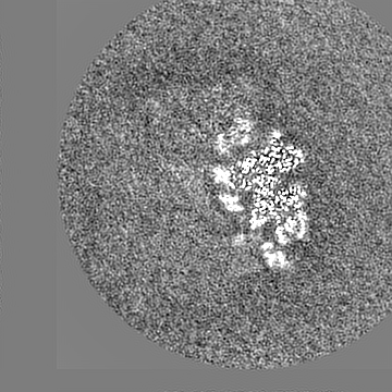
| Projections & slices | Image control
Images are generated by Spider. | ||||||||||||||||||||||||||||||||||||
| Voxel size | X=Y=Z: 1.37 Å | ||||||||||||||||||||||||||||||||||||
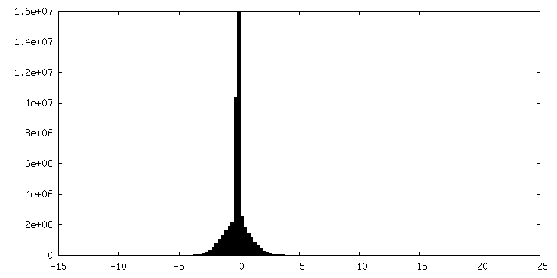
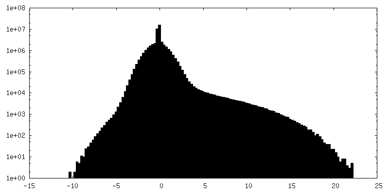
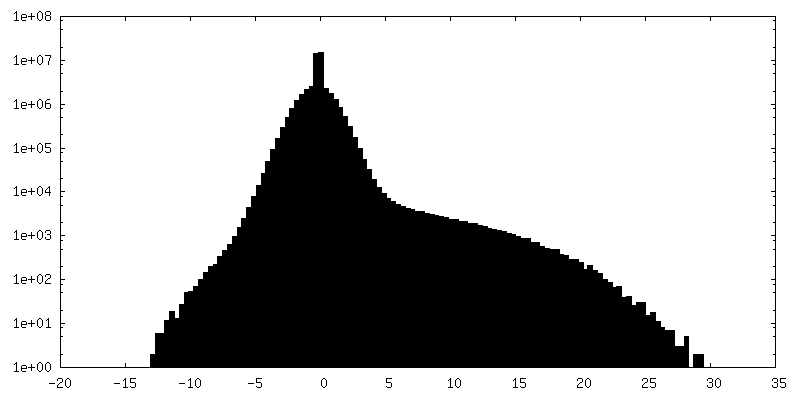
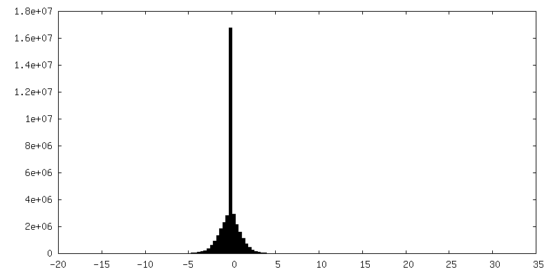
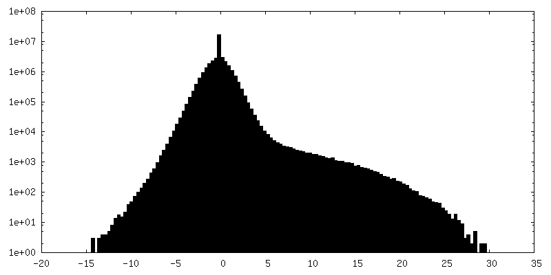
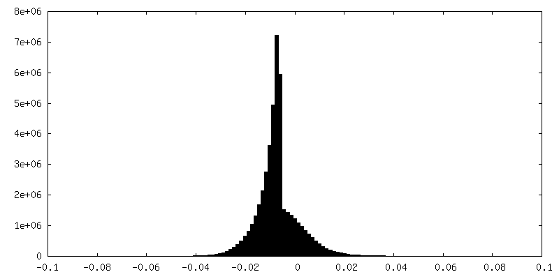
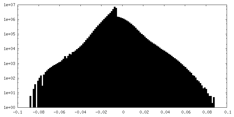
| Density |
| ||||||||||||||||||||||||||||||||||||
| Symmetry | Space group: 1 | ||||||||||||||||||||||||||||||||||||
| Details | EMDB XML:
|
 X (Sec.)
 Y (Row.)
 Z (Col.)


-Supplemental data
-Additional map: State 2, mt-monosome, LSU body
| File | emd_16560_additional_1.map | ||||||||||||
|---|---|---|---|---|---|---|---|---|---|---|---|---|---|
| Annotation | State 2, mt-monosome, LSU body | ||||||||||||
| Projections & Slices |
| ||||||||||||
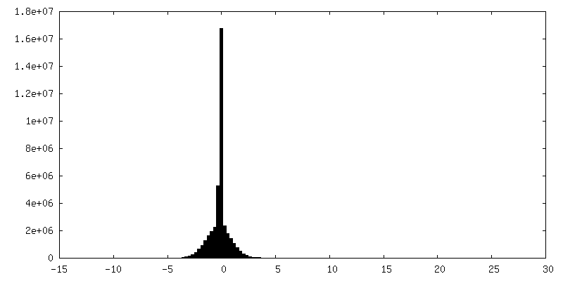
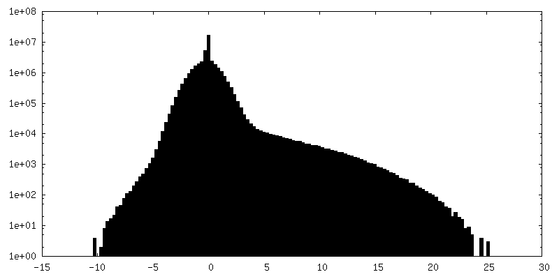
| Density Histograms |
-Additional map: State 2, mt-monosome, SSU body
| File | emd_16560_additional_2.map | ||||||||||||
|---|---|---|---|---|---|---|---|---|---|---|---|---|---|
| Annotation | State 2, mt-monosome, SSU body | ||||||||||||
| Projections & Slices |
| ||||||||||||
| Density Histograms |
-Additional map: State 2, mt-monosome, SSU head
| File | emd_16560_additional_3.map | ||||||||||||
|---|---|---|---|---|---|---|---|---|---|---|---|---|---|
| Annotation | State 2, mt-monosome, SSU head | ||||||||||||
| Projections & Slices |
| ||||||||||||
| Density Histograms |

-Additional map: State 2, mt-monosome, CP
| File | emd_16560_additional_4.map | ||||||||||||
|---|---|---|---|---|---|---|---|---|---|---|---|---|---|
| Annotation | State 2, mt-monosome, CP | ||||||||||||
| Projections & Slices |
| ||||||||||||
| Density Histograms |
-Half map: State 2, mt-monosome, halfmap 1
| File | emd_16560_half_map_1.map | ||||||||||||
|---|---|---|---|---|---|---|---|---|---|---|---|---|---|
| Annotation | State 2, mt-monosome, halfmap 1 | ||||||||||||
| Projections & Slices |
| ||||||||||||
| Density Histograms |
-Half map: State 2, mt-monosome, halfmap 2
| File | emd_16560_half_map_2.map | ||||||||||||
|---|---|---|---|---|---|---|---|---|---|---|---|---|---|
| Annotation | State 2, mt-monosome, halfmap 2 | ||||||||||||
| Projections & Slices |
| ||||||||||||
| Density Histograms |


- Sample components
-Entire : yeast mitochondrial ribosome- State2
| Entire | Name: yeast mitochondrial ribosome- State2 |
|---|---|
| Components |
|
-Supramolecule #1: yeast mitochondrial ribosome- State2
| Supramolecule | Name: yeast mitochondrial ribosome- State2 / type: complex / ID: 1 / Parent: 0 Details: Mitoribosomal particles from an mtg1-deletion strain were purified by sucrose cushion sedimentation of mitochondrial extracts prepared in the presence of 20% Mg2+ and 1% Triton X-100. This ...Details: Mitoribosomal particles from an mtg1-deletion strain were purified by sucrose cushion sedimentation of mitochondrial extracts prepared in the presence of 20% Mg2+ and 1% Triton X-100. This approach revealed the structures of novel mitoribosome assembly intermediates at resolutions 3.2 A. After processing, the structural characterization of the mtg1-deletion mitoribosome particles allowed us to define three major states: state 1, state 2, and state 3. |
|---|---|
| Source (natural) | Organism: |
-Experimental details
-Structure determination
| Method | cryo EM |
|---|---|
 Processing Processing | single particle reconstruction |
| Aggregation state | particle |
-Sample preparation
| Buffer | pH: 7.4 |
|---|---|
| Grid | Model: Quantifoil R2/2 / Material: COPPER / Support film - Material: CARBON / Support film - topology: CONTINUOUS / Pretreatment - Type: GLOW DISCHARGE |
| Vitrification | Cryogen name: ETHANE / Chamber humidity: 100 % / Chamber temperature: 277.15 K / Instrument: FEI VITROBOT MARK IV |
- Electron microscopy
| Microscope | FEI TITAN KRIOS |
|---|---|
| Image recording | Film or detector model: GATAN K2 SUMMIT (4k x 4k) / Detector mode: COUNTING / Average electron dose: 39.7 e/Å2 |
| Electron beam | Acceleration voltage: 300 kV / Electron source:  FIELD EMISSION GUN FIELD EMISSION GUN |
| Electron optics | C2 aperture diameter: 50.0 µm / Illumination mode: FLOOD BEAM / Imaging mode: BRIGHT FIELD / Cs: 2.7 mm / Nominal defocus max: 3.0 µm / Nominal defocus min: 1.2 µm |
| Experimental equipment |  Model: Titan Krios / Image courtesy: FEI Company |