 ムービー
ムービー コントローラー


+ データを開く

- 基本情報
基本情報
| 登録情報 |  | |||||||||
|---|---|---|---|---|---|---|---|---|---|---|
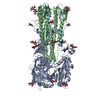
| タイトル | Reconstruction of a 13 nm wide Abeta(1-40) amyloid fibril | |||||||||
 マップデータ マップデータ | This is a reconstruction of an Abeta(1-40) amyloid fibril | |||||||||
 試料 試料 |
| |||||||||
 キーワード キーワード | Alzheimer's disease / amyloid / prion / protein folding | |||||||||
| 生物種 |  Homo sapiens (ヒト) Homo sapiens (ヒト) | |||||||||
| 手法 | らせん対称体再構成法 / クライオ電子顕微鏡法 / 解像度: 23.0 Å | |||||||||
 データ登録者 データ登録者 | Schmidt M / Sachse C / Richter W / Xu C / Fandrich M / Grigorieff N | |||||||||
 引用 引用 | ジャーナル: Proc Natl Acad Sci U S A / 年: 2009 タイトル: Comparison of Alzheimer Abeta(1-40) and Abeta(1-42) amyloid fibrils reveals similar protofilament structures. 著者: Matthias Schmidt / Carsten Sachse / Walter Richter / Chen Xu / Marcus Fändrich / Nikolaus Grigorieff /  要旨: We performed mass-per-length (MPL) measurements and electron cryomicroscopy (cryo-EM) with 3D reconstruction on an Abeta(1-42) amyloid fibril morphology formed under physiological pH conditions. The ...We performed mass-per-length (MPL) measurements and electron cryomicroscopy (cryo-EM) with 3D reconstruction on an Abeta(1-42) amyloid fibril morphology formed under physiological pH conditions. The data show that the examined Abeta(1-42) fibril morphology has only one protofilament, although two protofilaments were observed with a previously studied Abeta(1-40) fibril. The latter fibril was resolved at 8 A resolution showing pairs of beta-sheets at the cores of the two protofilaments making up a fibril. Detailed comparison of the Abeta(1-42) and Abeta(1-40) fibril structures reveals that they share an axial twofold symmetry and a similar protofilament structure. Furthermore, the MPL data indicate that the protofilaments of the examined Abeta(1-40) and Abeta(1-42) fibrils have the same number of Abeta molecules per cross-beta repeat. Based on this data and the previously studied Abeta(1-40) fibril structure, we describe a model for the arrangement of peptides within the Abeta(1-42) fibril. | |||||||||
| 履歴 |
|
- 構造の表示
構造の表示
| 構造ビューア | EMマップ:  SurfViewMolmilJmol/JSmol SurfViewMolmilJmol/JSmol |
|---|---|
| 添付画像 |
- ダウンロードとリンク
-EMDBアーカイブ
| マップデータ | emd_1650.map.gz | 58.5 KB | EMDBマップデータ形式 | |
|---|---|---|---|---|
| ヘッダ (付随情報) | emd-1650-v30.xmlemd-1650.xml | 9.8 KB 9.8 KB | 表示 表示 | EMDBヘッダ |
| 画像 | EMD-1650.tif | 37.8 KB | ||
| アーカイブディレクトリ |  http://ftp.pdbj.org/pub/emdb/structures/EMD-1650ftp://ftp.pdbj.org/pub/emdb/structures/EMD-1650 http://ftp.pdbj.org/pub/emdb/structures/EMD-1650ftp://ftp.pdbj.org/pub/emdb/structures/EMD-1650 | HTTPS FTP |
-関連構造データ


-リンク
| EMDBのページ | EMDB (EBI/PDBe) / EMDataResource |
|---|---|
| 「今月の分子」の関連する項目 |

-マップ
| ファイル | ダウンロード / ファイル: emd_1650.map.gz / 形式: CCP4 / 大きさ: 62.5 KB / タイプ: IMAGE STORED AS FLOATING POINT NUMBER (4 BYTES) | ||||||||||||||||||||||||||||||||||||
|---|---|---|---|---|---|---|---|---|---|---|---|---|---|---|---|---|---|---|---|---|---|---|---|---|---|---|---|---|---|---|---|---|---|---|---|---|---|
| 注釈 | This is a reconstruction of an Abeta(1-40) amyloid fibril | ||||||||||||||||||||||||||||||||||||
| 投影像・断面図 | 画像のコントロール
画像は Spider により作成 これらの図は立方格子座標系で作成されたものです | ||||||||||||||||||||||||||||||||||||
| ボクセルのサイズ | X=Y=Z: 4.8 Å | ||||||||||||||||||||||||||||||||||||
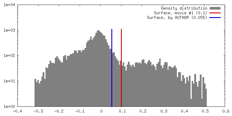
| 密度 |
| ||||||||||||||||||||||||||||||||||||
| 対称性 | 空間群: 1 | ||||||||||||||||||||||||||||||||||||
| 詳細 | EMDB XML:
|
 Z (Sec.)
 Y (Row.)
 X (Col.)


-添付データ
- 試料の構成要素
-全体 : Abeta(1-40) amyloid fibril
| 全体 | 名称: Abeta(1-40) amyloid fibril |
|---|---|
| 要素 |
|
-超分子 #1000: Abeta(1-40) amyloid fibril
| 超分子 | 名称: Abeta(1-40) amyloid fibril / タイプ: sample / ID: 1000 / 集合状態: Cross-beta structure / Number unique components: 1 |
|---|
-分子 #1: Abeta(1-40) peptide
| 分子 | 名称: Abeta(1-40) peptide / タイプ: protein_or_peptide / ID: 1 / Name.synonym: Alzheimer peptide / 集合状態: Cross-beta / 組換発現: Yes / データベース: NCBI |
|---|---|
| 由来(天然) | 生物種: Homo sapiens (ヒト) / 別称: Human |
| 分子量 | 理論値: 4.33 KDa |
-実験情報
-構造解析
| 手法 | クライオ電子顕微鏡法 |
|---|---|
 解析 解析 | らせん対称体再構成法 |
| 試料の集合状態 | helical array |
-試料調製
| 濃度 | 1 mg/mL |
|---|---|
| 緩衝液 | pH: 8.7 / 詳細: 50 mM sodium borate |
| グリッド | 詳細: 400 mesh copper grid |
| 凍結 | 凍結剤: ETHANE / チャンバー内湿度: 30 % / チャンバー内温度: 90 K / 装置: HOMEMADE PLUNGER / 詳細: Vitrification instrument: Manual plunger (Brandeis) / 手法: One-sided blotting for 5 seconds before plunging |
| 詳細 | Incubation for 4 days at 4 degC |
- 電子顕微鏡法
| 顕微鏡 | FEI TECNAI F30 |
|---|---|
| 温度 | 最低: 90 K / 最高: 90 K / 平均: 90 K |
| 撮影 | カテゴリ: FILM / フィルム・検出器のモデル: KODAK SO-163 FILM / デジタル化 - スキャナー: ZEISS SCAI / デジタル化 - サンプリング間隔: 7 µm / 実像数: 4 / 平均電子線量: 35 e/Å2 / Od range: 1.2 / ビット/ピクセル: 12 |
| Tilt angle min | 0 |
| Tilt angle max | 0 |
| 電子線 | 加速電圧: 300 kV / 電子線源:  FIELD EMISSION GUN FIELD EMISSION GUN |
| 電子光学系 | 倍率(補正後): 58090 / 照射モード: FLOOD BEAM / 撮影モード: BRIGHT FIELD / Cs: 2 mm / 最大 デフォーカス(公称値): 3.5 µm / 最小 デフォーカス(公称値): 2.0 µm / 倍率(公称値): 59000 |
| 試料ステージ | 試料ホルダー: Eucentric / 試料ホルダーモデル: GATAN LIQUID NITROGEN |
| 実験機器 |  モデル: Tecnai F30 / 画像提供: FEI Company |
-画像解析
| 詳細 | Fibrils were selected using BOXER |
|---|---|
| 最終 再構成 | 想定した対称性 - らせんパラメータ - 軸対称性: C2 (2回回転対称) アルゴリズム: OTHER / 解像度のタイプ: BY AUTHOR / 解像度: 23.0 Å / 解像度の算出法: FSC 0.5 CUT-OFF / ソフトウェア - 名称: Spider / 詳細: Final map was calculated from 4 fibrils |
| CTF補正 | 詳細: Each particle |
