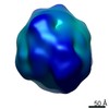
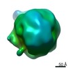
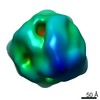
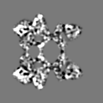
Journal: J Mol Biol / Year: 2009 Title: 10-A cryoEM structure and molecular model of the Myriapod (Scutigera) 6x6mer hemocyanin: understanding a giant oxygen transport protein. Authors: Jürgen Markl / Arne Moeller / Andreas G Martin / Judith Rheinbay / Wolfgang Gebauer / Frank Depoix / Abstract: Oxygen transport in Myriapoda is maintained by a unique 6x6mer hemocyanin, that is, 36 subunits arranged as six hexamers (1x6mers). In the sluggish diplopod Spirostreptus, the 1x6mers seem to operate ...Oxygen transport in Myriapoda is maintained by a unique 6x6mer hemocyanin, that is, 36 subunits arranged as six hexamers (1x6mers). In the sluggish diplopod Spirostreptus, the 1x6mers seem to operate as almost or fully independent allosteric units (h approximately 1.3; P(50) approximately 5 torr), whereas in the swift centipede Scutigera, they intensively cooperate allosterically (h approximately 10; P(50) approximately 50 torr). Here, we show the chemomechanical basis of this differential behavior as deduced from hybrid 6x6mer structures, obtained by single-particle cryo-electron microscopy of the Scutigera 6x6mer (10.0 A resolution according to the 0.5 criterion) and docking of homology-modeled subunits from Scutigera and two diplopods, Spirostreptus and Polydesmus. The Scutigera 6x6mer hemocyanin is a trigonal antiprism assembled from six smaller trigonal antiprisms (1x6mers), thereby exhibiting D3 point group symmetry. It can be described as two staggered 3x6mers or three oblique 2x6mers. Topologically, the 6x6mer is subdivided into six subunit zones, thereby exhibiting a mantle (24 subunits) and a core (12 subunits). The six hexamers are linked by 21 bridges, subdivided into five types: two within each 3x6mer and three between both 3x6mers. The molecular models of the 6x6mer reveal intriguing amino acid appositions at these inter-hexamer interfaces. Besides opportunities for salt bridges, we found pairs of carboxylate residues for possible bridging via a Ca(2+) or Mg(2+) ion. Moreover, we detected histidine clusters, notably in Scutigera, allowing us to advance hypotheses as to how the hexamers are allosterically coupled in centipede hemocyanin and why they act more independently in diplopod hemocyanin.
History
Deposition
Jun 22, 2009
-
Header (metadata) release
Apr 26, 2011
-
Map release
Apr 26, 2011
-
Update
Apr 26, 2011
-
Current status
Apr 26, 2011
Processing site: PDBe / Status: Released
-
Structure visualization
Movie
Surface view with section colored by density value
In the structure databanks used in Yorodumi, some data are registered as the other names, "COVID-19 virus" and "2019-nCoV". Here are the details of the virus and the list of structure data.
Jan 31, 2019. EMDB accession codes are about to change! (news from PDBe EMDB page)
EMDB accession codes are about to change! (news from PDBe EMDB page)
The allocation of 4 digits for EMDB accession codes will soon come to an end. Whilst these codes will remain in use, new EMDB accession codes will include an additional digit and will expand incrementally as the available range of codes is exhausted. The current 4-digit format prefixed with “EMD-” (i.e. EMD-XXXX) will advance to a 5-digit format (i.e. EMD-XXXXX), and so on. It is currently estimated that the 4-digit codes will be depleted around Spring 2019, at which point the 5-digit format will come into force.
The EM Navigator/Yorodumi systems omit the EMD- prefix.
Related info.:Q: What is EMD? / ID/Accession-code notation in Yorodumi/EM Navigator
Yorodumi is a browser for structure data from EMDB, PDB, SASBDB, etc.
This page is also the successor to EM Navigator detail page, and also detail information page/front-end page for Omokage search.
The word "yorodu" (or yorozu) is an old Japanese word meaning "ten thousand". "mi" (miru) is to see.
Related info.:EMDB / PDB / SASBDB / Comparison of 3 databanks / Yorodumi Search / Aug 31, 2016. New EM Navigator & Yorodumi / Yorodumi Papers / Jmol/JSmol / Function and homology information / Changes in new EM Navigator and Yorodumi
 Movie
Movie Controller

 Yorodumi
 Open data
 Basic information
 Map data
 Sample
 Keywords
 Scutigera coleoptrata (house centipede)
 Authors
 Citation
 Structure visualization
 Movie viewer
 Downloads & links
 em1628.png
 http://ftp.pdbj.org/pub/emdb/structures/EMD-1628
 Z (Sec.)
 Y (Row.)
 X (Col.)


 Sample components
 Processing
 Electron microscopy
 FIELD EMISSION GUN
