 ムービー
ムービー コントローラー


+ データを開く

- 基本情報
基本情報
| 登録情報 |  | |||||||||
|---|---|---|---|---|---|---|---|---|---|---|
| タイトル | Structure of CEACAM5 A3-B3 domain in Complex with Tusamitamab Fab | |||||||||
 マップデータ マップデータ | Cryo EM map for the complex | |||||||||
 試料 試料 |
| |||||||||
 キーワード キーワード | CEACAM5 / Tusamitamab / Cancer / cell adhesion / cryo-EM / small molecular weight / Fab / A3-B3 / human membrane protein | |||||||||
| 機能・相同性 |  機能・相同性情報 機能・相同性情報GPI anchor binding / homotypic cell-cell adhesion / negative regulation of myotube differentiation / Post-translational modification: synthesis of GPI-anchored proteins / heterophilic cell-cell adhesion via plasma membrane cell adhesion molecules / homophilic cell adhesion via plasma membrane adhesion molecules / negative regulation of anoikis / side of membrane / Cell surface interactions at the vascular wall / basolateral plasma membrane ...GPI anchor binding / homotypic cell-cell adhesion / negative regulation of myotube differentiation / Post-translational modification: synthesis of GPI-anchored proteins / heterophilic cell-cell adhesion via plasma membrane cell adhesion molecules / homophilic cell adhesion via plasma membrane adhesion molecules / negative regulation of anoikis / side of membrane / Cell surface interactions at the vascular wall / basolateral plasma membrane / apical plasma membrane / apoptotic process / negative regulation of apoptotic process / cell surface / protein homodimerization activity / extracellular exosome / extracellular region / identical protein binding / membrane / plasma membrane 類似検索 - 分子機能 | |||||||||
| 生物種 |  Homo sapiens (ヒト) / Homo sapiens (ヒト) /  | |||||||||
| 手法 | 単粒子再構成法 / クライオ電子顕微鏡法 / 解像度: 3.11 Å | |||||||||
 データ登録者 データ登録者 | Kumar A / Bertrand T / Rapisarda C / Rak A | |||||||||
| 資金援助 |  フランス, 1件 フランス, 1件
| |||||||||
 引用 引用 | ジャーナル: Nat Commun / 年: 2024 タイトル: Structural insights into epitope-paratope interactions of a monoclonal antibody targeting CEACAM5-expressing tumors. 著者: Anand Kumar / Francis Duffieux / Marie Gagnaire / Chiara Rapisarda / Thomas Bertrand / Alexey Rak / 要旨: Carcinoembryonic antigen-related cell adhesion molecules (CEACAMs) are overexpressed in some tumor types. The antibody-drug conjugate tusamitamab ravtansine specifically recognizes the A3-B3 domains ...Carcinoembryonic antigen-related cell adhesion molecules (CEACAMs) are overexpressed in some tumor types. The antibody-drug conjugate tusamitamab ravtansine specifically recognizes the A3-B3 domains of human CEACAM5 (hCEACAM5). To understand this specificity, here we map the epitope-paratope interface between the A3-B3 domains of hCEACAM5 (hCEACAM5) and the antigen-binding fragment of tusamitamab (tusa Fab). We use hydrogen/deuterium exchange mass spectrometry to identify the tusa Fab paratope, which involves heavy chain (HC) residues 101-109 and light chain residues 48-54 and 88-104. Using surface plasmon resonance, we demonstrate that alanine variants of HC residues 96-108 abolish binding to hCEACAM5, suggesting that these residues are critical for tusa-Fab-antigen complex formation. The cryogenic electron microscopy structure of the hCEACAM5- tusa Fab complex (3.11 Å overall resolution) reveals a discontinuous epitope involving residues in the A3-B3 domains and an N-linked mannose at residue Asn612. Conformational constraints on the epitope-paratope interface enable tusamitamab to target hCEACAM5 and distinguish CEACAM5 from other CEACAMs. #1: ジャーナル: Res Sq / 年: 2023タイトル: Structural insights into epitope-paratope interactions of monoclonal antibody targeting CEACAM5-expressing tumors 著者: Rak A / Kumar A / Duffi F / Gagnaire M / Rapisarda C / Bertrand T | |||||||||
| 履歴 |
|
- 構造の表示
構造の表示
| 添付画像 |
|---|

- ダウンロードとリンク
-EMDBアーカイブ
| マップデータ | emd_16279.map.gz | 58.3 MB | EMDBマップデータ形式 | |
|---|---|---|---|---|
| ヘッダ (付随情報) | emd-16279-v30.xmlemd-16279.xml | 20.2 KB 20.2 KB | 表示 表示 | EMDBヘッダ |
| 画像 |  emd_16279.png emd_16279.png | 80.5 KB | ||
| Filedesc metadata | emd-16279.cif.gz | 6.5 KB | ||
| その他 | emd_16279_half_map_1.map.gzemd_16279_half_map_2.map.gz | 59.5 MB 59.5 MB | ||
| アーカイブディレクトリ |  http://ftp.pdbj.org/pub/emdb/structures/EMD-16279ftp://ftp.pdbj.org/pub/emdb/structures/EMD-16279 http://ftp.pdbj.org/pub/emdb/structures/EMD-16279ftp://ftp.pdbj.org/pub/emdb/structures/EMD-16279 | HTTPS FTP |
-関連構造データ
-リンク
| EMDBのページ | EMDB (EBI/PDBe) / EMDataResource |
|---|---|
| 「今月の分子」の関連する項目 |


-マップ
| ファイル | ダウンロード / ファイル: emd_16279.map.gz / 形式: CCP4 / 大きさ: 64 MB / タイプ: IMAGE STORED AS FLOATING POINT NUMBER (4 BYTES) | ||||||||||||||||||||||||||||||||||||
|---|---|---|---|---|---|---|---|---|---|---|---|---|---|---|---|---|---|---|---|---|---|---|---|---|---|---|---|---|---|---|---|---|---|---|---|---|---|
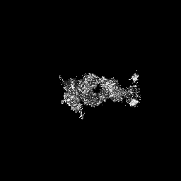
| 注釈 | Cryo EM map for the complex | ||||||||||||||||||||||||||||||||||||
| 投影像・断面図 | 画像のコントロール
画像は Spider により作成 | ||||||||||||||||||||||||||||||||||||
| ボクセルのサイズ | X=Y=Z: 1.16 Å | ||||||||||||||||||||||||||||||||||||
| 密度 |
| ||||||||||||||||||||||||||||||||||||
| 対称性 | 空間群: 1 | ||||||||||||||||||||||||||||||||||||
| 詳細 | EMDB XML:
|
 X (Sec.)
 Y (Row.)
 Z (Col.)


-添付データ
-ハーフマップ: Half map A
| ファイル | emd_16279_half_map_1.map | ||||||||||||
|---|---|---|---|---|---|---|---|---|---|---|---|---|---|
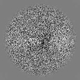
| 注釈 | Half map A | ||||||||||||
| 投影像・断面図 |
| ||||||||||||
| 密度ヒストグラム |


-ハーフマップ: Half map B
| ファイル | emd_16279_half_map_2.map | ||||||||||||
|---|---|---|---|---|---|---|---|---|---|---|---|---|---|
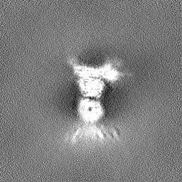
| 注釈 | Half map B | ||||||||||||
| 投影像・断面図 |
| ||||||||||||
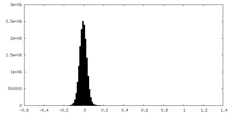
| 密度ヒストグラム |


- 試料の構成要素
-全体 : hCEACAM5-A3B3-Tusamitamab Fab complex
| 全体 | 名称: hCEACAM5-A3B3-Tusamitamab Fab complex |
|---|---|
| 要素 |
|
-超分子 #1: hCEACAM5-A3B3-Tusamitamab Fab complex
| 超分子 | 名称: hCEACAM5-A3B3-Tusamitamab Fab complex / タイプ: complex / ID: 1 / 親要素: 0 / 含まれる分子: #1-#3 詳細: Human CEACAM5 a3-B3 domain in the comlex with the Tusamitamab Fab |
|---|---|
| 由来(天然) | 生物種: Homo sapiens (ヒト) |
| 分子量 | 理論値: 70 KDa |
-分子 #1: Tusamitamab Fab heavy Chain
| 分子 | 名称: Tusamitamab Fab heavy Chain / タイプ: protein_or_peptide / ID: 1 / コピー数: 1 / 光学異性体: LEVO |
|---|---|
| 由来(天然) | 生物種: |
| 分子量 | 理論値: 24.833652 KDa |
| 組換発現 | 生物種: Homo sapiens (ヒト) |
| 配列 | 文字列: EVQLQESGPG LVKPGGSLSL SCAASGFVFS SYDMSWVRQT PERGLEWVAY ISSGGGITYA PSTVKGRFTV SRDNAKNTLY LQMNSLTSE DTAVYYCAAH YFGSSGPFAY WGQGTLVTVS SASTKGPSVF PLAPSSKSTS GGTAALGCLV KDYFPEPVTV S WNSGALTS ...文字列: EVQLQESGPG LVKPGGSLSL SCAASGFVFS SYDMSWVRQT PERGLEWVAY ISSGGGITYA PSTVKGRFTV SRDNAKNTLY LQMNSLTSE DTAVYYCAAH YFGSSGPFAY WGQGTLVTVS SASTKGPSVF PLAPSSKSTS GGTAALGCLV KDYFPEPVTV S WNSGALTS GVHTFPAVLQ SSGLYSLSSV VTVPSSSLGT QTYICNVNHK PSNTKVDKKV EPKSCDKTHT HHHHHH |
-分子 #2: Tusamitamab Light Chain
| 分子 | 名称: Tusamitamab Light Chain / タイプ: protein_or_peptide / ID: 2 / コピー数: 1 / 光学異性体: LEVO |
|---|---|
| 由来(天然) | 生物種: |
| 分子量 | 理論値: 23.497072 KDa |
| 組換発現 | 生物種: Homo sapiens (ヒト) |
| 配列 | 文字列: DIQMTQSPAS LSASVGDRVT ITCRASENIF SYLAWYQQKP GKSPKLLVYN TRTLAEGVPS RFSGSGSGTD FSLTISSLQP EDFATYYCQ HHYGTPFTFG SGTKLEIKRT VAAPSVFIFP PSDEQLKSGT ASVVCLLNNF YPREAKVQWK VDNALQSGNS Q ESVTEQDS ...文字列: DIQMTQSPAS LSASVGDRVT ITCRASENIF SYLAWYQQKP GKSPKLLVYN TRTLAEGVPS RFSGSGSGTD FSLTISSLQP EDFATYYCQ HHYGTPFTFG SGTKLEIKRT VAAPSVFIFP PSDEQLKSGT ASVVCLLNNF YPREAKVQWK VDNALQSGNS Q ESVTEQDS KDSTYSLSST LTLSKADYEK HKVYACEVTH QGLSSPVTKS FNRGEC |
-分子 #3: Carcinoembryonic antigen-related cell adhesion molecule 5
| 分子 | 名称: Carcinoembryonic antigen-related cell adhesion molecule 5 タイプ: protein_or_peptide / ID: 3 / 詳細: A3-B3 domain / コピー数: 1 / 光学異性体: LEVO |
|---|---|
| 由来(天然) | 生物種: Homo sapiens (ヒト) |
| 分子量 | 理論値: 20.883041 KDa |
| 組換発現 | 生物種: Homo sapiens (ヒト) |
| 配列 | 文字列: ELPKPSISSN NSKPVEDKDA VAFTCEPEAQ NTTYLWWVNG QSLPVSPRLQ LSNGNRTLTL FNVTRNDARA YVCGIQNSVS ANRSDPVTL DVLYGPDTPI ISPPDSSYLS GANLNLSCHS ASNPSPQYSW RINGIPQQHT QVLFIAKITP NNNGTYACFV S NLATGRNN ...文字列: ELPKPSISSN NSKPVEDKDA VAFTCEPEAQ NTTYLWWVNG QSLPVSPRLQ LSNGNRTLTL FNVTRNDARA YVCGIQNSVS ANRSDPVTL DVLYGPDTPI ISPPDSSYLS GANLNLSCHS ASNPSPQYSW RINGIPQQHT QVLFIAKITP NNNGTYACFV S NLATGRNN SIVKSITVSA SGTSPGLSAH HHHHH UniProtKB: Cell adhesion molecule CEACAM5 |
-分子 #6: 2-acetamido-2-deoxy-beta-D-glucopyranose
| 分子 | 名称: 2-acetamido-2-deoxy-beta-D-glucopyranose / タイプ: ligand / ID: 6 / コピー数: 3 / 式: NAG |
|---|---|
| 分子量 | 理論値: 221.208 Da |
| Chemical component information |  ChemComp-NAG: |
-実験情報
-構造解析
| 手法 | クライオ電子顕微鏡法 |
|---|---|
 解析 解析 | 単粒子再構成法 |
| 試料の集合状態 | particle |
-試料調製
| 濃度 | 0.87 mg/mL |
|---|---|
| 緩衝液 | pH: 7.4 / 構成要素 - 濃度: 1.0 X / 構成要素 - 名称: D-PBS / 詳細: Dulbeccos phosphate buffered saline |
| グリッド | モデル: UltrAuFoil R0./1 / 材質: GOLD / メッシュ: 300 / 前処理 - タイプ: GLOW DISCHARGE / 前処理 - 時間: 40 sec. |
| 凍結 | 凍結剤: ETHANE / チャンバー内湿度: 100 % / チャンバー内温度: 277.15 K / 装置: FEI VITROBOT MARK IV |
- 電子顕微鏡法
| 顕微鏡 | TFS GLACIOS |
|---|---|
| 撮影 | フィルム・検出器のモデル: FEI FALCON IV (4k x 4k) 撮影したグリッド数: 1 / 実像数: 5072 / 平均露光時間: 4.72 sec. / 平均電子線量: 62.0 e/Å2 |
| 電子線 | 加速電圧: 200 kV / 電子線源:  FIELD EMISSION GUN FIELD EMISSION GUN |
| 電子光学系 | C2レンズ絞り径: 30.0 µm / 照射モード: OTHER / 撮影モード: DARK FIELD / Cs: 2.7 mm / 最大 デフォーカス(公称値): 2.2 µm / 最小 デフォーカス(公称値): 0.6 µm / 倍率(公称値): 240000 |
| 試料ステージ | ホルダー冷却材: NITROGEN |
+画像解析
-原子モデル構築 1
| 精密化 | プロトコル: AB INITIO MODEL |
|---|---|
| 得られたモデル |  PDB-8bw0: |
