 Movie
Movie Controller

[English] 日本語
 Yorodumi
- EMDB-16223: Cryo-EM structure of the catalytic domain tetramer of N-terminall... -
+ Open data

- Basic information
| Entry |  | |||||||||
|---|---|---|---|---|---|---|---|---|---|---|
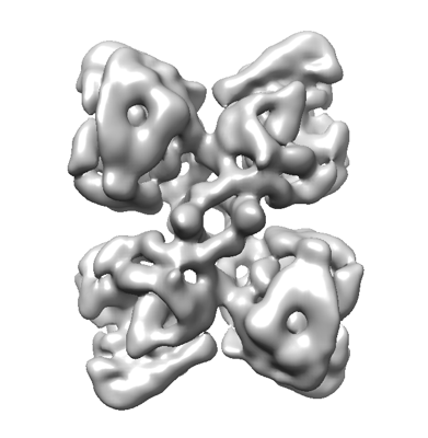
| Title | Cryo-EM structure of the catalytic domain tetramer of N-terminally truncated human tryptophan hydroxylase 2 | |||||||||
 Map data Map data | ||||||||||
 Sample Sample |
| |||||||||
 Keywords Keywords | Serotonin biosynthesis / Tryptophan hydroxylase / aromatic amino acid hydroxylases / OXIDOREDUCTASE | |||||||||
| Biological species |  Homo sapiens (human) Homo sapiens (human) | |||||||||
| Method | single particle reconstruction / cryo EM / Resolution: 3.9 Å | |||||||||
 Authors Authors | Zhang Z / Vedel IM / Skawinska NT / Harris P / Stark H / Peters GHJ | |||||||||
| Funding support |  Denmark, 1 items Denmark, 1 items
| |||||||||
 Citation Citation | Journal: Structure / Year: 2023 Title: Structural characterization of human tryptophan hydroxylase 2 reveals that L-Phe is superior to L-Trp as the regulatory domain ligand. Authors: Ida M Vedel / Andreas Prestel / Zhenwei Zhang / Natalia T Skawinska / Holger Stark / Pernille Harris / Birthe B Kragelund / Günther H J Peters /  Abstract: Tryptophan hydroxylase 2 (TPH2) catalyzes the rate-limiting step in serotonin biosynthesis in the brain. Consequently, regulation of TPH2 is relevant for serotonin-related diseases, yet the ...Tryptophan hydroxylase 2 (TPH2) catalyzes the rate-limiting step in serotonin biosynthesis in the brain. Consequently, regulation of TPH2 is relevant for serotonin-related diseases, yet the regulatory mechanism of TPH2 is poorly understood and structural and dynamical insights are missing. We use NMR spectroscopy to determine the structure of a 47 N-terminally truncated variant of the regulatory domain (RD) dimer of human TPH2 in complex with L-Phe, and show that L-Phe is the superior RD ligand compared with the natural substrate, L-Trp. Using cryo-EM, we obtain a low-resolution structure of a similarly truncated variant of the complete tetrameric enzyme with dimerized RDs. The cryo-EM two-dimensional (2D) class averages additionally indicate that the RDs are dynamic in the tetramer and likely exist in a monomer-dimer equilibrium. Our results provide structural information on the RD as an isolated domain and in the TPH2 tetramer, which will facilitate future elucidation of TPH2's regulatory mechanism. | |||||||||
| History |
|
- Structure visualization
| Supplemental images |
|---|
- Downloads & links
-EMDB archive
| Map data | emd_16223.map.gz | 139 MB |  EMDB map data format EMDB map data format | |
|---|---|---|---|---|
| Header (meta data) | emd-16223-v30.xmlemd-16223.xml | 12 KB 12 KB | Display Display | EMDB header |
| Images |  emd_16223.png emd_16223.png | 70.7 KB | ||
| Others | emd_16223_half_map_1.map.gzemd_16223_half_map_2.map.gz | 139 MB 139 MB | ||
| Archive directory |  http://ftp.pdbj.org/pub/emdb/structures/EMD-16223ftp://ftp.pdbj.org/pub/emdb/structures/EMD-16223 http://ftp.pdbj.org/pub/emdb/structures/EMD-16223ftp://ftp.pdbj.org/pub/emdb/structures/EMD-16223 | HTTPS FTP |
-Related structure data


-Links
| EMDB pages | EMDB (EBI/PDBe) / EMDataResource |
|---|
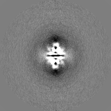
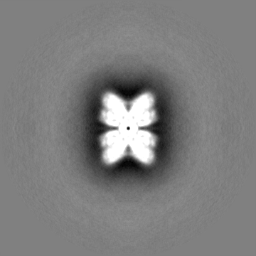
-Map
| File | Download / File: emd_16223.map.gz / Format: CCP4 / Size: 178 MB / Type: IMAGE STORED AS FLOATING POINT NUMBER (4 BYTES) | ||||||||||||||||||||||||||||||||||||
|---|---|---|---|---|---|---|---|---|---|---|---|---|---|---|---|---|---|---|---|---|---|---|---|---|---|---|---|---|---|---|---|---|---|---|---|---|---|
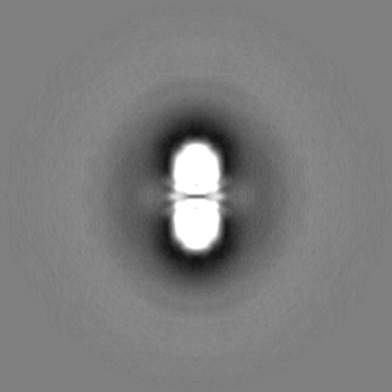
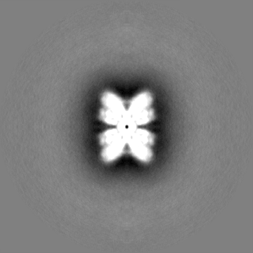
| Projections & slices | Image control
Images are generated by Spider. | ||||||||||||||||||||||||||||||||||||
| Voxel size | X=Y=Z: 1.06 Å | ||||||||||||||||||||||||||||||||||||
| Density |
| ||||||||||||||||||||||||||||||||||||
| Symmetry | Space group: 1 | ||||||||||||||||||||||||||||||||||||
| Details | EMDB XML:
|
 Z (Sec.)
 Y (Row.)
 X (Col.)


-Supplemental data
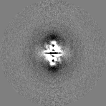
-Half map: #2
| File | emd_16223_half_map_1.map | ||||||||||||
|---|---|---|---|---|---|---|---|---|---|---|---|---|---|
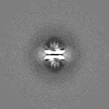
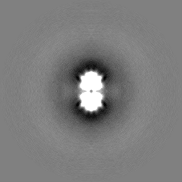
| Projections & Slices |
| ||||||||||||
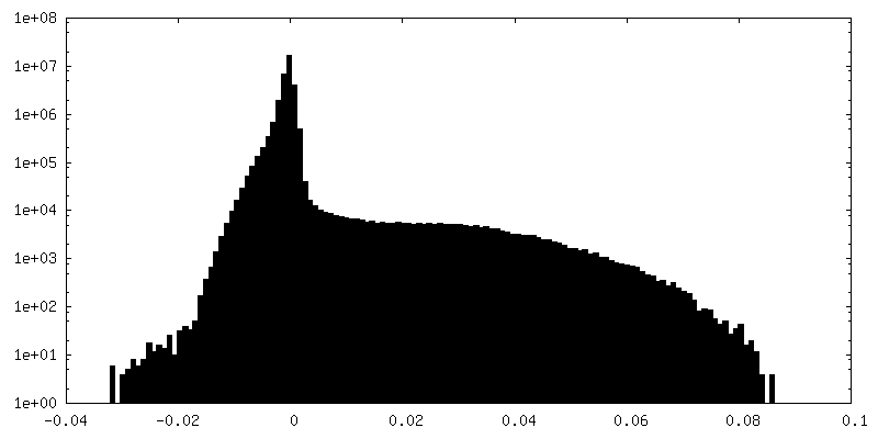
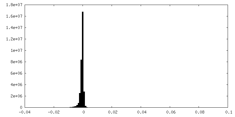
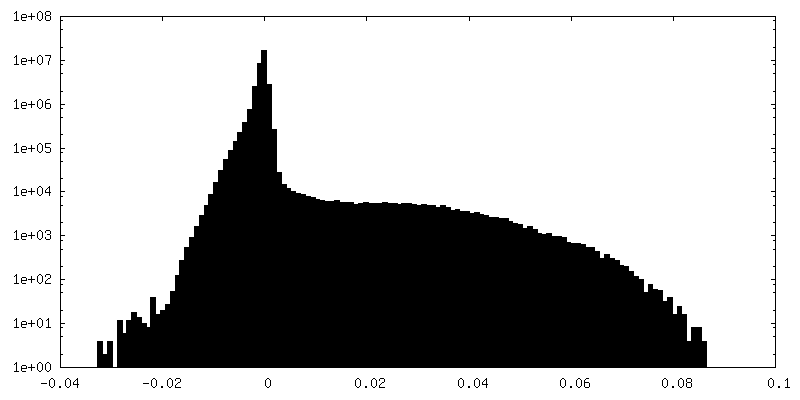
| Density Histograms |


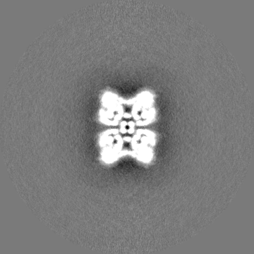
-Half map: #1
| File | emd_16223_half_map_2.map | ||||||||||||
|---|---|---|---|---|---|---|---|---|---|---|---|---|---|
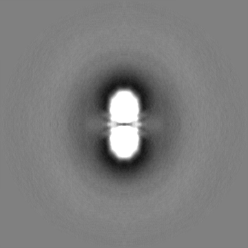
| Projections & Slices |
| ||||||||||||
| Density Histograms |

- Sample components
-Entire : 47 N-terminally truncated human tryptophan hydroxylase 2
| Entire | Name: 47 N-terminally truncated human tryptophan hydroxylase 2 |
|---|---|
| Components |
|
-Supramolecule #1: 47 N-terminally truncated human tryptophan hydroxylase 2
| Supramolecule | Name: 47 N-terminally truncated human tryptophan hydroxylase 2 type: complex / ID: 1 / Parent: 0 |
|---|---|
| Source (natural) | Organism: Homo sapiens (human) |
-Experimental details
-Structure determination
| Method | cryo EM |
|---|---|
 Processing Processing | single particle reconstruction |
| Aggregation state | particle |
-Sample preparation
| Buffer | pH: 7 |
|---|---|
| Vitrification | Cryogen name: ETHANE |
- Electron microscopy
| Microscope | FEI TITAN KRIOS |
|---|---|
| Image recording | Film or detector model: FEI FALCON III (4k x 4k) / Detector mode: INTEGRATING / Average electron dose: 60.0 e/Å2 |
| Electron beam | Acceleration voltage: 300 kV / Electron source:  FIELD EMISSION GUN FIELD EMISSION GUN |
| Electron optics | Illumination mode: FLOOD BEAM / Imaging mode: BRIGHT FIELD / Nominal defocus max: 4.0 µm / Nominal defocus min: 1.0 µm |
| Experimental equipment |  Model: Titan Krios / Image courtesy: FEI Company |
-Image processing
| Startup model | Type of model: OTHER |
|---|---|
| Final reconstruction | Resolution.type: BY AUTHOR / Resolution: 3.9 Å / Resolution method: FSC 0.143 CUT-OFF / Number images used: 197917 |
| Initial angle assignment | Type: MAXIMUM LIKELIHOOD |
| Final angle assignment | Type: MAXIMUM LIKELIHOOD |
