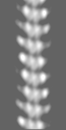
 Movie
Movie Controller

[English] 日本語
 Yorodumi
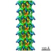
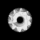
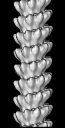
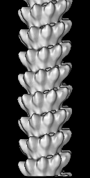
- EMDB-1622: Helical reconstruction of Respiratory Syncytial Virus N-RNA helices -
+ Open data

- Basic information
| Entry | Database: EMDB / ID: EMD-1622 | |||||||||
|---|---|---|---|---|---|---|---|---|---|---|
| Title | Helical reconstruction of Respiratory Syncytial Virus N-RNA helices | |||||||||
 Map data Map data | Helical reconstruction of Respiratory Syncytial Virus N-RNA nucleocapsid-like structure. (Related to PDB entry 2wj8) | |||||||||
 Sample Sample |
| |||||||||
 Keywords Keywords | paramyxovirus / virus / nucleocapsid / RNP / nucleoprotein | |||||||||
| Biological species |  Respiratory syncytial virus Respiratory syncytial virus | |||||||||
| Method | helical reconstruction / cryo EM / negative staining / Resolution: 26.0 Å | |||||||||
 Authors Authors | MacLellan K / Yeo RP / Bhella D | |||||||||
 Citation Citation | Journal: Science / Year: 2009 Title: Crystal structure of a nucleocapsid-like nucleoprotein-RNA complex of respiratory syncytial virus. Authors: Rajiv G Tawar / Stéphane Duquerroy / Clemens Vonrhein / Paloma F Varela / Laurence Damier-Piolle / Nathalie Castagné / Kirsty MacLellan / Hugues Bedouelle / Gérard Bricogne / David Bhella ...Authors: Rajiv G Tawar / Stéphane Duquerroy / Clemens Vonrhein / Paloma F Varela / Laurence Damier-Piolle / Nathalie Castagné / Kirsty MacLellan / Hugues Bedouelle / Gérard Bricogne / David Bhella / Jean-François Eléouët / Félix A Rey /  Abstract: The respiratory syncytial virus (RSV) is an important human pathogen, yet neither a vaccine nor effective therapies are available to treat infection. To help elucidate the replication mechanism of ...The respiratory syncytial virus (RSV) is an important human pathogen, yet neither a vaccine nor effective therapies are available to treat infection. To help elucidate the replication mechanism of this RNA virus, we determined the three-dimensional (3D) crystal structure at 3.3 A resolution of a decameric, annular ribonucleoprotein complex of the RSV nucleoprotein (N) bound to RNA. This complex mimics one turn of the viral helical nucleocapsid complex, which serves as template for viral RNA synthesis. The RNA wraps around the protein ring, with seven nucleotides contacting each N subunit, alternating rows of four and three stacked bases that are exposed and buried within a protein groove, respectively. Combined with electron microscopy data, this structure provides a detailed model for the RSV nucleocapsid, in which the bases are accessible for readout by the viral polymerase. Furthermore, the nucleoprotein structure highlights possible key sites for drug targeting. | |||||||||
| History |
|
- Structure visualization
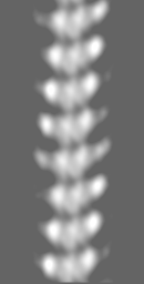
| Movie |
 Movie viewer Movie viewer |
|---|---|
| Structure viewer | EM map: SurfViewMolmilJmol/JSmol |
| Supplemental images |
 UCSF Chimera
- Downloads & links
-EMDB archive
| Map data | emd_1622.map.gz | 3.5 MB | EMDB map data format | |
|---|---|---|---|---|
| Header (meta data) | emd-1622-v30.xmlemd-1622.xml | 10.1 KB 10.1 KB | Display Display | EMDB header |
| Images |  emd_1622.png emd_1622.png | 1.2 MB | ||
| Archive directory |  http://ftp.pdbj.org/pub/emdb/structures/EMD-1622ftp://ftp.pdbj.org/pub/emdb/structures/EMD-1622 http://ftp.pdbj.org/pub/emdb/structures/EMD-1622ftp://ftp.pdbj.org/pub/emdb/structures/EMD-1622 | HTTPS FTP |
-Related structure data

-Links
| EMDB pages | EMDB (EBI/PDBe) / EMDataResource |
|---|
-Map
| File | Download / File: emd_1622.map.gz / Format: CCP4 / Size: 16.1 MB / Type: IMAGE STORED AS FLOATING POINT NUMBER (4 BYTES) | ||||||||||||||||||||||||||||||||||||||||||||||||||||||||||||||||||||
|---|---|---|---|---|---|---|---|---|---|---|---|---|---|---|---|---|---|---|---|---|---|---|---|---|---|---|---|---|---|---|---|---|---|---|---|---|---|---|---|---|---|---|---|---|---|---|---|---|---|---|---|---|---|---|---|---|---|---|---|---|---|---|---|---|---|---|---|---|---|
| Annotation | Helical reconstruction of Respiratory Syncytial Virus N-RNA nucleocapsid-like structure. (Related to PDB entry 2wj8) | ||||||||||||||||||||||||||||||||||||||||||||||||||||||||||||||||||||
| Projections & slices | Image control
Images are generated by Spider. generated in cubic-lattice coordinate | ||||||||||||||||||||||||||||||||||||||||||||||||||||||||||||||||||||
| Voxel size | X=Y=Z: 2.18 Å | ||||||||||||||||||||||||||||||||||||||||||||||||||||||||||||||||||||
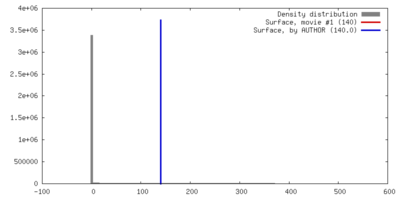
| Density |
| ||||||||||||||||||||||||||||||||||||||||||||||||||||||||||||||||||||
| Symmetry | Space group: 1 | ||||||||||||||||||||||||||||||||||||||||||||||||||||||||||||||||||||
| Details | EMDB XML:
CCP4 map header:
| ||||||||||||||||||||||||||||||||||||||||||||||||||||||||||||||||||||
 Z (Sec.)
 Y (Row.)
 X (Col.)


-Supplemental data
- Sample components
-Entire : Respiratory Syncytial Virus Nucleoprotein-RNA
| Entire | Name: Respiratory Syncytial Virus Nucleoprotein-RNA |
|---|---|
| Components |
|
-Supramolecule #1000: Respiratory Syncytial Virus Nucleoprotein-RNA
| Supramolecule | Name: Respiratory Syncytial Virus Nucleoprotein-RNA / type: sample / ID: 1000 / Details: Nucleoprotein was expressed in insect cells / Number unique components: 1 |
|---|
-Macromolecule #1: Nucleocapsid protein
| Macromolecule | Name: Nucleocapsid protein / type: protein_or_peptide / ID: 1 / Name.synonym: nucleoprotein / Oligomeric state: helical / Recombinant expression: Yes |
|---|---|
| Source (natural) | Organism: Respiratory syncytial virus / synonym: RSV / Cell: SF21 |
| Molecular weight | Theoretical: 43 MDa |
| Recombinant expression | Organism: unidentified baculovirus |
-Experimental details
-Structure determination
| Method | negative staining, cryo EM |
|---|---|
 Processing Processing | helical reconstruction |
| Aggregation state | filament |
-Sample preparation
| Concentration | 0.1 mg/mL |
|---|---|
| Buffer | pH: 7.4 / Details: Phosphate buffered saline |
| Staining | Type: NEGATIVE Details: Cryo-negative staining 5 ul of protein suspension at an approximate concentration of 0.2 mg/ml was loaded onto a freshly glow-discharged Quantifoil holey carbon support film for ...Details: Cryo-negative staining 5 ul of protein suspension at an approximate concentration of 0.2 mg/ml was loaded onto a freshly glow-discharged Quantifoil holey carbon support film for approximately 10 seconds. The grid was then transferred to a droplet of 20% (w/v) ammonium molybdate solution (pH 7.4) for approximately 10 seconds, blotted for 2-3 seconds and plunged into a bath of liquid nitrogen cooled ethane slush |
| Grid | Details: 400 mesh quantifoil |
| Vitrification | Cryogen name: ETHANE / Instrument: OTHER / Method: blot for 2 seconds, wait for 2 seconds plunge |
- Electron microscopy
| Microscope | JEOL 1200EXII |
|---|---|
| Alignment procedure | Legacy - Astigmatism: objective astigmatism corrected at 200k x |
| Image recording | Category: FILM / Film or detector model: KODAK SO-163 FILM / Digitization - Scanner: NIKON COOLSCAN / Digitization - Sampling interval: 2.18 µm / Average electron dose: 10 e/Å2 / Bits/pixel: 16 |
| Electron beam | Acceleration voltage: 120 kV / Electron source: LAB6 |
| Electron optics | Calibrated magnification: 29200 / Illumination mode: FLOOD BEAM / Imaging mode: BRIGHT FIELD / Cs: 3.4 mm / Nominal magnification: 30000 |
| Sample stage | Specimen holder: side entry / Specimen holder model: OTHER |
-Image processing
| Final reconstruction | Applied symmetry - Helical parameters - Δz: 7 Å Applied symmetry - Helical parameters - Δ&Phi: 36.8 ° Algorithm: OTHER / Resolution.type: BY AUTHOR / Resolution: 26.0 Å / Resolution method: OTHER / Software - Name: SPIDER, HELICALS, HELICALI |
|---|
