Inactivation, recovery and regulation of the phototransduction cascade / methionyl aminopeptidase / initiator methionyl aminopeptidase activity / maturation of SSU-rRNA from tricistronic rRNA transcript (SSU-rRNA, LSU-rRNA,5S) / regulation of amino acid metabolic process / negative regulation of glucose mediated signaling pathway / translational readthrough / mTORC1-mediated signalling / Protein hydroxylation / PELO:HBS1L and ABCE1 dissociate a ribosome on a non-stop mRNA ...Inactivation, recovery and regulation of the phototransduction cascade / methionyl aminopeptidase / initiator methionyl aminopeptidase activity / maturation of SSU-rRNA from tricistronic rRNA transcript (SSU-rRNA, LSU-rRNA,5S) / regulation of amino acid metabolic process / negative regulation of glucose mediated signaling pathway / translational readthrough / mTORC1-mediated signalling / Protein hydroxylation / PELO:HBS1L and ABCE1 dissociate a ribosome on a non-stop mRNA / pre-mRNA 5'-splice site binding / GDP-dissociation inhibitor activity / cytosolic large ribosomal subunit assembly / nonfunctional rRNA decay / Formation of the ternary complex, and subsequently, the 43S complex / Translation initiation complex formation / Ribosomal scanning and start codon recognition / response to cycloheximide / ribosome-associated ubiquitin-dependent protein catabolic process / cleavage in ITS2 between 5.8S rRNA and LSU-rRNA of tricistronic rRNA transcript (SSU-rRNA, 5.8S rRNA, LSU-rRNA) / positive regulation of protein kinase activity / Major pathway of rRNA processing in the nucleolus and cytosol / mRNA destabilization / metalloaminopeptidase activity / SRP-dependent cotranslational protein targeting to membrane / GTP hydrolysis and joining of the 60S ribosomal subunit / Formation of a pool of free 40S subunits / preribosome, large subunit precursor / Nonsense Mediated Decay (NMD) independent of the Exon Junction Complex (EJC) / Nonsense Mediated Decay (NMD) enhanced by the Exon Junction Complex (EJC) / negative regulation of mRNA splicing, via spliceosome / L13a-mediated translational silencing of Ceruloplasmin expression / negative regulation of translational frameshifting / translational elongation / ribosomal large subunit export from nucleus / G-protein alpha-subunit binding / 90S preribosome / endonucleolytic cleavage to generate mature 3'-end of SSU-rRNA from (SSU-rRNA, 5.8S rRNA, LSU-rRNA) / ribosomal subunit export from nucleus / regulation of translational fidelity / maturation of LSU-rRNA / protein-RNA complex assembly / endonucleolytic cleavage in ITS1 to separate SSU-rRNA from 5.8S rRNA and LSU-rRNA from tricistronic rRNA transcript (SSU-rRNA, 5.8S rRNA, LSU-rRNA) / translation regulator activity / rescue of stalled cytosolic ribosome / positive regulation of apoptotic signaling pathway / cellular response to amino acid starvation / protein kinase C binding / cytosolic ribosome / ribosomal large subunit biogenesis / maturation of LSU-rRNA from tricistronic rRNA transcript (SSU-rRNA, 5.8S rRNA, LSU-rRNA) / macroautophagy / maturation of SSU-rRNA from tricistronic rRNA transcript (SSU-rRNA, 5.8S rRNA, LSU-rRNA) / maturation of SSU-rRNA / translational initiation / small-subunit processome / protein processing / maintenance of translational fidelity / modification-dependent protein catabolic process / cytoplasmic stress granule / protein tag activity / rRNA processing / ribosomal small subunit assembly / ribosome binding / ribosome biogenesis / ribosomal small subunit biogenesis / 5S rRNA binding / ribosomal large subunit assembly / small ribosomal subunit / small ribosomal subunit rRNA binding / large ribosomal subunit rRNA binding / cytosolic small ribosomal subunit / cytosolic large ribosomal subunit / cytoplasmic translation / negative regulation of translation / rRNA binding / structural constituent of ribosome / protein ubiquitination / ribosome / translation / G protein-coupled receptor signaling pathway / ribonucleoprotein complex / negative regulation of gene expression / response to antibiotic / mRNA binding / ubiquitin protein ligase binding / nucleolus / mitochondrion / RNA binding / zinc ion binding / nucleoplasm / metal ion binding / nucleus / cytosol / cytoplasm Similarity search - Function
MYND-like zinc finger / zf-MYND-like zinc finger, mRNA-binding / Zinc finger C6H2-type profile. / Methionine aminopeptidase subfamily 1 signature. / Peptidase M24A, methionine aminopeptidase, subfamily 1 / Peptidase M24, methionine aminopeptidase / Peptidase M24 / Metallopeptidase family M24 / Creatinase/aminopeptidase-like / : ...MYND-like zinc finger / zf-MYND-like zinc finger, mRNA-binding / Zinc finger C6H2-type profile. / Methionine aminopeptidase subfamily 1 signature. / Peptidase M24A, methionine aminopeptidase, subfamily 1 / Peptidase M24, methionine aminopeptidase / Peptidase M24 / Metallopeptidase family M24 / Creatinase/aminopeptidase-like / : / Ribosomal protein L1, conserved site / Ribosomal protein L1 signature. / Ribosomal protein L1 / Ribosomal protein L1, 3-layer alpha/beta-sandwich / : / Ribosomal protein S26e signature. / Ribosomal protein L41 / Ribosomal protein L41 / Ribosomal protein L13e, conserved site / Ribosomal protein L13e signature. / Ribosomal protein S21e, conserved site / Ribosomal protein S21e signature. / : / Ribosomal protein S12e signature. / Ribosomal protein L29e / Ribosomal L29e protein family / Ribosomal protein S26e / Ribosomal protein S26e superfamily / Ribosomal protein S26e / Ribosomal protein S12e / Ribosomal protein L1-like / Ribosomal protein L1/ribosomal biogenesis protein / Ribosomal protein L1p/L10e family / Small (40S) ribosomal subunit Asc1/RACK1 / Ribosomal protein L22e / Ribosomal protein L22e superfamily / Ribosomal L22e protein family / Ribosomal protein L27e, conserved site / Ribosomal protein L27e signature. / Ribosomal protein L13e / Ribosomal protein L13e / Ribosomal protein S5, eukaryotic/archaeal / : / Ribosomal protein L38e / Ribosomal protein L38e superfamily / Ribosomal L38e protein family / Ribosomal protein L6e signature. / Ribosomal protein S19e, conserved site / Ribosomal protein S19e signature. / Ribosomal protein S21e / Ribosomal protein S21e superfamily / Ribosomal protein S21e / Ribosomal protein L19, eukaryotic / 60S ribosomal protein L18a/ L20, eukaryotes / Ribosomal protein S2, eukaryotic / Ribosomal protein L10e, conserved site / Ribosomal protein L10e signature. / 40S Ribosomal protein S10 / Ribosomal protein L18/L18-A/B/e, conserved site / Ribosomal protein L18e signature. / Ribosomal protein L19/L19e conserved site / Ribosomal protein L19e signature. / Ribosomal protein L44e signature. / Ribosomal protein L24e, conserved site / Ribosomal protein L24e signature. / S27a-like superfamily / Plectin/S10, N-terminal / Plectin/S10 domain / Ribosomal protein L10e / Ribosomal protein L34e, conserved site / Ribosomal protein L34e signature. / Ribosomal protein L5 eukaryotic, C-terminal / Ribosomal L18 C-terminal region / Ribosomal protein L23/L25, N-terminal / Ribosomal protein L23, N-terminal domain / Ribosomal protein S10, eukaryotic/archaeal / Ribosomal protein S30 / Ribosomal protein S30 / Ribosomal protein L30e signature 1. / Ribosomal L40e family / Ribosomal protein L36e signature. / 50S ribosomal protein L18Ae/60S ribosomal protein L20 and L18a / Ribosomal protein L35Ae, conserved site / Ribosomal protein L35Ae signature. / Eukaryotic Ribosomal Protein L27, KOW domain / : / Ribosomal protein S25 / Ribosomal protein 50S-L18Ae/60S-L20/60S-L18A / Ribosomal proteins 50S-L18Ae/60S-L20/60S-L18A / S25 ribosomal protein / : / Ribosomal_L40e / Ribosomal protein L27e / Ribosomal protein L40e / Ribosomal protein L40e superfamily / Ribosomal protein L27e superfamily / Ribosomal L27e protein family / Ribosomal protein L44e / Ribosomal Protein L6, KOW domain / Ribosomal protein L44 Similarity search - Domain/homology
Rps5p / RPS22A isoform 1 / RPL38 isoform 1 / RPL10 isoform 1 / RPS29A isoform 1 / RPS20 isoform 1 / RPS2 isoform 1 / 60S ribosomal protein L29 / RPL11B isoform 1 / 40S ribosomal protein S25 ...Rps5p / RPS22A isoform 1 / RPL38 isoform 1 / RPL10 isoform 1 / RPS29A isoform 1 / RPS20 isoform 1 / RPS2 isoform 1 / 60S ribosomal protein L29 / RPL11B isoform 1 / 40S ribosomal protein S25 / 40S ribosomal protein S26 / RPL5 isoform 1 / RPL32 isoform 1 / 40S ribosomal protein S3 / RPL4A isoform 1 / Large ribosomal subunit protein uL3 / RPS28A isoform 1 / 40S ribosomal protein S12 / RPL9A isoform 1 / 60S ribosomal protein L41 / Small ribosomal subunit protein uS2A / Small ribosomal subunit protein eS1A / Small ribosomal subunit protein uS4A / Large ribosomal subunit protein uL15 / Small ribosomal subunit protein eS17A / Large ribosomal subunit protein eL24A / Large ribosomal subunit protein uL23 / Large ribosomal subunit protein eL39 / Large ribosomal subunit protein uL30A / Large ribosomal subunit protein eL6B / Large ribosomal subunit protein uL22A / Large ribosomal subunit protein uL24A / Large ribosomal subunit protein eL33A / Large ribosomal subunit protein eL36A / Large ribosomal subunit protein eL15A / Large ribosomal subunit protein eL22A / Small ribosomal subunit protein uS15 / Ubiquitin-ribosomal protein eS31 fusion protein / Small ribosomal subunit protein eS19A / Small ribosomal subunit protein eS21A / Large ribosomal subunit protein eL27A / Large ribosomal subunit protein eL31A / Ubiquitin-ribosomal protein eL40A fusion protein / Large ribosomal subunit protein eL20A / Large ribosomal subunit protein eL43A / Large ribosomal subunit protein eL42A / Small ribosomal subunit protein uS12A / Small ribosomal subunit protein eS24A / Small ribosomal subunit protein eS30A / Small ribosomal subunit protein eS4A / Small ribosomal subunit protein eS6A / Small ribosomal subunit protein eS8B / Large ribosomal subunit protein uL14A / Large ribosomal subunit protein uL1A / Large ribosomal subunit protein uL2A / Small ribosomal subunit protein uS17A / Large ribosomal subunit protein eL18A / Small ribosomal subunit protein uS9A / Small ribosomal subunit protein uS13A / Large ribosomal subunit protein eL19A / Large ribosomal subunit protein uL29A / Large ribosomal subunit protein eL30 / Large ribosomal subunit protein eL8A / Large ribosomal subunit protein uL13A / Small ribosomal subunit protein eS7A / Small ribosomal subunit protein eS27A / Large ribosomal subunit protein eL14A / Small ribosomal subunit protein RACK1 / Small ribosomal subunit protein uS11B / Large ribosomal subunit protein eL37A / Large ribosomal subunit protein eL34A / Methionine aminopeptidase 1 / Small ribosomal subunit protein uS19 / Large ribosomal subunit protein eL21A / Small ribosomal subunit protein eS10A / Large ribosomal subunit protein eL13A Similarity search - Component
Biological species
Saccharomyces cerevisiae (brewer's yeast)
Method
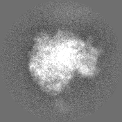
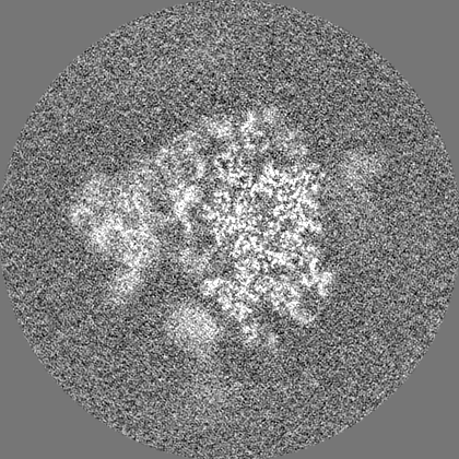
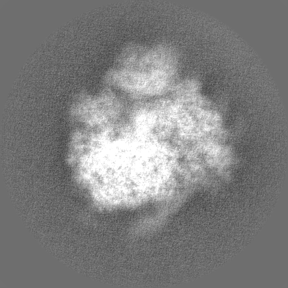
single particle reconstruction / cryo EM / Resolution: 3.9 Å
Journal: PLoS Biol / Year: 2023 Title: The dynamic architecture of Map1- and NatB-ribosome complexes coordinates the sequential modifications of nascent polypeptide chains. Authors: Alexandra G Knorr / Timur Mackens-Kiani / Joanna Musial / Otto Berninghausen / Thomas Becker / Birgitta Beatrix / Roland Beckmann / Abstract: Cotranslational modification of the nascent polypeptide chain is one of the first events during the birth of a new protein. In eukaryotes, methionine aminopeptidases (MetAPs) cleave off the starter ...Cotranslational modification of the nascent polypeptide chain is one of the first events during the birth of a new protein. In eukaryotes, methionine aminopeptidases (MetAPs) cleave off the starter methionine, whereas N-acetyl-transferases (NATs) catalyze N-terminal acetylation. MetAPs and NATs compete with other cotranslationally acting chaperones, such as ribosome-associated complex (RAC), protein targeting and translocation factors (SRP and Sec61) for binding sites at the ribosomal tunnel exit. Yet, whereas well-resolved structures for ribosome-bound RAC, SRP and Sec61, are available, structural information on the mode of ribosome interaction of eukaryotic MetAPs or of the five cotranslationally active NATs is only available for NatA. Here, we present cryo-EM structures of yeast Map1 and NatB bound to ribosome-nascent chain complexes. Map1 is mainly associated with the dynamic rRNA expansion segment ES27a, thereby kept at an ideal position below the tunnel exit to act on the emerging substrate nascent chain. For NatB, we observe two copies of the NatB complex. NatB-1 binds directly below the tunnel exit, again involving ES27a, and NatB-2 is located below the second universal adapter site (eL31 and uL22). The binding mode of the two NatB complexes on the ribosome differs but overlaps with that of NatA and Map1, implying that NatB binds exclusively to the tunnel exit. We further observe that ES27a adopts distinct conformations when bound to NatA, NatB, or Map1, together suggesting a contribution to the coordination of a sequential activity of these factors on the emerging nascent chain at the ribosomal exit tunnel.
In the structure databanks used in Yorodumi, some data are registered as the other names, "COVID-19 virus" and "2019-nCoV". Here are the details of the virus and the list of structure data.
Jan 31, 2019. EMDB accession codes are about to change! (news from PDBe EMDB page)
EMDB accession codes are about to change! (news from PDBe EMDB page)
The allocation of 4 digits for EMDB accession codes will soon come to an end. Whilst these codes will remain in use, new EMDB accession codes will include an additional digit and will expand incrementally as the available range of codes is exhausted. The current 4-digit format prefixed with “EMD-” (i.e. EMD-XXXX) will advance to a 5-digit format (i.e. EMD-XXXXX), and so on. It is currently estimated that the 4-digit codes will be depleted around Spring 2019, at which point the 5-digit format will come into force.
The EM Navigator/Yorodumi systems omit the EMD- prefix.
Related info.:Q: What is EMD? / ID/Accession-code notation in Yorodumi/EM Navigator
Yorodumi is a browser for structure data from EMDB, PDB, SASBDB, etc.
This page is also the successor to EM Navigator detail page, and also detail information page/front-end page for Omokage search.
The word "yorodu" (or yorozu) is an old Japanese word meaning "ten thousand". "mi" (miru) is to see.
Related info.:EMDB / PDB / SASBDB / Comparison of 3 databanks / Yorodumi Search / Aug 31, 2016. New EM Navigator & Yorodumi / Yorodumi Papers / Jmol/JSmol / Function and homology information / Changes in new EM Navigator and Yorodumi
 Movie
Movie Controller


 Open data
 Basic information
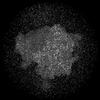
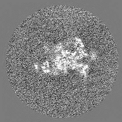
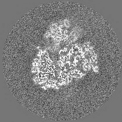
 Map data
 Sample
 Keywords
 Function and homology information
 Authors
 Germany, 1 items
  Citation
 Structure visualization
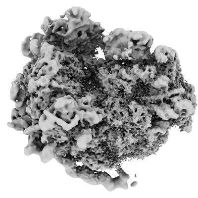
 Downloads & links
 emd_16182.png
 http://ftp.pdbj.org/pub/emdb/structures/EMD-16182


 Z (Sec.)
 Y (Row.)
 X (Col.)


 Sample components
 Processing
 Electron microscopy
 FIELD EMISSION GUN


