Movie
Movie Controller

[English] 日本語
 Yorodumi
- EMDB-16091: Cryo-EM structure of beta-galactosidase at 3.3 A resolution plung... -
+ Open data

- Basic information
| Entry |  | ||||||||||||
|---|---|---|---|---|---|---|---|---|---|---|---|---|---|
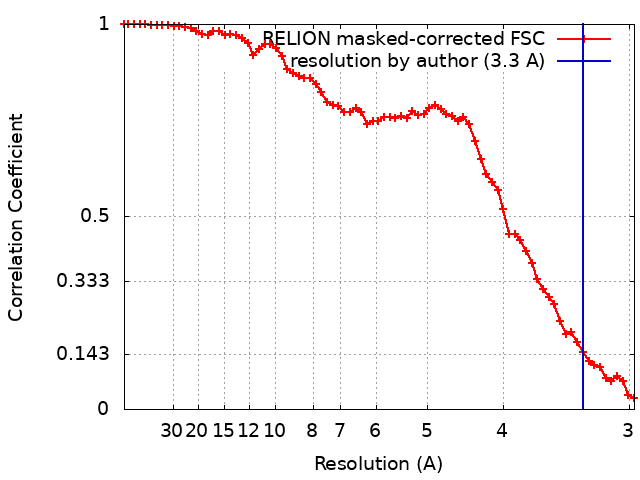
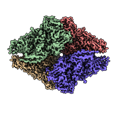
| Title | Cryo-EM structure of beta-galactosidase at 3.3 A resolution plunged 5 ms after mixing with apoferritin | ||||||||||||
 Map data Map data | |||||||||||||
 Sample Sample |
| ||||||||||||
 Keywords Keywords | Glycosyl hydrolase / HYDROLASE | ||||||||||||
| Function / homology |  Function and homology information Function and homology informationalkali metal ion binding / lactose catabolic process / beta-galactosidase complex / beta-galactosidase / beta-galactosidase activity / carbohydrate binding / magnesium ion binding / identical protein binding Similarity search - Function | ||||||||||||
| Biological species |  | ||||||||||||
| Method | single particle reconstruction / cryo EM / Resolution: 3.3 Å | ||||||||||||
 Authors Authors | Torino S / Dhurandhar M / Efremov R | ||||||||||||
| Funding support | European Union,  Belgium, 3 items Belgium, 3 items
| ||||||||||||
 Citation Citation | Journal: Nat Methods / Year: 2023 Title: Time-resolved cryo-EM using a combination of droplet microfluidics with on-demand jetting. Authors: Stefania Torino / Mugdha Dhurandhar / Annelore Stroobants / Raf Claessens / Rouslan G Efremov / Abstract: Single-particle cryogenic electron microscopy (cryo-EM) allows reconstruction of high-resolution structures of proteins in different conformations. Protein function often involves transient ...Single-particle cryogenic electron microscopy (cryo-EM) allows reconstruction of high-resolution structures of proteins in different conformations. Protein function often involves transient functional conformations, which can be resolved using time-resolved cryo-EM (trEM). In trEM, reactions are arrested after a defined delay time by rapid vitrification of protein solution on the EM grid. Despite the increasing interest in trEM among the cryo-EM community, making trEM samples with a time resolution below 100 ms remains challenging. Here we report the design and the realization of a time-resolved cryo-plunger that combines a droplet-based microfluidic mixer with a laser-induced generator of microjets that allows rapid reaction initiation and plunge-freezing of cryo-EM grids. Using this approach, a time resolution of 5 ms was achieved and the protein density map was reconstructed to a resolution of 2.1 Å. trEM experiments on GroEL:GroES chaperonin complex resolved the kinetics of the complex formation and visualized putative short-lived conformations of GroEL-ATP complex. | ||||||||||||
| History |
|
- Structure visualization
| Supplemental images |
|---|
- Downloads & links
-EMDB archive
| Map data | emd_16091.map.gz | 20.8 MB | EMDB map data format | |
|---|---|---|---|---|
| Header (meta data) | emd-16091-v30.xmlemd-16091.xml | 20.9 KB 20.9 KB | Display Display | EMDB header |
| FSC (resolution estimation) | emd_16091_fsc.xml | 6.4 KB | Display | FSC data file |
| Images |  emd_16091.png emd_16091.png | 150.4 KB | ||
| Masks | emd_16091_msk_1.map | 22.2 MB | Mask map | |
| Filedesc metadata | emd-16091.cif.gz | 7.1 KB | ||
| Others | emd_16091_half_map_1.map.gzemd_16091_half_map_2.map.gz | 16.1 MB 16.1 MB | ||
| Archive directory |  http://ftp.pdbj.org/pub/emdb/structures/EMD-16091ftp://ftp.pdbj.org/pub/emdb/structures/EMD-16091 http://ftp.pdbj.org/pub/emdb/structures/EMD-16091ftp://ftp.pdbj.org/pub/emdb/structures/EMD-16091 | HTTPS FTP |
-Related structure data
| Related structure data |  8bk7MC  8bk8C  8bk9C  8bkaC  8bkbC  8bkgC  8bkzC  8bl2C  8bl7C  8blcC  8bldC  8bleC  8blfC  8blyC  8bm0C  8bm1C  8bmdC  8bmoC  8bmtC M: atomic model generated by this map C: citing same article ( |
|---|---|
| Similar structure data |


-Links
| EMDB pages | EMDB (EBI/PDBe) / EMDataResource |
|---|---|
| Related items in Molecule of the Month |


-Map
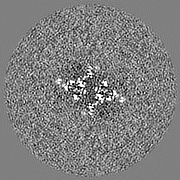
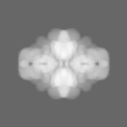
| File | Download / File: emd_16091.map.gz / Format: CCP4 / Size: 22.2 MB / Type: IMAGE STORED AS FLOATING POINT NUMBER (4 BYTES) | ||||||||||||||||||||||||||||||||||||
|---|---|---|---|---|---|---|---|---|---|---|---|---|---|---|---|---|---|---|---|---|---|---|---|---|---|---|---|---|---|---|---|---|---|---|---|---|---|
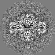
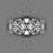
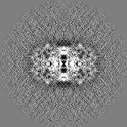
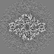
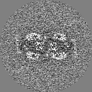
| Projections & slices | Image control
Images are generated by Spider. | ||||||||||||||||||||||||||||||||||||
| Voxel size | X=Y=Z: 1.486 Å | ||||||||||||||||||||||||||||||||||||
| Density |
| ||||||||||||||||||||||||||||||||||||
| Symmetry | Space group: 1 | ||||||||||||||||||||||||||||||||||||
| Details | EMDB XML:
|
 Z (Sec.)
 Y (Row.)
 X (Col.)


-Supplemental data
-Mask #1
| File | emd_16091_msk_1.map | ||||||||||||
|---|---|---|---|---|---|---|---|---|---|---|---|---|---|
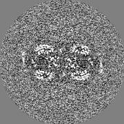
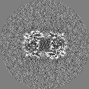
| Projections & Slices |
| ||||||||||||
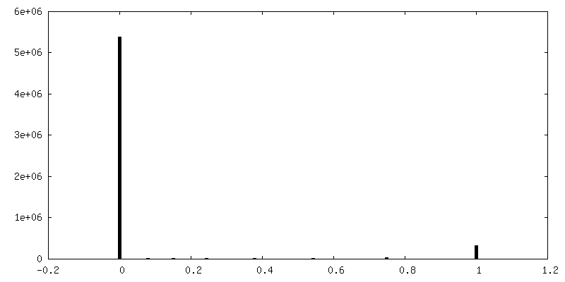
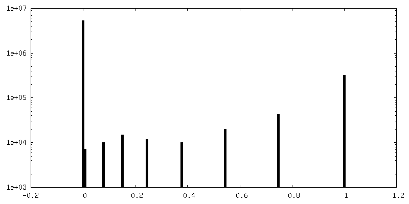
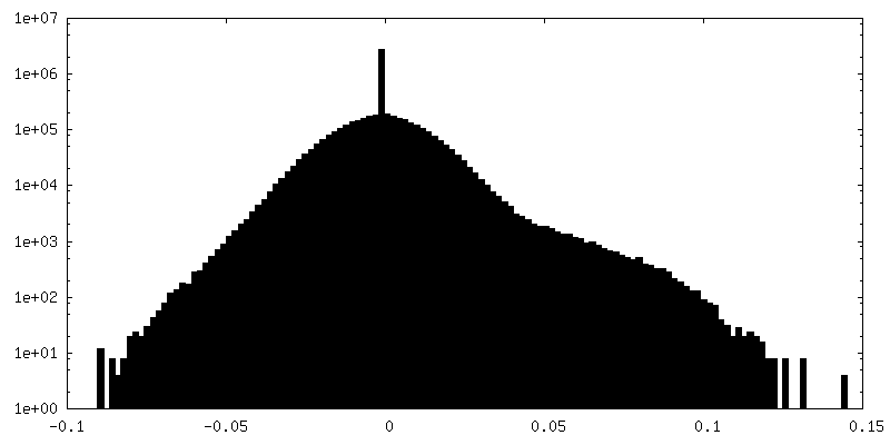
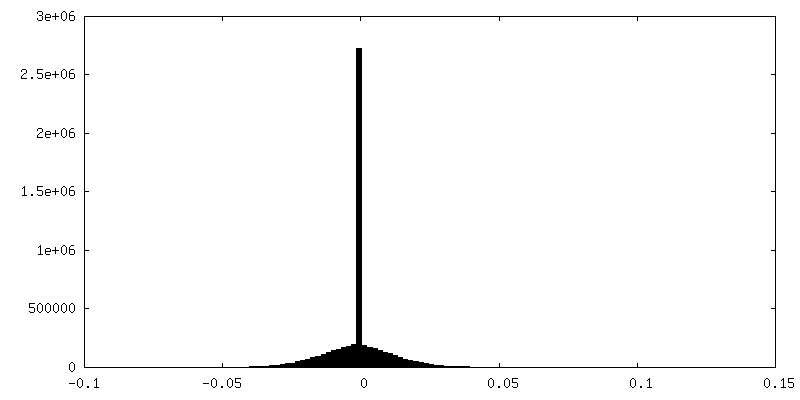
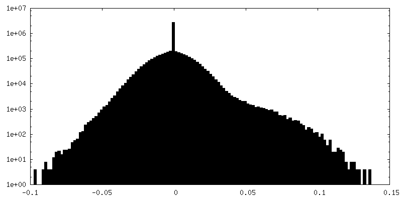
| Density Histograms |


-Half map: #2
| File | emd_16091_half_map_1.map | ||||||||||||
|---|---|---|---|---|---|---|---|---|---|---|---|---|---|
| Projections & Slices |
| ||||||||||||
| Density Histograms |


-Half map: #1
| File | emd_16091_half_map_2.map | ||||||||||||
|---|---|---|---|---|---|---|---|---|---|---|---|---|---|
| Projections & Slices |
| ||||||||||||
| Density Histograms |
- Sample components
-Entire : Beta-galactosidase from E.Coli
| Entire | Name: Beta-galactosidase from E.Coli |
|---|---|
| Components |
|
-Supramolecule #1: Beta-galactosidase from E.Coli
| Supramolecule | Name: Beta-galactosidase from E.Coli / type: complex / ID: 1 / Parent: 0 / Macromolecule list: all |
|---|---|
| Source (natural) | Organism: |
-Macromolecule #1: Beta-galactosidase
| Macromolecule | Name: Beta-galactosidase / type: protein_or_peptide / ID: 1 / Number of copies: 4 / Enantiomer: LEVO / EC number: beta-galactosidase |
|---|---|
| Source (natural) | Organism: |
| Molecular weight | Theoretical: 117.488375 KDa |
| Recombinant expression | Organism: |
| Sequence | String: MTMITDSLAV VLQRRDWENP GVTQLNRLAA HPPFASWRNS EEARTDRPSQ QLRSLNGEWR FAWFPAPEAV PESWLECDLP EADTVVVPS NWQMHGYDAP IYTNVTYPIT VNPPFVPTEN PTGCYSLTFN VDESWLQEGQ TRIIFDGVNS AFHLWCNGRW V GYGQDSRL ...String: MTMITDSLAV VLQRRDWENP GVTQLNRLAA HPPFASWRNS EEARTDRPSQ QLRSLNGEWR FAWFPAPEAV PESWLECDLP EADTVVVPS NWQMHGYDAP IYTNVTYPIT VNPPFVPTEN PTGCYSLTFN VDESWLQEGQ TRIIFDGVNS AFHLWCNGRW V GYGQDSRL PSEFDLSAFL RAGENRLAVM VLRWSDGSYL EDQDMWRMSG IFRDVSLLHK PTTQISDFHV ATRFNDDFSR AV LEAEVQM CGELRDYLRV TVSLWQGETQ VASGTAPFGG EIIDERGGYA DRVTLRLNVE NPKLWSAEIP NLYRAVVELH TAD GTLIEA EACDVGFREV RIENGLLLLN GKPLLIRGVN RHEHHPLHGQ VMDEQTMVQD ILLMKQNNFN AVRCSHYPNH PLWY TLCDR YGLYVVDEAN IETHGMVPMN RLTDDPRWLP AMSERVTRMV QRDRNHPSVI IWSLGNESGH GANHDALYRW IKSVD PSRP VQYEGGGADT TATDIICPMY ARVDEDQPFP AVPKWSIKKW LSLPGETRPL ILCEYAHAMG NSLGGFAKYW QAFRQY PRL QGGFVWDWVD QSLIKYDENG NPWSAYGGDF GDTPNDRQFC MNGLVFADRT PHPALTEAKH QQQFFQFRLS GQTIEVT SE YLFRHSDNEL LHWMVALDGK PLASGEVPLD VAPQGKQLIE LPELPQPESA GQLWLTVRVV QPNATAWSEA GHISAWQQ W RLAENLSVTL PAASHAIPHL TTSEMDFCIE LGNKRWQFNR QSGFLSQMWI GDKKQLLTPL RDQFTRAPLD NDIGVSEAT RIDPNAWVER WKAAGHYQAE AALLQCTADT LADAVLITTA HAWQHQGKTL FISRKTYRID GSGQMAITVD VEVASDTPHP ARIGLNCQL AQVAERVNWL GLGPQENYPD RLTAACFDRW DLPLSDMYTP YVFPSENGLR CGTRELNYGP HQWRGDFQFN I SRYSQQQL METSHRHLLH AEEGTWLNID GFHMGIGGDD SWSPSVSAEF QLSAGRYHYQ LVWCQKGHHH HHH UniProtKB: Beta-galactosidase |
-Experimental details
-Structure determination
| Method | cryo EM |
|---|---|
 Processing Processing | single particle reconstruction |
| Aggregation state | particle |
-Sample preparation
| Concentration | 2.7 mg/mL | |||||||||||||||
|---|---|---|---|---|---|---|---|---|---|---|---|---|---|---|---|---|
| Buffer | pH: 8 Component:
Details: contains Amaranth dye (acid red 27) 32 mM | |||||||||||||||
| Grid | Model: Quantifoil R2/1 / Material: COPPER / Mesh: 300 / Support film - Material: CARBON / Support film - topology: CONTINUOUS / Support film - Film thickness: 3 | |||||||||||||||
| Vitrification | Cryogen name: ETHANE | |||||||||||||||
| Details | Beta-galactosidase monodisperse in buffer |
- Electron microscopy
| Microscope | JEOL CRYO ARM 300 |
|---|---|
| Specialist optics | Energy filter - Name: In-column Omega Filter / Energy filter - Slit width: 20 eV |
| Image recording | Film or detector model: GATAN K3 (6k x 4k) / Number grids imaged: 2 / Number real images: 1733 / Average exposure time: 2.796 sec. / Average electron dose: 59.0 e/Å2 |
| Electron beam | Acceleration voltage: 300 kV / Electron source:  FIELD EMISSION GUN FIELD EMISSION GUN |
| Electron optics | Illumination mode: FLOOD BEAM / Imaging mode: BRIGHT FIELD / Cs: 2.55 mm / Nominal defocus max: 3.5 µm / Nominal defocus min: 0.5 µm / Nominal magnification: 60000 |
| Sample stage | Specimen holder model: JEOL CRYOSPECPORTER / Cooling holder cryogen: NITROGEN |