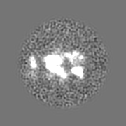
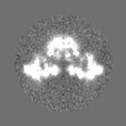
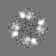
ジャーナル: PLoS Biol / 年: 2009 タイトル: Coordinated movement of cytoplasmic and transmembrane domains of RyR1 upon gating. 著者: Montserrat Samsó / Wei Feng / Isaac N Pessah / P D Allen / 要旨: Ryanodine receptor type 1 (RyR1) produces spatially and temporally defined Ca2+ signals in several cell types. How signals received in the cytoplasmic domain are transmitted to the ion gate and how ...Ryanodine receptor type 1 (RyR1) produces spatially and temporally defined Ca2+ signals in several cell types. How signals received in the cytoplasmic domain are transmitted to the ion gate and how the channel gates are unknown. We used EGTA or neuroactive PCB 95 to stabilize the full closed or open states of RyR1. Single-channel measurements in the presence of FKBP12 indicate that PCB 95 inverts the thermodynamic stability of RyR1 and locks it in a long-lived open state whose unitary current is indistinguishable from the native open state. We analyzed two datasets of 15,625 and 18,527 frozen-hydrated RyR1-FKBP12 particles in the closed and open conformations, respectively, by cryo-electron microscopy. Their corresponding three-dimensional structures at 10.2 A resolution refine the structure surrounding the ion pathway previously identified in the closed conformation: two right-handed bundles emerging from the putative ion gate (the cytoplasmic "inner branches" and the transmembrane "inner helices"). Furthermore, six of the identifiable transmembrane segments of RyR1 have similar organization to those of the mammalian Kv1.2 potassium channel. Upon gating, the distal cytoplasmic domains move towards the transmembrane domain while the central cytoplasmic domains move away from it, and also away from the 4-fold axis. Along the ion pathway, precise relocation of the inner helices and inner branches results in an approximately 4 A diameter increase of the ion gate. Whereas the inner helices of the K+ channels and of the RyR1 channel cross-correlate best with their corresponding open/closed states, the cytoplasmic inner branches, which are not observed in the K+ channels, appear to have at least as important a role as the inner helices for RyR1 gating. We propose a theoretical model whereby the inner helices, the inner branches, and the h1 densities together create an efficient novel gating mechanism for channel opening by relaxing two right-handed bundle structures along a common 4-fold axis.
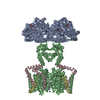
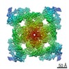
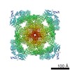
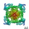
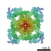
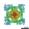
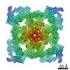
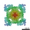
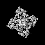
全体 : ryanodine receptor 1 (RyR1) with FKBP12 bound
全体
名称: ryanodine receptor 1 (RyR1) with FKBP12 bound
要素
試料: ryanodine receptor 1 (RyR1) with FKBP12 bound
タンパク質・ペプチド: ryanodine receptor isoform 1
-
超分子 #1000: ryanodine receptor 1 (RyR1) with FKBP12 bound
超分子
名称: ryanodine receptor 1 (RyR1) with FKBP12 bound / タイプ: sample / ID: 1000 集合状態: RyR1 forms a homotetramer and one FKBP12 binds to each RyR1 subunit Number unique components: 2
 ムービー
ムービー コントローラー


 データを開く
 基本情報
基本情報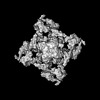
 マップデータ
 試料
試料 キーワード
キーワード 機能・相同性情報
 データ登録者
 引用
引用
 構造の表示
構造の表示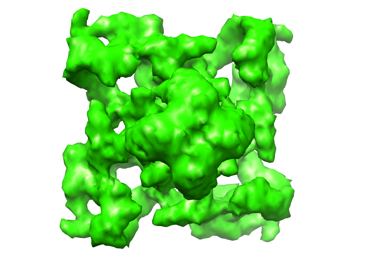
 ダウンロードとリンク
 1607.png
 http://ftp.pdbj.org/pub/emdb/structures/EMD-1607


 Z (Sec.)
 Y (Row.)
 X (Col.)


 試料の構成要素
 解析
解析 電子顕微鏡法
 FIELD EMISSION GUN