+ Open data
- Basic information
| Entry |  | ||||||||||||
|---|---|---|---|---|---|---|---|---|---|---|---|---|---|
| Title | pre-60S with human 5S RNP | ||||||||||||
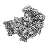
 Map data Map data | pre-60S with human 5S RNP Pre-60S particle from S. cerevisiae with Rpf2, Rrs1, uL18/L5 and uL5/L11 from human (hs). Particles were obtained by split-tag affinity purification of hsuL5 and hsRpf2. | ||||||||||||
 Sample Sample |
| ||||||||||||
 Keywords Keywords | 5S RNP / Ribosome biogenesis / RIBOSOME | ||||||||||||
| Biological species |  Homo sapiens (human) Homo sapiens (human) | ||||||||||||
| Method | single particle reconstruction / cryo EM / Resolution: 3.7 Å | ||||||||||||
 Authors Authors | Castillo N / Thoms M / Flemming D / Hammaren HM / Buschauer R / Ameismeier M / Bassler J / Beck M / Beckmann R / Hurt E | ||||||||||||
| Funding support |  Germany, European Union, 3 items Germany, European Union, 3 items
| ||||||||||||
 Citation Citation |  Journal: Nat Struct Mol Biol / Year: 2023 Journal: Nat Struct Mol Biol / Year: 2023Title: Structure of nascent 5S RNPs at the crossroad between ribosome assembly and MDM2-p53 pathways. Authors: Nestor Miguel Castillo Duque de Estrada / Matthias Thoms / Dirk Flemming / Henrik M Hammaren / Robert Buschauer / Michael Ameismeier / Jochen Baßler / Martin Beck / Roland Beckmann / Ed Hurt /  Abstract: The 5S ribonucleoprotein (RNP) is assembled from its three components (5S rRNA, Rpl5/uL18 and Rpl11/uL5) before being incorporated into the pre-60S subunit. However, when ribosome synthesis is ...The 5S ribonucleoprotein (RNP) is assembled from its three components (5S rRNA, Rpl5/uL18 and Rpl11/uL5) before being incorporated into the pre-60S subunit. However, when ribosome synthesis is disturbed, a free 5S RNP can enter the MDM2-p53 pathway to regulate cell cycle and apoptotic signaling. Here we reconstitute and determine the cryo-electron microscopy structure of the conserved hexameric 5S RNP with fungal or human factors. This reveals how the nascent 5S rRNA associates with the initial nuclear import complex Syo1-uL18-uL5 and, upon further recruitment of the nucleolar factors Rpf2 and Rrs1, develops into the 5S RNP precursor that can assemble into the pre-ribosome. In addition, we elucidate the structure of another 5S RNP intermediate, carrying the human ubiquitin ligase Mdm2, which unravels how this enzyme can be sequestered from its target substrate p53. Our data provide molecular insight into how the 5S RNP can mediate between ribosome biogenesis and cell proliferation. | ||||||||||||
| History |
|
- Structure visualization
| Supplemental images |
|---|
- Downloads & links
-EMDB archive
| Map data |  emd_16040.map.gz emd_16040.map.gz | 21.4 MB |  EMDB map data format EMDB map data format | |
|---|---|---|---|---|
| Header (meta data) |  emd-16040-v30.xml emd-16040-v30.xml emd-16040.xml emd-16040.xml | 17.1 KB 17.1 KB | Display Display |  EMDB header EMDB header |
| Images |  emd_16040.png emd_16040.png | 100.4 KB | ||
| Others |  emd_16040_additional_1.map.gz emd_16040_additional_1.map.gz emd_16040_additional_2.map.gz emd_16040_additional_2.map.gz emd_16040_half_map_1.map.gz emd_16040_half_map_1.map.gz emd_16040_half_map_2.map.gz emd_16040_half_map_2.map.gz | 450.6 MB 237.6 MB 442.4 MB 442.4 MB | ||
| Archive directory |  http://ftp.pdbj.org/pub/emdb/structures/EMD-16040 http://ftp.pdbj.org/pub/emdb/structures/EMD-16040 ftp://ftp.pdbj.org/pub/emdb/structures/EMD-16040 ftp://ftp.pdbj.org/pub/emdb/structures/EMD-16040 | HTTPS FTP |
-Validation report
| Summary document |  emd_16040_validation.pdf.gz emd_16040_validation.pdf.gz | 838 KB | Display |  EMDB validaton report EMDB validaton report |
|---|---|---|---|---|
| Full document |  emd_16040_full_validation.pdf.gz emd_16040_full_validation.pdf.gz | 837.6 KB | Display | |
| Data in XML |  emd_16040_validation.xml.gz emd_16040_validation.xml.gz | 18.6 KB | Display | |
| Data in CIF |  emd_16040_validation.cif.gz emd_16040_validation.cif.gz | 22.3 KB | Display | |
| Arichive directory |  https://ftp.pdbj.org/pub/emdb/validation_reports/EMD-16040 https://ftp.pdbj.org/pub/emdb/validation_reports/EMD-16040 ftp://ftp.pdbj.org/pub/emdb/validation_reports/EMD-16040 ftp://ftp.pdbj.org/pub/emdb/validation_reports/EMD-16040 | HTTPS FTP |
-Related structure data
- Links
Links
| EMDB pages |  EMDB (EBI/PDBe) / EMDB (EBI/PDBe) /  EMDataResource EMDataResource |
|---|
- Map
Map
| File |  Download / File: emd_16040.map.gz / Format: CCP4 / Size: 476.8 MB / Type: IMAGE STORED AS FLOATING POINT NUMBER (4 BYTES) Download / File: emd_16040.map.gz / Format: CCP4 / Size: 476.8 MB / Type: IMAGE STORED AS FLOATING POINT NUMBER (4 BYTES) | ||||||||||||||||||||||||||||||||||||
|---|---|---|---|---|---|---|---|---|---|---|---|---|---|---|---|---|---|---|---|---|---|---|---|---|---|---|---|---|---|---|---|---|---|---|---|---|---|
| Annotation | pre-60S with human 5S RNP Pre-60S particle from S. cerevisiae with Rpf2, Rrs1, uL18/L5 and uL5/L11 from human (hs). Particles were obtained by split-tag affinity purification of hsuL5 and hsRpf2. | ||||||||||||||||||||||||||||||||||||
| Projections & slices | Image control
Images are generated by Spider. | ||||||||||||||||||||||||||||||||||||
| Voxel size | X=Y=Z: 1.084 Å | ||||||||||||||||||||||||||||||||||||
| Density |
| ||||||||||||||||||||||||||||||||||||
| Symmetry | Space group: 1 | ||||||||||||||||||||||||||||||||||||
| Details | EMDB XML:
|
-Supplemental data
-Additional map: pre-60S with human 5S RNP (Homogeneous Refinement, sharpened map)
| File | emd_16040_additional_1.map | ||||||||||||
|---|---|---|---|---|---|---|---|---|---|---|---|---|---|
| Annotation | pre-60S with human 5S RNP (Homogeneous Refinement, sharpened map) | ||||||||||||
| Projections & Slices |
| ||||||||||||
| Density Histograms |
-Additional map: pre-60S with human 5S RNP (Homogeneous Refinement)
| File | emd_16040_additional_2.map | ||||||||||||
|---|---|---|---|---|---|---|---|---|---|---|---|---|---|
| Annotation | pre-60S with human 5S RNP (Homogeneous Refinement) | ||||||||||||
| Projections & Slices |
| ||||||||||||
| Density Histograms |
-Half map: pre-60S with human 5S RNP (half map A, Homogeneous Refinement)
| File | emd_16040_half_map_1.map | ||||||||||||
|---|---|---|---|---|---|---|---|---|---|---|---|---|---|
| Annotation | pre-60S with human 5S RNP (half map A, Homogeneous Refinement) | ||||||||||||
| Projections & Slices |
| ||||||||||||
| Density Histograms |
-Half map: pre-60S with human 5S RNP (half map B, Homogeneous Refinement)
| File | emd_16040_half_map_2.map | ||||||||||||
|---|---|---|---|---|---|---|---|---|---|---|---|---|---|
| Annotation | pre-60S with human 5S RNP (half map B, Homogeneous Refinement) | ||||||||||||
| Projections & Slices |
| ||||||||||||
| Density Histograms |
- Sample components
-Entire : pre-60S with human 5S RNP
| Entire | Name: pre-60S with human 5S RNP |
|---|---|
| Components |
|
-Supramolecule #1: pre-60S with human 5S RNP
| Supramolecule | Name: pre-60S with human 5S RNP / type: complex / ID: 1 / Parent: 0 |
|---|---|
| Source (natural) | Organism:  Homo sapiens (human) Homo sapiens (human) |
-Experimental details
-Structure determination
| Method | cryo EM |
|---|---|
 Processing Processing | single particle reconstruction |
| Aggregation state | particle |
- Sample preparation
| Buffer | pH: 7.5 |
|---|---|
| Vitrification | Cryogen name: ETHANE |
- Electron microscopy
| Microscope | FEI TITAN KRIOS |
|---|---|
| Image recording | Film or detector model: FEI FALCON II (4k x 4k) / Average electron dose: 25.0 e/Å2 |
| Electron beam | Acceleration voltage: 300 kV / Electron source:  FIELD EMISSION GUN FIELD EMISSION GUN |
| Electron optics | Illumination mode: SPOT SCAN / Imaging mode: BRIGHT FIELD / Nominal defocus max: 3.7 µm / Nominal defocus min: 0.65 µm |
| Experimental equipment |  Model: Titan Krios / Image courtesy: FEI Company |
- Image processing
| Startup model | Type of model: EMDB MAP EMDB ID: |
|---|---|
| Final reconstruction | Resolution.type: BY AUTHOR / Resolution: 3.7 Å / Resolution method: FSC 0.143 CUT-OFF / Number images used: 34299 |
| Initial angle assignment | Type: MAXIMUM LIKELIHOOD |
| Final angle assignment | Type: MAXIMUM LIKELIHOOD |
 Movie
Movie Controller


 Z (Sec.)
 Y (Row.)
 X (Col.)


