 Movie
Movie Controller


+ Open data

- Basic information
| Entry |  | |||||||||
|---|---|---|---|---|---|---|---|---|---|---|
| Title | Ionotropic glutamate receptor K3 with glutamate and BPAM | |||||||||
 Map data Map data | GluK3 glutamate BPAM | |||||||||
 Sample Sample |
| |||||||||
 Keywords Keywords | ionotropic glutamate receptor / MEMBRANE PROTEIN | |||||||||
| Biological species |  Homo sapiens (human) Homo sapiens (human) | |||||||||
| Method | single particle reconstruction / cryo EM / negative staining / Resolution: 6.61 Å | |||||||||
 Authors Authors | Gajhede M / Boesen T | |||||||||
| Funding support |  Denmark, 2 items Denmark, 2 items
| |||||||||
 Citation Citation | Journal: FEBS J / Year: 2024 Title: Small-molecule positive allosteric modulation of homomeric kainate receptors GluK1-3: development of screening assays and insight into GluK3 structure. Authors: Yasmin Bay / Raminta Venskutonytė / Stine M Frantsen / Thor S Thorsen / Maria Musgaard / Karla Frydenvang / Pierre Francotte / Bernard Pirotte / Philip C Biggin / Anders S Kristensen / ...Authors: Yasmin Bay / Raminta Venskutonytė / Stine M Frantsen / Thor S Thorsen / Maria Musgaard / Karla Frydenvang / Pierre Francotte / Bernard Pirotte / Philip C Biggin / Anders S Kristensen / Thomas Boesen / Darryl S Pickering / Michael Gajhede / Jette S Kastrup /   Abstract: The kainate receptors GluK1-3 (glutamate receptor ionotropic, kainate receptors 1-3) belong to the family of ionotropic glutamate receptors and are essential for fast excitatory neurotransmission in ...The kainate receptors GluK1-3 (glutamate receptor ionotropic, kainate receptors 1-3) belong to the family of ionotropic glutamate receptors and are essential for fast excitatory neurotransmission in the brain, and are associated with neurological and psychiatric diseases. How these receptors can be modulated by small-molecule agents is not well understood, especially for GluK3. We show that the positive allosteric modulator BPAM344 can be used to establish robust calcium-sensitive fluorescence-based assays to test agonists, antagonists, and positive allosteric modulators of GluK1-3. The half-maximal effective concentration (EC) of BPAM344 for potentiating the response of 100 μm kainate was determined to be 26.3 μm for GluK1, 75.4 μm for GluK2, and 639 μm for GluK3. Domoate was found to be a potent agonist for GluK1 and GluK2, with an EC of 0.77 and 1.33 μm, respectively, upon co-application of 150 μm BPAM344. At GluK3, domoate acts as a very weak agonist or antagonist with a half-maximal inhibitory concentration (IC) of 14.5 μm, in presence of 500 μm BPAM344 and 100 μm kainate for competition binding. Using H523A-mutated GluK3, we determined the first dimeric structure of the ligand-binding domain by X-ray crystallography, allowing location of BPAM344, as well as zinc-, sodium-, and chloride-ion binding sites at the dimer interface. Molecular dynamics simulations support the stability of the ion sites as well as the involvement of Asp761, Asp790, and Glu797 in the binding of zinc ions. Using electron microscopy, we show that, in presence of glutamate and BPAM344, full-length GluK3 adopts a dimer-of-dimers arrangement. | |||||||||
| History |
|
- Structure visualization
| Supplemental images |
|---|

- Downloads & links
-EMDB archive
| Map data | emd_15986.map.gz | 1.8 MB |  EMDB map data format EMDB map data format | |
|---|---|---|---|---|
| Header (meta data) | emd-15986-v30.xmlemd-15986.xml | 14 KB 14 KB | Display Display | EMDB header |
| FSC (resolution estimation) | emd_15986_fsc.xml | 3.3 KB | Display | FSC data file |
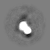
| Images | 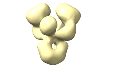 emd_15986.png emd_15986.png | 33.9 KB | ||
| Filedesc metadata | emd-15986.cif.gz | 5.1 KB | ||
| Others | emd_15986_half_map_1.map.gzemd_15986_half_map_2.map.gz | 3.5 MB 3.5 MB | ||
| Archive directory |  http://ftp.pdbj.org/pub/emdb/structures/EMD-15986ftp://ftp.pdbj.org/pub/emdb/structures/EMD-15986 http://ftp.pdbj.org/pub/emdb/structures/EMD-15986ftp://ftp.pdbj.org/pub/emdb/structures/EMD-15986 | HTTPS FTP |
-Related structure data


-Links
| EMDB pages | EMDB (EBI/PDBe) / EMDataResource |
|---|
-Map
| File | Download / File: emd_15986.map.gz / Format: CCP4 / Size: 3.8 MB / Type: IMAGE STORED AS FLOATING POINT NUMBER (4 BYTES) | ||||||||||||||||||||||||||||||||||||
|---|---|---|---|---|---|---|---|---|---|---|---|---|---|---|---|---|---|---|---|---|---|---|---|---|---|---|---|---|---|---|---|---|---|---|---|---|---|
| Annotation | GluK3 glutamate BPAM | ||||||||||||||||||||||||||||||||||||
| Projections & slices | Image control
Images are generated by Spider. | ||||||||||||||||||||||||||||||||||||
| Voxel size | X=Y=Z: 3.14 Å | ||||||||||||||||||||||||||||||||||||
| Density |
| ||||||||||||||||||||||||||||||||||||
| Symmetry | Space group: 1 | ||||||||||||||||||||||||||||||||||||
| Details | EMDB XML:
|
 Z (Sec.)
 Y (Row.)
 X (Col.)


-Supplemental data
-Half map: Half map A
| File | emd_15986_half_map_1.map | ||||||||||||
|---|---|---|---|---|---|---|---|---|---|---|---|---|---|
| Annotation | Half map A | ||||||||||||
| Projections & Slices |
| ||||||||||||
| Density Histograms |


-Half map: Half map B
| File | emd_15986_half_map_2.map | ||||||||||||
|---|---|---|---|---|---|---|---|---|---|---|---|---|---|
| Annotation | Half map B | ||||||||||||
| Projections & Slices |
| ||||||||||||
| Density Histograms |


- Sample components
-Entire : Complex of GRIK3_HUMAN with glutamate and BPAM
| Entire | Name: Complex of GRIK3_HUMAN with glutamate and BPAM |
|---|---|
| Components |
|
-Supramolecule #1: Complex of GRIK3_HUMAN with glutamate and BPAM
| Supramolecule | Name: Complex of GRIK3_HUMAN with glutamate and BPAM / type: complex / ID: 1 / Parent: 0 / Macromolecule list: all |
|---|---|
| Source (natural) | Organism: Homo sapiens (human) |
-Macromolecule #1: GRIK3_HUMAN
| Macromolecule | Name: GRIK3_HUMAN / type: protein_or_peptide / ID: 1 / Enantiomer: DEXTRO |
|---|---|
| Source (natural) | Organism: Homo sapiens (human) |
| Sequence | String: MTAPWRRLRS LVWEYWAGLL VCAFWIPDSR GMPHVIRIGG IFEYADGPNA QVMNAEEHAF RFSANIINRN RTLLPNTTLT YDIQRIHFHD SFEATKKACD QLALGVVAIF GPSQGSCTNA VQSICNALEV PHIQLRWKHH PLDNKDTFYV NLYPDYASLS HAILDLVQYL ...String: MTAPWRRLRS LVWEYWAGLL VCAFWIPDSR GMPHVIRIGG IFEYADGPNA QVMNAEEHAF RFSANIINRN RTLLPNTTLT YDIQRIHFHD SFEATKKACD QLALGVVAIF GPSQGSCTNA VQSICNALEV PHIQLRWKHH PLDNKDTFYV NLYPDYASLS HAILDLVQYL KWRSATVVYD DSTGLIRLQE LIMAPSRYNI RLKIRQLPID SDDSRPLLKE MKRGREFRII FDCSHTMAAQ ILKQAMAMGM MTEYYHFIFT TLDLYALDLE PYRYSGVNLT GFRILNVDNP HVSAIVEKWS MERLQAAPRS ESGLLDGVMM TDAALLYDAV HIVSVCYQRA PQMTVNSLQC HRHKAWRFGG RFMNFIKEAQ WEGLTGRIVF NKTSGLRTDF DLDIISLKED GLEKVGVWSP ADGLNITEVA KGRGPNVTDS LTNRSLIVTT VLEEPFVMFR KSDRTLYGND RFEGYCIDLL KELAHILGFS YEIRLVEDGK YGAQDDKGQW NGMVKELIDH KADLAVAPLT ITHVREKAID FSKPFMTLGV SILYRKPNGT NPSVFSFLNP LSPDIWMYVL LAYLGVSCVL FVIARFSPYE WYDAHPCNPG SEVVENNFTL LNSFWFGMGS LMQQGSELMP KALSTRIIGG IWWFFTLIII SSYTANLAAF LTVERMESPI DSADDLAKQT KIEYGAVKDG ATMTFFKKSK ISTFEKMWAF MSSKPSALVK NNEEGIQRAL TADYALLMES TTIEYVTQRN CNLTQIGGLI DSKGYGIGTP MGSPYRDKIT IAILQLQEED KLHIMKEKWW RGSGCPEEEN KEASALGIQK IGGIFIVLAA GLVLSVLVAV GEFVYKLRKT AEREQRSFCS TVADEIRFSL TCQRRVKHKP QPPMMVKTDA VINMHTFNDR RLPGKDSMAC STSLAPVFP |
-Experimental details
-Structure determination
| Method | negative staining, cryo EM |
|---|---|
 Processing Processing | single particle reconstruction |
| Aggregation state | particle |
-Sample preparation
| Buffer | pH: 8 |
|---|---|
| Staining | Type: NEGATIVE / Material: Uranyl formate |
| Vitrification | Cryogen name: ETHANE |
- Electron microscopy
| Microscope | FEI TECNAI SPIRIT |
|---|---|
| Image recording | Film or detector model: OTHER / Average electron dose: 60.0 e/Å2 |
| Electron beam | Acceleration voltage: 120 kV / Electron source:  FIELD EMISSION GUN FIELD EMISSION GUN |
| Electron optics | Illumination mode: FLOOD BEAM / Imaging mode: BRIGHT FIELD / Nominal defocus max: 5.0 µm / Nominal defocus min: 1.2 µm |
| Experimental equipment |  Model: Tecnai Spirit / Image courtesy: FEI Company |

