- EMDB-15921: Open conformation of the complex of DNA ligase I on PCNA and DNA ... -
+
Open data
ID or keywords:
Loading...
-
Basic information
Entry
Database: EMDB / ID: EMD-15921
Title
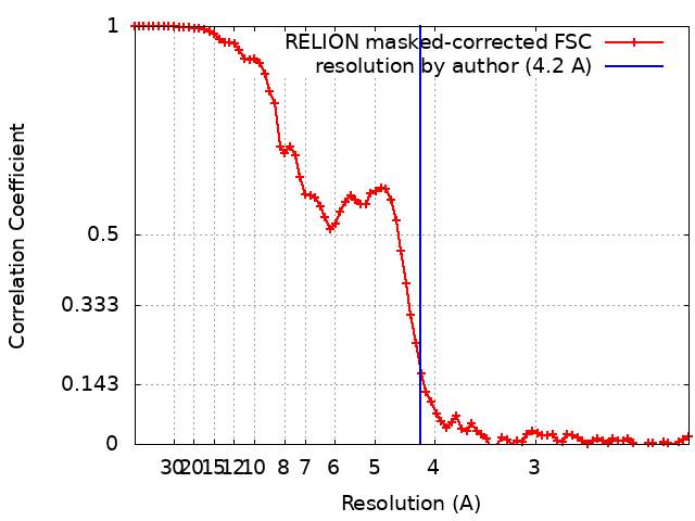
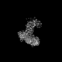
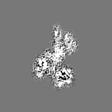
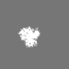
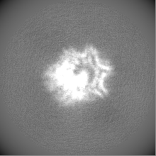
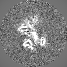
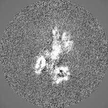
Open conformation of the complex of DNA ligase I on PCNA and DNA in the presence of ATP
Map data
Sample
Complex: complex of DNA ligase I on PCNA and DNA in the presence of ATP
Protein or peptide: DNA ligase 1
Protein or peptide: Proliferating cell nuclear antigen
Keywords
DNA / Replication / Complex / Ligase / PCNA / Ligation / Okazaki fragment maturation
Function / homology
Function and homology information
Okazaki fragment processing involved in mitotic DNA replication / DNA ligase activity / DNA ligase (ATP) / dinucleotide insertion or deletion binding / DNA ligase (ATP) activity / PCNA-p21 complex / telomere maintenance via semi-conservative replication / mitotic telomere maintenance via semi-conservative replication / purine-specific mismatch base pair DNA N-glycosylase activity / nuclear lamina ...Okazaki fragment processing involved in mitotic DNA replication / DNA ligase activity / DNA ligase (ATP) / dinucleotide insertion or deletion binding / DNA ligase (ATP) activity / PCNA-p21 complex / telomere maintenance via semi-conservative replication / mitotic telomere maintenance via semi-conservative replication / purine-specific mismatch base pair DNA N-glycosylase activity / nuclear lamina / Polymerase switching / Regulation of MITF-M-dependent genes involved in DNA replication, damage repair and senescence / Processive synthesis on the lagging strand / PCNA complex / MutLalpha complex binding / Removal of the Flap Intermediate / Telomere C-strand (Lagging Strand) Synthesis / lagging strand elongation / Mismatch repair (MMR) directed by MSH2:MSH3 (MutSbeta) / Mismatch repair (MMR) directed by MSH2:MSH6 (MutSalpha) / Transcription of E2F targets under negative control by DREAM complex / Polymerase switching on the C-strand of the telomere / replisome / mitotic DNA replication / Processive synthesis on the C-strand of the telomere / response to L-glutamate / Removal of the Flap Intermediate from the C-strand / response to dexamethasone / DNA biosynthetic process / histone acetyltransferase binding / Early Phase of HIV Life Cycle / leading strand elongation / DNA polymerase processivity factor activity / G1/S-Specific Transcription / nuclear replication fork / SUMOylation of DNA replication proteins / replication fork processing / POLB-Dependent Long Patch Base Excision Repair / DNA repair-dependent chromatin remodeling / PCNA-Dependent Long Patch Base Excision Repair / anatomical structure morphogenesis / response to cadmium ion / estrous cycle / mismatch repair / base-excision repair, gap-filling / cyclin-dependent protein kinase holoenzyme complex / DNA polymerase binding / translesion synthesis / epithelial cell differentiation / TP53 Regulates Transcription of Genes Involved in G2 Cell Cycle Arrest / liver regeneration / positive regulation of DNA replication / nuclear estrogen receptor binding / positive regulation of DNA repair / Translesion synthesis by REV1 / Translesion synthesis by POLK / replication fork / Translesion synthesis by POLI / Gap-filling DNA repair synthesis and ligation in GG-NER / Termination of translesion DNA synthesis / Translesion Synthesis by POLH / base-excision repair / receptor tyrosine kinase binding / Recognition of DNA damage by PCNA-containing replication complex / HDR through Homologous Recombination (HRR) / cellular response to hydrogen peroxide / Dual Incision in GG-NER / cellular response to UV / Dual incision in TC-NER / Gap-filling DNA repair synthesis and ligation in TC-NER / response to estradiol / heart development / E3 ubiquitin ligases ubiquitinate target proteins / chromatin organization / DNA recombination / damaged DNA binding / chromosome, telomeric region / cell division / DNA repair / chromatin binding / centrosome / chromatin / protein-containing complex binding / enzyme binding / DNA binding / extracellular exosome / nucleoplasm / ATP binding / metal ion binding / identical protein binding / nucleus Similarity search - Function
: / DNA ligase, ATP-dependent / DNA ligase, ATP-dependent, N-terminal / DNA ligase, ATP-dependent, N-terminal domain superfamily / DNA ligase N terminus / ATP-dependent DNA ligase AMP-binding site. / ATP-dependent DNA ligase signature 2. / DNA ligase, ATP-dependent, C-terminal / ATP dependent DNA ligase C terminal region / DNA ligase, ATP-dependent, conserved site ...: / DNA ligase, ATP-dependent / DNA ligase, ATP-dependent, N-terminal / DNA ligase, ATP-dependent, N-terminal domain superfamily / DNA ligase N terminus / ATP-dependent DNA ligase AMP-binding site. / ATP-dependent DNA ligase signature 2. / DNA ligase, ATP-dependent, C-terminal / ATP dependent DNA ligase C terminal region / DNA ligase, ATP-dependent, conserved site / ATP-dependent DNA ligase family profile. / DNA ligase, ATP-dependent, central / ATP dependent DNA ligase domain / Proliferating cell nuclear antigen signature 2. / Proliferating cell nuclear antigen, PCNA, C-terminal / Proliferating cell nuclear antigen, C-terminal domain / Proliferating cell nuclear antigen, PCNA, conserved site / Proliferating cell nuclear antigen signature 1. / Proliferating cell nuclear antigen, PCNA / Proliferating cell nuclear antigen, PCNA, N-terminal / Proliferating cell nuclear antigen, N-terminal domain / : / Nucleic acid-binding, OB-fold Similarity search - Domain/homology
Journal: Nat Commun / Year: 2022 Title: Mechanism of human Lig1 regulation by PCNA in Okazaki fragment sealing. Authors: Kerry Blair / Muhammad Tehseen / Vlad-Stefan Raducanu / Taha Shahid / Claudia Lancey / Fahad Rashid / Ramon Crehuet / Samir M Hamdan / Alfredo De Biasio / Abstract: During lagging strand synthesis, DNA Ligase 1 (Lig1) cooperates with the sliding clamp PCNA to seal the nicks between Okazaki fragments generated by Pol δ and Flap endonuclease 1 (FEN1). We present ...During lagging strand synthesis, DNA Ligase 1 (Lig1) cooperates with the sliding clamp PCNA to seal the nicks between Okazaki fragments generated by Pol δ and Flap endonuclease 1 (FEN1). We present several cryo-EM structures combined with functional assays, showing that human Lig1 recruits PCNA to nicked DNA using two PCNA-interacting motifs (PIPs) located at its disordered N-terminus (PIP) and DNA binding domain (PIP). Once Lig1 and PCNA assemble as two-stack rings encircling DNA, PIP is released from PCNA and only PIP is required for ligation to facilitate the substrate handoff from FEN1. Consistently, we observed that PCNA forms a defined complex with FEN1 and nicked DNA, and it recruits Lig1 to an unoccupied monomer creating a toolbelt that drives the transfer of DNA to Lig1. Collectively, our results provide a structural model on how PCNA regulates FEN1 and Lig1 during Okazaki fragments maturation.
In the structure databanks used in Yorodumi, some data are registered as the other names, "COVID-19 virus" and "2019-nCoV". Here are the details of the virus and the list of structure data.
Jan 31, 2019. EMDB accession codes are about to change! (news from PDBe EMDB page)
EMDB accession codes are about to change! (news from PDBe EMDB page)
The allocation of 4 digits for EMDB accession codes will soon come to an end. Whilst these codes will remain in use, new EMDB accession codes will include an additional digit and will expand incrementally as the available range of codes is exhausted. The current 4-digit format prefixed with “EMD-” (i.e. EMD-XXXX) will advance to a 5-digit format (i.e. EMD-XXXXX), and so on. It is currently estimated that the 4-digit codes will be depleted around Spring 2019, at which point the 5-digit format will come into force.
The EM Navigator/Yorodumi systems omit the EMD- prefix.
Related info.:Q: What is EMD? / ID/Accession-code notation in Yorodumi/EM Navigator
Yorodumi is a browser for structure data from EMDB, PDB, SASBDB, etc.
This page is also the successor to EM Navigator detail page, and also detail information page/front-end page for Omokage search.
The word "yorodu" (or yorozu) is an old Japanese word meaning "ten thousand". "mi" (miru) is to see.
Related info.:EMDB / PDB / SASBDB / Comparison of 3 databanks / Yorodumi Search / Aug 31, 2016. New EM Navigator & Yorodumi / Yorodumi Papers / Jmol/JSmol / Function and homology information / Changes in new EM Navigator and Yorodumi
 Movie
Movie Controller

 Yorodumi
 Open data
 Basic information
 Map data
 Sample
 Keywords
 Function and homology information
 Homo sapiens (human)
 Authors
 United Kingdom, 1 items
  Citation

 Structure visualization
 Downloads & links
 emd_15921.png
 http://ftp.pdbj.org/pub/emdb/structures/EMD-15921


 Z (Sec.)
 Y (Row.)
 X (Col.)


 Sample components
 Processing
 Electron microscopy
 FIELD EMISSION GUN