Biotechnology and Biological Sciences Research Council (BBSRC)
BB/M000265/1
英国
Wellcome Trust
nr29785
英国
Royal Society
URF/R1/19154
英国
Biotechnology and Biological Sciences Research Council (BBSRC)
BB/V006630/1
英国
引用
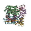
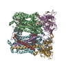
ジャーナル: Proc Natl Acad Sci U S A / 年: 2023 タイトル: Cryo-EM structure of the four-subunit cytochrome complex in styrene maleic acid nanodiscs. 著者: David J K Swainsbury / Frederick R Hawkings / Elizabeth C Martin / Sabina Musiał / Jack H Salisbury / Philip J Jackson / David A Farmer / Matthew P Johnson / C Alistair Siebert / Andrew ...著者: David J K Swainsbury / Frederick R Hawkings / Elizabeth C Martin / Sabina Musiał / Jack H Salisbury / Philip J Jackson / David A Farmer / Matthew P Johnson / C Alistair Siebert / Andrew Hitchcock / C Neil Hunter / 要旨: Cytochrome complexes are ubiquinol:cytochrome oxidoreductases, and as such, they are centrally important components of respiratory and photosynthetic electron transfer chains in many species of ...Cytochrome complexes are ubiquinol:cytochrome oxidoreductases, and as such, they are centrally important components of respiratory and photosynthetic electron transfer chains in many species of bacteria and in mitochondria. The minimal complex has three catalytic components, which are cytochrome , cytochrome , and the Rieske iron-sulfur subunit, but the function of mitochondrial cytochrome complexes is modified by up to eight supernumerary subunits. The cytochrome complex from the purple phototrophic bacterium has a single supernumerary subunit called subunit IV, which is absent from current structures of the complex. In this work we use the styrene-maleic acid copolymer to purify the cytochrome complex in native lipid nanodiscs, which retains the labile subunit IV, annular lipids, and natively bound quinones. The catalytic activity of the four-subunit cytochrome complex is threefold higher than that of the complex lacking subunit IV. To understand the role of subunit IV, we determined the structure of the four-subunit complex at 2.9 Å using single particle cryogenic electron microscopy. The structure shows the position of the transmembrane domain of subunit IV, which lies across the transmembrane helices of the Rieske and cytochrome subunits. We observe a quinone at the Q quinone-binding site and show that occupancy of this site is linked to conformational changes in the Rieske head domain during catalysis. Twelve lipids were structurally resolved, making contacts with the Rieske and cytochrome subunits, with some spanning both of the two monomers that make up the dimeric complex.
凍結剤: ETHANE / チャンバー内湿度: 80 % / チャンバー内温度: 298 K / 装置: LEICA EM GP 詳細: 15 ul of sample was applied to the grid, incubated for 30 s, blotted for 4 s then plunged in liquid ethane..
詳細
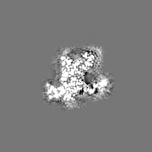
Monodisperse particles consisting of four-subunit cyt b-c1 solubilised in styrene maleic acid nanodiscs
Images were motion corrected using Motiocorr 2 within RELION using 5 x 5 patches.
粒子像選択
選択した数: 4060135 詳細: Particles were picked using crYOLO 1.7.5 using a model trained on this dataset
CTF補正
ソフトウェア - 名称: CTFFIND (ver. 4.1.14) / ソフトウェア - 詳細: Run within RELION 3.1 / タイプ: PHASE FLIPPING AND AMPLITUDE CORRECTION
初期モデル
モデルのタイプ: INSILICO MODEL
最終 再構成
使用したクラス数: 1 / 想定した対称性 - 点群: C1 (非対称) / アルゴリズム: FOURIER SPACE / 解像度のタイプ: BY AUTHOR / 解像度: 3.75 Å / 解像度の算出法: FSC 0.5 CUT-OFF / ソフトウェア - 名称: RELION (ver. 3.1) 詳細: Refinement performed without alignment from particles within a single class after focussed refinement 使用した粒子像数: 72118
初期 角度割当
タイプ: MAXIMUM LIKELIHOOD / ソフトウェア - 名称: RELION (ver. 3.1)
最終 角度割当
タイプ: MAXIMUM LIKELIHOOD / ソフトウェア - 名称: RELION (ver. 3.1)
最終 3次元分類
クラス数: 10 / ソフトウェア - 名称: RELION (ver. 3.1) ソフトウェア - 詳細: Alignment free masked classification on previous consensus reconstruction 詳細: One high quality class was selected for final refinement
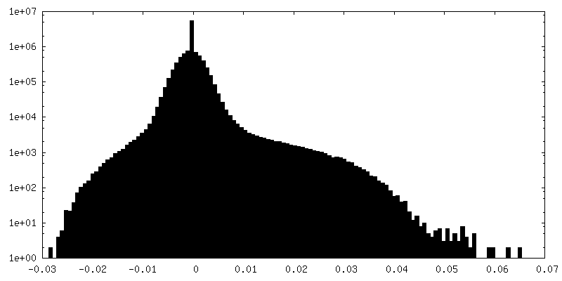
FSC曲線 (解像度の算出)
-
原子モデル構築 1
精密化
空間: REAL
得られたモデル
PDB-8asj: Four subunit cytochrome b-c1 complex from Rhodobacter sphaeroides in native nanodiscs - focussed refinement in the b-c conformation
 ムービー
ムービー コントローラー


 データを開く
 基本情報
基本情報
 マップデータ
 試料
試料 キーワード
キーワード 機能・相同性情報
 Cereibacter sphaeroides (バクテリア) /
  データ登録者
 英国, 5件
  引用
引用 構造の表示
構造の表示
 ダウンロードとリンク
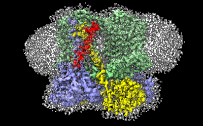 emd_15617.png
 http://ftp.pdbj.org/pub/emdb/structures/EMD-15617


 Z (Sec.)
 Y (Row.)
 X (Col.)


 試料の構成要素


 解析
解析 電子顕微鏡法
 FIELD EMISSION GUN