 ムービー
ムービー コントローラー


+ データを開く

- 基本情報
基本情報
| 登録情報 | データベース: EMDB / ID: EMD-1537 | |||||||||
|---|---|---|---|---|---|---|---|---|---|---|
| タイトル | 2D Arrays of F-actin Cross-linked by Villin | |||||||||
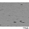
 マップデータ マップデータ | tomogram of F-actin crosslinked with villin on a 2D lipid monolayer | |||||||||
 試料 試料 |
| |||||||||
 キーワード キーワード | Cytoskeleton / actin / electron tomography / microvilli / image processing / lipid monolayer | |||||||||
| 生物種 |   | |||||||||
| 手法 | 電子線トモグラフィー法 / クライオ電子顕微鏡法 / ネガティブ染色法 / 解像度: 30.0 Å | |||||||||
 データ登録者 データ登録者 | Hampton CM / Liu J / Taylor DW / DeRosier DJ / Taylor KA | |||||||||
 引用 引用 | ジャーナル: Structure / 年: 2008 タイトル: The 3D structure of villin as an unusual F-Actin crosslinker. 著者: Cheri M Hampton / Jun Liu / Dianne W Taylor / David J DeRosier / Kenneth A Taylor /  要旨: Villin is an F-actin nucleating, crosslinking, severing, and capping protein within the gelsolin superfamily. We have used electron tomography of 2D arrays of villin-crosslinked F-actin to generate ...Villin is an F-actin nucleating, crosslinking, severing, and capping protein within the gelsolin superfamily. We have used electron tomography of 2D arrays of villin-crosslinked F-actin to generate 3D images revealing villin's crosslinking structure. In these polar arrays, neighboring filaments are spaced 125.9 +/- 7.1 A apart, offset axially by 17 A, with one villin crosslink per actin crossover. More than 6500 subvolumes containing a single villin crosslink and the neighboring actin filaments were aligned and classified to produce 3D subvolume averages. Placement of a complete villin homology model into the average density reveals that full-length villin binds to different sites on F-actin from those used by other actin-binding proteins and villin's close homolog gelsolin. | |||||||||
| 履歴 |
|
- 構造の表示
構造の表示
| ムービー |
 ムービービューア ムービービューア |
|---|---|
| 構造ビューア | EMマップ: SurfViewMolmilJmol/JSmol |
| 添付画像 |
 UCSF Chimera
- ダウンロードとリンク
-EMDBアーカイブ
| マップデータ | emd_1537.map.gz | 519.8 MB | EMDBマップデータ形式 | |
|---|---|---|---|---|
| ヘッダ (付随情報) | emd-1537-v30.xmlemd-1537.xml | 8.8 KB 8.8 KB | 表示 表示 | EMDBヘッダ |
| 画像 |  emd_1537.png emd_1537.png | 379.4 KB | ||
| アーカイブディレクトリ |  http://ftp.pdbj.org/pub/emdb/structures/EMD-1537ftp://ftp.pdbj.org/pub/emdb/structures/EMD-1537 http://ftp.pdbj.org/pub/emdb/structures/EMD-1537ftp://ftp.pdbj.org/pub/emdb/structures/EMD-1537 | HTTPS FTP |
-関連構造データ


-リンク
| EMDBのページ | EMDB (EBI/PDBe) / EMDataResource |
|---|
-マップ
| ファイル | ダウンロード / ファイル: emd_1537.map.gz / 形式: CCP4 / 大きさ: 547.8 MB / タイプ: IMAGE STORED AS FLOATING POINT NUMBER (4 BYTES) | ||||||||||||||||||||||||||||||||||||||||||||||||||||||||||||||||||||
|---|---|---|---|---|---|---|---|---|---|---|---|---|---|---|---|---|---|---|---|---|---|---|---|---|---|---|---|---|---|---|---|---|---|---|---|---|---|---|---|---|---|---|---|---|---|---|---|---|---|---|---|---|---|---|---|---|---|---|---|---|---|---|---|---|---|---|---|---|---|
| 注釈 | tomogram of F-actin crosslinked with villin on a 2D lipid monolayer | ||||||||||||||||||||||||||||||||||||||||||||||||||||||||||||||||||||
| 投影像・断面図 | 画像のコントロール
画像は Spider により作成 これらの図は立方格子座標系で作成されたものです | ||||||||||||||||||||||||||||||||||||||||||||||||||||||||||||||||||||
| ボクセルのサイズ | X=Y=Z: 5.76 Å | ||||||||||||||||||||||||||||||||||||||||||||||||||||||||||||||||||||
| 密度 |
| ||||||||||||||||||||||||||||||||||||||||||||||||||||||||||||||||||||
| 対称性 | 空間群: 1 | ||||||||||||||||||||||||||||||||||||||||||||||||||||||||||||||||||||
| 詳細 | EMDB XML:
CCP4マップ ヘッダ情報:
| ||||||||||||||||||||||||||||||||||||||||||||||||||||||||||||||||||||
 Z (Sec.)
 Y (Row.)
 X (Col.)


-添付データ
- 試料の構成要素
-全体 : Rabbit F-actin cross-linked with chicken villin
| 全体 | 名称: Rabbit F-actin cross-linked with chicken villin |
|---|---|
| 要素 |
|
-超分子 #1000: Rabbit F-actin cross-linked with chicken villin
| 超分子 | 名称: Rabbit F-actin cross-linked with chicken villin / タイプ: sample / ID: 1000 / 詳細: 2D rafts are formed on a lipid monolayer / Number unique components: 2 |
|---|
-超分子 #1: Cytoskeleton
| 超分子 | 名称: Cytoskeleton / タイプ: organelle_or_cellular_component / ID: 1 / Name.synonym: Actin / 組換発現: No / データベース: NCBI |
|---|---|
| 由来(天然) | 生物種: |
| 分子量 | 実験値: 42 MDa |
-超分子 #2: Cytoskeleton
| 超分子 | 名称: Cytoskeleton / タイプ: organelle_or_cellular_component / ID: 2 / Name.synonym: Villin / 集合状態: Monomer / 組換発現: No / データベース: NCBI |
|---|---|
| 由来(天然) | 生物種: |
| 分子量 | 実験値: 95 MDa |
-実験情報
-構造解析
| 手法 | ネガティブ染色法, クライオ電子顕微鏡法 |
|---|---|
 解析 解析 | 電子線トモグラフィー法 |
| 試料の集合状態 | 2D array |
-試料調製
| 染色 | タイプ: NEGATIVE / 詳細: Uranyl acetate |
|---|---|
| グリッド | 詳細: 200 mesh holey carbon film |
| 凍結 | 凍結剤: ETHANE / 装置: HOMEMADE PLUNGER / 詳細: Vitrification instrument: Plunge freeze |
| 詳細 | Crystals grown on a lipid-monolayer |
- 電子顕微鏡法
| 顕微鏡 | FEI/PHILIPS CM300FEG/T |
|---|---|
| 日付 | 2005年5月19日 |
| 撮影 | カテゴリ: CCD / フィルム・検出器のモデル: GENERIC CCD (2k x 2k) |
| 電子線 | 加速電圧: 300 kV / 電子線源: LAB6 |
| 電子光学系 | 照射モード: SPOT SCAN / 撮影モード: BRIGHT FIELD / Cs: 2.0 mm / 倍率(公称値): 24000 |
| 試料ステージ | 試料ホルダー: High tilt / 試料ホルダーモデル: OTHER |
-画像解析
| 最終 再構成 | アルゴリズム: OTHER / 解像度のタイプ: BY AUTHOR / 解像度: 30.0 Å / 解像度の算出法: OTHER / ソフトウェア - 名称: Protomo / 詳細: Tomogram is from a single tilt series / 使用した粒子像数: 60 |
|---|---|
| 結晶パラメータ | 面群: P 2 |
