 Movie
Movie Controller


+ Open data

- Basic information
| Entry | Database: EMDB / ID: EMD-1496 | |||||||||
|---|---|---|---|---|---|---|---|---|---|---|
| Title | 2D Arrays of F-actin Cross-linked by Villin | |||||||||
 Map data Map data | Tomogram of F-actin crosslinked by villin | |||||||||
 Sample Sample |
| |||||||||
 Keywords Keywords | cytoskeleton / actin / electron tomography / microvilli / image processing / lipid monolayer | |||||||||
| Biological species |   | |||||||||
| Method | electron tomography / cryo EM / Resolution: 30.0 Å | |||||||||
 Authors Authors | Hampton CM / Liu J / Taylor DW / DeRosier DJ / Taylor KA | |||||||||
 Citation Citation | Journal: Structure / Year: 2008 Title: The 3D structure of villin as an unusual F-Actin crosslinker. Authors: Cheri M Hampton / Jun Liu / Dianne W Taylor / David J DeRosier / Kenneth A Taylor /  Abstract: Villin is an F-actin nucleating, crosslinking, severing, and capping protein within the gelsolin superfamily. We have used electron tomography of 2D arrays of villin-crosslinked F-actin to generate ...Villin is an F-actin nucleating, crosslinking, severing, and capping protein within the gelsolin superfamily. We have used electron tomography of 2D arrays of villin-crosslinked F-actin to generate 3D images revealing villin's crosslinking structure. In these polar arrays, neighboring filaments are spaced 125.9 +/- 7.1 A apart, offset axially by 17 A, with one villin crosslink per actin crossover. More than 6500 subvolumes containing a single villin crosslink and the neighboring actin filaments were aligned and classified to produce 3D subvolume averages. Placement of a complete villin homology model into the average density reveals that full-length villin binds to different sites on F-actin from those used by other actin-binding proteins and villin's close homolog gelsolin. | |||||||||
| History |
|
- Structure visualization
| Movie |
 Movie viewer Movie viewer |
|---|---|
| Structure viewer | EM map: SurfViewMolmilJmol/JSmol |
| Supplemental images |
 UCSF Chimera
- Downloads & links
-EMDB archive
| Map data | emd_1496.map.gz | 780.8 MB | EMDB map data format | |
|---|---|---|---|---|
| Header (meta data) | emd-1496-v30.xmlemd-1496.xml | 8.3 KB 8.3 KB | Display Display | EMDB header |
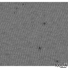
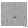
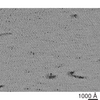
| Images |  1496.gif 1496.gif | 158.9 KB | ||
| Archive directory |  http://ftp.pdbj.org/pub/emdb/structures/EMD-1496ftp://ftp.pdbj.org/pub/emdb/structures/EMD-1496 http://ftp.pdbj.org/pub/emdb/structures/EMD-1496ftp://ftp.pdbj.org/pub/emdb/structures/EMD-1496 | HTTPS FTP |
-Related structure data


-Links
| EMDB pages | EMDB (EBI/PDBe) / EMDataResource |
|---|
-Map
| File | Download / File: emd_1496.map.gz / Format: CCP4 / Size: 822.5 MB / Type: IMAGE STORED AS FLOATING POINT NUMBER (4 BYTES) | ||||||||||||||||||||||||||||||||||||||||||||||||||||||||||||||||||||
|---|---|---|---|---|---|---|---|---|---|---|---|---|---|---|---|---|---|---|---|---|---|---|---|---|---|---|---|---|---|---|---|---|---|---|---|---|---|---|---|---|---|---|---|---|---|---|---|---|---|---|---|---|---|---|---|---|---|---|---|---|---|---|---|---|---|---|---|---|---|
| Annotation | Tomogram of F-actin crosslinked by villin | ||||||||||||||||||||||||||||||||||||||||||||||||||||||||||||||||||||
| Projections & slices | Image control
Images are generated by Spider. generated in cubic-lattice coordinate | ||||||||||||||||||||||||||||||||||||||||||||||||||||||||||||||||||||
| Voxel size | X=Y=Z: 5.76 Å | ||||||||||||||||||||||||||||||||||||||||||||||||||||||||||||||||||||
| Density |
| ||||||||||||||||||||||||||||||||||||||||||||||||||||||||||||||||||||
| Symmetry | Space group: 1 | ||||||||||||||||||||||||||||||||||||||||||||||||||||||||||||||||||||
| Details | EMDB XML:
CCP4 map header:
| ||||||||||||||||||||||||||||||||||||||||||||||||||||||||||||||||||||
 Z (Sec.)
 Y (Row.)
 X (Col.)


-Supplemental data
- Sample components
-Entire : rabbit F-actin cross-linked with chicken villin
| Entire | Name: rabbit F-actin cross-linked with chicken villin |
|---|---|
| Components |
|
-Supramolecule #1000: rabbit F-actin cross-linked with chicken villin
| Supramolecule | Name: rabbit F-actin cross-linked with chicken villin / type: sample / ID: 1000 / Details: 2D rafts are formed on a lipid monolayer / Number unique components: 2 |
|---|
-Supramolecule #1: cytoskeleton
| Supramolecule | Name: cytoskeleton / type: organelle_or_cellular_component / ID: 1 / Name.synonym: F-actin / Recombinant expression: No / Database: NCBI |
|---|---|
| Source (natural) | Organism: |
| Molecular weight | Experimental: 42 MDa |
-Supramolecule #2: cytoskeleton
| Supramolecule | Name: cytoskeleton / type: organelle_or_cellular_component / ID: 2 / Name.synonym: villin / Oligomeric state: monomer / Recombinant expression: No / Database: NCBI |
|---|---|
| Source (natural) | Organism: |
| Molecular weight | Experimental: 95 MDa |
-Experimental details
-Structure determination
| Method | cryo EM |
|---|---|
 Processing Processing | electron tomography |
| Aggregation state | 2D array |
-Sample preparation
| Vitrification | Cryogen name: ETHANE / Instrument: HOMEMADE PLUNGER / Details: Vitrification instrument: plunge freeze |
|---|
- Electron microscopy
| Microscope | FEI/PHILIPS CM300FEG/T |
|---|---|
| Date | May 19, 2005 |
| Image recording | Category: CCD / Film or detector model: GENERIC CCD (2k x 2k) |
| Electron beam | Acceleration voltage: 300 kV / Electron source: LAB6 |
| Electron optics | Illumination mode: SPOT SCAN / Imaging mode: BRIGHT FIELD / Cs: 2.0 mm |
| Sample stage | Specimen holder: high tilt / Specimen holder model: OTHER |
-Image processing
| Final reconstruction | Algorithm: OTHER / Resolution.type: BY AUTHOR / Resolution: 30.0 Å / Resolution method: OTHER / Software - Name: protomo / Details: tomogram is from a single tilt series / Number images used: 60 |
|---|---|
| Crystal parameters | Plane group: P 2 |
