[English] 日本語
 Yorodumi
- EMDB-15357: RNA polymerase at U-rich pause bound to RNA putL triple mutant - ... -
+ Open data
- Basic information
| Entry |  | |||||||||||||||
|---|---|---|---|---|---|---|---|---|---|---|---|---|---|---|---|---|
| Title | RNA polymerase at U-rich pause bound to RNA putL triple mutant - pause prone, closed clamp state | |||||||||||||||
 Map data Map data | ||||||||||||||||
 Sample Sample |
| |||||||||||||||
 Keywords Keywords | RNA polymerase / transcriptional pausing / transcription termination / regulatory RNA / TRANSCRIPTION | |||||||||||||||
| Function / homology |  Function and homology information Function and homology informationsigma factor antagonist complex / RNA polymerase complex / submerged biofilm formation / cellular response to cell envelope stress / regulation of DNA-templated transcription initiation / sigma factor activity / bacterial-type flagellum assembly / bacterial-type RNA polymerase core enzyme binding / cytosolic DNA-directed RNA polymerase complex / bacterial-type flagellum-dependent cell motility ...sigma factor antagonist complex / RNA polymerase complex / submerged biofilm formation / cellular response to cell envelope stress / regulation of DNA-templated transcription initiation / sigma factor activity / bacterial-type flagellum assembly / bacterial-type RNA polymerase core enzyme binding / cytosolic DNA-directed RNA polymerase complex / bacterial-type flagellum-dependent cell motility / nitrate assimilation / DNA-directed RNA polymerase complex / regulation of DNA-templated transcription elongation / transcription elongation factor complex / transcription antitermination / cell motility / DNA-templated transcription initiation / ribonucleoside binding / DNA-directed RNA polymerase / DNA-directed RNA polymerase activity / response to heat / protein-containing complex assembly / intracellular iron ion homeostasis / protein dimerization activity / response to antibiotic / negative regulation of DNA-templated transcription / DNA-templated transcription / magnesium ion binding / DNA binding / zinc ion binding / membrane / cytoplasm / cytosol Similarity search - Function | |||||||||||||||
| Biological species |  | |||||||||||||||
| Method | single particle reconstruction / cryo EM / Resolution: 4.1 Å | |||||||||||||||
 Authors Authors | Dey S / Weixlbaumer A | |||||||||||||||
| Funding support | European Union,  France, 4 items France, 4 items
| |||||||||||||||
 Citation Citation |  Journal: Mol Cell / Year: 2022 Journal: Mol Cell / Year: 2022Title: Structural insights into RNA-mediated transcription regulation in bacteria. Authors: Sanjay Dey / Claire Batisse / Jinal Shukla / Michael W Webster / Maria Takacs / Charlotte Saint-André / Albert Weixlbaumer /  Abstract: RNA can regulate its own synthesis without auxiliary proteins. For example, U-rich RNA sequences signal RNA polymerase (RNAP) to pause transcription and are required for transcript release at ...RNA can regulate its own synthesis without auxiliary proteins. For example, U-rich RNA sequences signal RNA polymerase (RNAP) to pause transcription and are required for transcript release at intrinsic terminators in all kingdoms of life. In contrast, the regulatory RNA putL suppresses pausing and termination in cis. However, how nascent RNA modulates its own synthesis remains largely unknown. We present cryo-EM reconstructions of RNAP captured during transcription of putL variants or an unrelated sequence at a U-rich pause site. Our results suggest how putL suppresses pausing and promotes its synthesis. We demonstrate that transcribing a U-rich sequence, a ubiquitous trigger of intrinsic termination, promotes widening of the RNAP nucleic-acid-binding channel. Widening destabilizes RNAP interactions with DNA and RNA to facilitate transcript dissociation reminiscent of intrinsic transcription termination. Surprisingly, RNAP remains bound to DNA after transcript release. Our results provide the structural framework to understand RNA-mediated intrinsic transcription termination. | |||||||||||||||
| History |
|
- Structure visualization
| Supplemental images |
|---|
- Downloads & links
-EMDB archive
| Map data |  emd_15357.map.gz emd_15357.map.gz | 168.1 MB |  EMDB map data format EMDB map data format | |
|---|---|---|---|---|
| Header (meta data) |  emd-15357-v30.xml emd-15357-v30.xml emd-15357.xml emd-15357.xml | 33.6 KB 33.6 KB | Display Display |  EMDB header EMDB header |
| Images |  emd_15357.png emd_15357.png | 77.2 KB | ||
| Filedesc metadata |  emd-15357.cif.gz emd-15357.cif.gz | 10.4 KB | ||
| Others |  emd_15357_additional_1.map.gz emd_15357_additional_1.map.gz emd_15357_half_map_1.map.gz emd_15357_half_map_1.map.gz emd_15357_half_map_2.map.gz emd_15357_half_map_2.map.gz | 88.7 MB 165.4 MB 165.4 MB | ||
| Archive directory |  http://ftp.pdbj.org/pub/emdb/structures/EMD-15357 http://ftp.pdbj.org/pub/emdb/structures/EMD-15357 ftp://ftp.pdbj.org/pub/emdb/structures/EMD-15357 ftp://ftp.pdbj.org/pub/emdb/structures/EMD-15357 | HTTPS FTP |
-Validation report
| Summary document |  emd_15357_validation.pdf.gz emd_15357_validation.pdf.gz | 1.2 MB | Display |  EMDB validaton report EMDB validaton report |
|---|---|---|---|---|
| Full document |  emd_15357_full_validation.pdf.gz emd_15357_full_validation.pdf.gz | 1.2 MB | Display | |
| Data in XML |  emd_15357_validation.xml.gz emd_15357_validation.xml.gz | 15.1 KB | Display | |
| Data in CIF |  emd_15357_validation.cif.gz emd_15357_validation.cif.gz | 17.8 KB | Display | |
| Arichive directory |  https://ftp.pdbj.org/pub/emdb/validation_reports/EMD-15357 https://ftp.pdbj.org/pub/emdb/validation_reports/EMD-15357 ftp://ftp.pdbj.org/pub/emdb/validation_reports/EMD-15357 ftp://ftp.pdbj.org/pub/emdb/validation_reports/EMD-15357 | HTTPS FTP |
-Related structure data
| Related structure data | 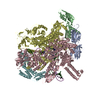 8ad1MC 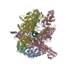 8abyC 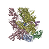 8abzC  8ac0C 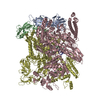 8ac1C 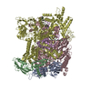 8ac2C 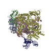 8acpC M: atomic model generated by this map C: citing same article ( |
|---|---|
| Similar structure data | Similarity search - Function & homology  F&H Search F&H Search |
- Links
Links
| EMDB pages |  EMDB (EBI/PDBe) / EMDB (EBI/PDBe) /  EMDataResource EMDataResource |
|---|---|
| Related items in Molecule of the Month |
- Map
Map
| File |  Download / File: emd_15357.map.gz / Format: CCP4 / Size: 178 MB / Type: IMAGE STORED AS FLOATING POINT NUMBER (4 BYTES) Download / File: emd_15357.map.gz / Format: CCP4 / Size: 178 MB / Type: IMAGE STORED AS FLOATING POINT NUMBER (4 BYTES) | ||||||||||||||||||||||||||||||||||||
|---|---|---|---|---|---|---|---|---|---|---|---|---|---|---|---|---|---|---|---|---|---|---|---|---|---|---|---|---|---|---|---|---|---|---|---|---|---|
| Projections & slices | Image control
Images are generated by Spider. | ||||||||||||||||||||||||||||||||||||
| Voxel size | X=Y=Z: 0.862 Å | ||||||||||||||||||||||||||||||||||||
| Density |
| ||||||||||||||||||||||||||||||||||||
| Symmetry | Space group: 1 | ||||||||||||||||||||||||||||||||||||
| Details | EMDB XML:
|
-Supplemental data
-Additional map: #1
| File | emd_15357_additional_1.map | ||||||||||||
|---|---|---|---|---|---|---|---|---|---|---|---|---|---|
| Projections & Slices |
| ||||||||||||
| Density Histograms |
-Half map: #2
| File | emd_15357_half_map_1.map | ||||||||||||
|---|---|---|---|---|---|---|---|---|---|---|---|---|---|
| Projections & Slices |
| ||||||||||||
| Density Histograms |
-Half map: #1
| File | emd_15357_half_map_2.map | ||||||||||||
|---|---|---|---|---|---|---|---|---|---|---|---|---|---|
| Projections & Slices |
| ||||||||||||
| Density Histograms |
- Sample components
+Entire : Escherichia coli RNA polymerase putL triple mutant complex - paus...
+Supramolecule #1: Escherichia coli RNA polymerase putL triple mutant complex - paus...
+Macromolecule #1: Non-template DNA
+Macromolecule #3: Template DNA
+Macromolecule #2: RNA putL triple mutant
+Macromolecule #4: DNA-directed RNA polymerase subunit omega
+Macromolecule #5: RNA polymerase sigma factor RpoD
+Macromolecule #6: DNA-directed RNA polymerase subunit alpha
+Macromolecule #7: DNA-directed RNA polymerase subunit beta
+Macromolecule #8: DNA-directed RNA polymerase subunit beta'
+Macromolecule #9: MAGNESIUM ION
+Macromolecule #10: ZINC ION
-Experimental details
-Structure determination
| Method | cryo EM |
|---|---|
 Processing Processing | single particle reconstruction |
| Aggregation state | particle |
- Sample preparation
| Concentration | 12 mg/mL |
|---|---|
| Buffer | pH: 8 Details: 20 mM Tris-glutamate pH 8.0, 50 mM K-glutamate, 10 mM Mg-glutamate, 0.001 mM ZnCl2, 2mM DTT |
| Grid | Model: Quantifoil / Material: GOLD / Mesh: 300 / Support film - Material: GOLD / Support film - topology: HOLEY / Pretreatment - Type: PLASMA CLEANING / Pretreatment - Time: 30 sec. / Pretreatment - Atmosphere: OTHER |
| Vitrification | Cryogen name: ETHANE / Chamber humidity: 95 % / Chamber temperature: 283 K / Instrument: FEI VITROBOT MARK IV Details: Quantifoil UltrAuFoil R1.2/1.3 300 mesh holey gold grids were plasma cleaned on a Model 1070 (Fischione Instruments) for 30 sec at 70% power and with an 80% Argon and 20% Oxygen mixture ...Details: Quantifoil UltrAuFoil R1.2/1.3 300 mesh holey gold grids were plasma cleaned on a Model 1070 (Fischione Instruments) for 30 sec at 70% power and with an 80% Argon and 20% Oxygen mixture prior to the application of 0.004 ml of sample. Grids were plunge frozen into liquid ethane using a Vitrobot mark IV (FEI) with 95% chamber humidity at 283K.. |
| Details | The co-transcriptionally halted complexes for cryo-EM analysis were prepared in vitro by mixing E. coli RNAP holoenzyme with templates containing the DNA base analogue isoG. The RNAP ECs halted at the U-rich pause (G93). The complexes were prepared in 0.03 ml reactions and contained: 0.02 mM of E. coli RNAP, 0.080 mM of E. coli Sigma70, 0.02 mM of template DNA, 20 mM Tris-glutamate pH 8.0, 50 mM K-glutamate, 10 mM Mg-glutamate, 0.001 mM ZnCl2, 2mM DTT and 6 mM of each ATP, GTP, CTP and UTP. Initially, all the components except NTPs were added and incubated for 10 min at 310K. To allow transcription elongation, NTPs were added and the sample was incubated for 20 to 40 min at 310K. The final products were purified on a Superose 6 Increase 3.2/300 gel filtration column equilibrated in Glutamate buffer. Samples were concentrated to 10-12 mg/mL using an Amicon Ultra 0.5mL centrifugal filter unit (30 KDa MWCO). Before grid freezing, 8 mM of CHAPSO was added to freshly prepared sample to overcome preferred particle orientation. |
- Electron microscopy
| Microscope | FEI TITAN KRIOS |
|---|---|
| Image recording | Film or detector model: GATAN K3 (6k x 4k) / Number grids imaged: 1 / Average exposure time: 2.2 sec. / Average electron dose: 52.0 e/Å2 |
| Electron beam | Acceleration voltage: 300 kV / Electron source:  FIELD EMISSION GUN FIELD EMISSION GUN |
| Electron optics | Illumination mode: FLOOD BEAM / Imaging mode: BRIGHT FIELD / Nominal defocus max: 2.5 µm / Nominal defocus min: 0.8 µm |
| Sample stage | Specimen holder model: FEI TITAN KRIOS AUTOGRID HOLDER |
| Experimental equipment |  Model: Titan Krios / Image courtesy: FEI Company |
+ Image processing
-Atomic model buiding 1
| Initial model | PDB ID: Chain - Source name: PDB / Chain - Initial model type: experimental model |
|---|---|
| Refinement | Space: REAL / Protocol: FLEXIBLE FIT / Target criteria: map cross correlation |
| Output model |  PDB-8ad1: |
 Movie
Movie Controller


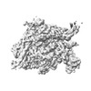


 Z (Sec.)
 Y (Row.)
 X (Col.)


