Movie
Movie Controller

[English] 日本語
 Yorodumi
- EMDB-15351: Pol alpha local refinement. Human replisome bound by Pol Alpha, e... -
+ Open data

- Basic information
| Entry |  | |||||||||
|---|---|---|---|---|---|---|---|---|---|---|
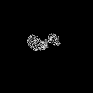
| Title | Pol alpha local refinement. Human replisome bound by Pol Alpha, engaged on a fork DNA substrate containing a 60 nt lagging strand. | |||||||||
 Map data Map data | Local refinement of Pol Alpha. Human replisome bound by Pol Alpha, engaged on a fork DNA substrate containing a 60 nt lagging strand. | |||||||||
 Sample Sample |
| |||||||||
 Keywords Keywords | Replication / helicase / polymerase / pol alpha / priming | |||||||||
| Biological species |  | |||||||||
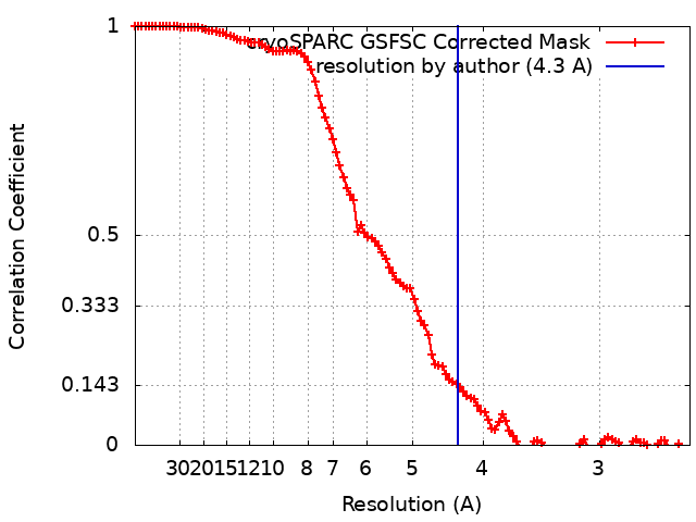
| Method | single particle reconstruction / cryo EM / Resolution: 4.3 Å | |||||||||
 Authors Authors | Jones ML / Yeeles JTP | |||||||||
| Funding support |  United Kingdom, 1 items United Kingdom, 1 items
| |||||||||
 Citation Citation | Journal: Mol.Cell / Year: 2023 Title: How Pol a-primase is targeted to replisomes to prime eukaryotic DNA replication Authors: Jones ML / Aria V / Baris Y / Yeeles JTP | |||||||||
| History |
|
- Structure visualization
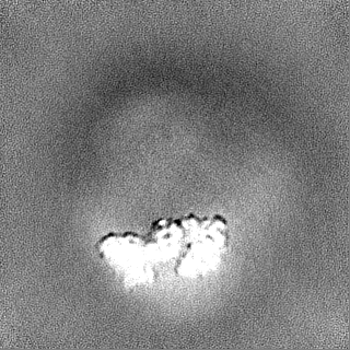
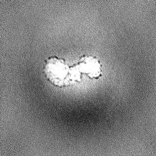
| Supplemental images |
|---|

- Downloads & links
-EMDB archive
| Map data | emd_15351.map.gz | 62.3 MB |  EMDB map data format EMDB map data format | |
|---|---|---|---|---|
| Header (meta data) | emd-15351-v30.xmlemd-15351.xml | 11.5 KB 11.5 KB | Display Display | EMDB header |
| FSC (resolution estimation) | emd_15351_fsc.xml | 10.6 KB | Display | FSC data file |
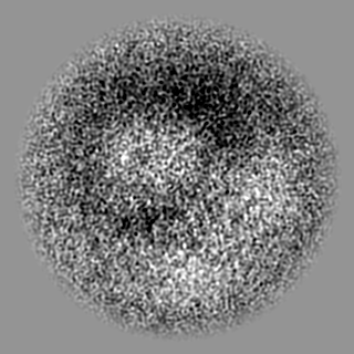
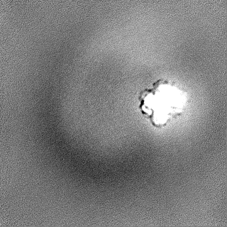
| Images | 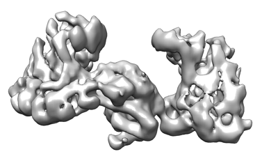 emd_15351.png emd_15351.png | 57.3 KB | ||
| Others | emd_15351_half_map_1.map.gzemd_15351_half_map_2.map.gz | 115.9 MB 115.9 MB | ||
| Archive directory |  http://ftp.pdbj.org/pub/emdb/structures/EMD-15351ftp://ftp.pdbj.org/pub/emdb/structures/EMD-15351 http://ftp.pdbj.org/pub/emdb/structures/EMD-15351ftp://ftp.pdbj.org/pub/emdb/structures/EMD-15351 | HTTPS FTP |
-Validation report
| Summary document | emd_15351_validation.pdf.gz | 792.5 KB | Display | EMDB validaton report |
|---|---|---|---|---|
| Full document | emd_15351_full_validation.pdf.gz | 792.1 KB | Display | |
| Data in XML | emd_15351_validation.xml.gz | 19.1 KB | Display | |
| Data in CIF | emd_15351_validation.cif.gz | 24.5 KB | Display | |
| Arichive directory | https://ftp.pdbj.org/pub/emdb/validation_reports/EMD-15351ftp://ftp.pdbj.org/pub/emdb/validation_reports/EMD-15351 | HTTPS FTP |
-Related structure data
| Related structure data |
|---|
-Links
| EMDB pages | EMDB (EBI/PDBe) / EMDataResource |
|---|
-Map
| File | Download / File: emd_15351.map.gz / Format: CCP4 / Size: 125 MB / Type: IMAGE STORED AS FLOATING POINT NUMBER (4 BYTES) | ||||||||||||||||||||||||||||||||||||
|---|---|---|---|---|---|---|---|---|---|---|---|---|---|---|---|---|---|---|---|---|---|---|---|---|---|---|---|---|---|---|---|---|---|---|---|---|---|
| Annotation | Local refinement of Pol Alpha. Human replisome bound by Pol Alpha, engaged on a fork DNA substrate containing a 60 nt lagging strand. | ||||||||||||||||||||||||||||||||||||
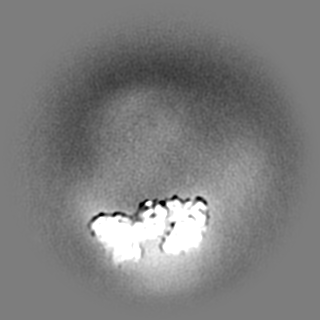
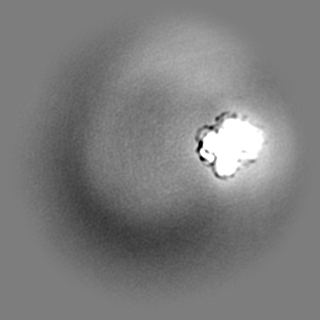
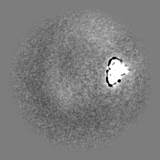
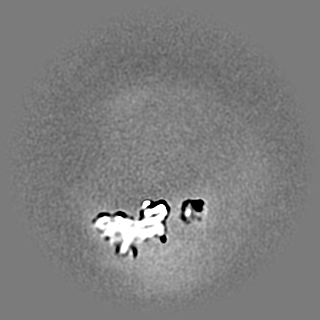
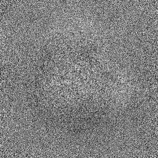
| Projections & slices | Image control
Images are generated by Spider. | ||||||||||||||||||||||||||||||||||||
| Voxel size | X=Y=Z: 1.2363 Å | ||||||||||||||||||||||||||||||||||||
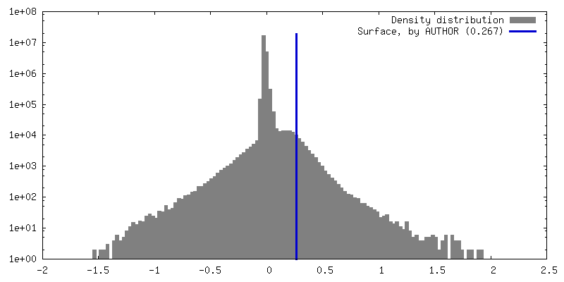
| Density |
| ||||||||||||||||||||||||||||||||||||
| Symmetry | Space group: 1 | ||||||||||||||||||||||||||||||||||||
| Details | EMDB XML:
|
 Z (Sec.)
 Y (Row.)
 X (Col.)


-Supplemental data
-Half map: Half map B
| File | emd_15351_half_map_1.map | ||||||||||||
|---|---|---|---|---|---|---|---|---|---|---|---|---|---|
| Annotation | Half map B | ||||||||||||
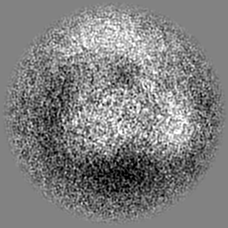
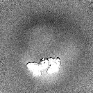
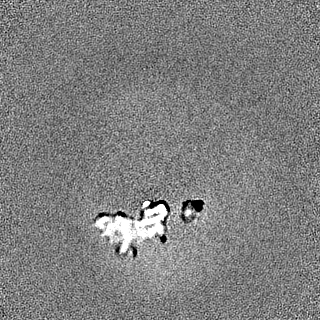
| Projections & Slices |
| ||||||||||||
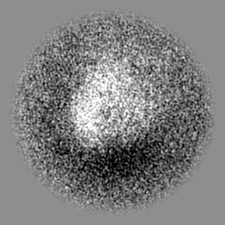
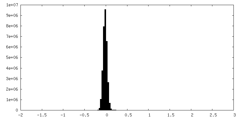
| Density Histograms |


-Half map: Half map A
| File | emd_15351_half_map_2.map | ||||||||||||
|---|---|---|---|---|---|---|---|---|---|---|---|---|---|
| Annotation | Half map A | ||||||||||||
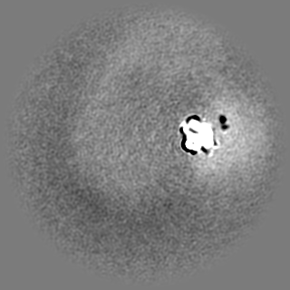
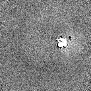
| Projections & Slices |
| ||||||||||||
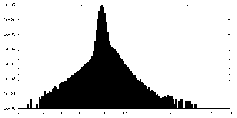
| Density Histograms |

- Sample components
-Entire : Replisome - pol alpha complex
| Entire | Name: Replisome - pol alpha complex |
|---|---|
| Components |
|
-Supramolecule #1: Replisome - pol alpha complex
| Supramolecule | Name: Replisome - pol alpha complex / type: complex / ID: 1 / Parent: 0 Details: S. cerevisiae pol alpha bound to the core replisome engaged with a fork DNA substrate containing a 60 nucleotide lagging strand. |
|---|---|
| Source (natural) | Organism: |
-Experimental details
-Structure determination
| Method | cryo EM |
|---|---|
 Processing Processing | single particle reconstruction |
| Aggregation state | particle |
-Sample preparation
| Buffer | pH: 7.6 |
|---|---|
| Vitrification | Cryogen name: ETHANE |
- Electron microscopy
| Microscope | FEI TITAN KRIOS |
|---|---|
| Image recording | Film or detector model: GATAN K3 (6k x 4k) / Average electron dose: 40.184 e/Å2 |
| Electron beam | Acceleration voltage: 300 kV / Electron source:  FIELD EMISSION GUN FIELD EMISSION GUN |
| Electron optics | Illumination mode: FLOOD BEAM / Imaging mode: BRIGHT FIELD / Nominal defocus max: 3.5 µm / Nominal defocus min: 1.5 µm |
| Experimental equipment |  Model: Titan Krios / Image courtesy: FEI Company |