 Movie
Movie Controller

[English] 日本語
 Yorodumi
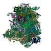
- EMDB-15228: RQT-bound 80S ribosome from S. cerevisiae (C1, raw consensus map) -
+ Open data

- Basic information
| Entry |  | ||||||||||||
|---|---|---|---|---|---|---|---|---|---|---|---|---|---|
| Title | RQT-bound 80S ribosome from S. cerevisiae (C1, raw consensus map) | ||||||||||||
 Map data Map data | refined map of the RQTc1 ribsome | ||||||||||||
 Sample Sample |
| ||||||||||||
 Keywords Keywords | collision / RNA binding / RQT / RQC / RIBOSOME | ||||||||||||
| Biological species |  | ||||||||||||
| Method | single particle reconstruction / cryo EM / Resolution: 2.4 Å | ||||||||||||
 Authors Authors | Best KM / Ikeuchi K / Kater L / Best DM / Musial J / Matsuo Y / Berninghausen O / Becker T / Inada T / Beckmann R | ||||||||||||
| Funding support | European Union,  Germany, 3 items Germany, 3 items
| ||||||||||||
 Citation Citation | Journal: Acta Crystallogr D Struct Biol / Year: 2018 Title: Real-space refinement in PHENIX for cryo-EM and crystallography. Authors: Pavel V Afonine / Billy K Poon / Randy J Read / Oleg V Sobolev / Thomas C Terwilliger / Alexandre Urzhumtsev / Paul D Adams /    Abstract: This article describes the implementation of real-space refinement in the phenix.real_space_refine program from the PHENIX suite. The use of a simplified refinement target function enables very fast ...This article describes the implementation of real-space refinement in the phenix.real_space_refine program from the PHENIX suite. The use of a simplified refinement target function enables very fast calculation, which in turn makes it possible to identify optimal data-restraint weights as part of routine refinements with little runtime cost. Refinement of atomic models against low-resolution data benefits from the inclusion of as much additional information as is available. In addition to standard restraints on covalent geometry, phenix.real_space_refine makes use of extra information such as secondary-structure and rotamer-specific restraints, as well as restraints or constraints on internal molecular symmetry. The re-refinement of 385 cryo-EM-derived models available in the Protein Data Bank at resolutions of 6 Å or better shows significant improvement of the models and of the fit of these models to the target maps. | ||||||||||||
| History |
|
- Structure visualization
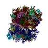
| Supplemental images |
|---|

- Downloads & links
-EMDB archive
| Map data | emd_15228.map.gz | 334.9 MB |  EMDB map data format EMDB map data format | |
|---|---|---|---|---|
| Header (meta data) | emd-15228-v30.xmlemd-15228.xml | 37.2 KB 37.2 KB | Display Display | EMDB header |
| FSC (resolution estimation) | emd_15228_fsc.xml | 18.3 KB | Display | FSC data file |
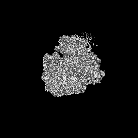
| Images | 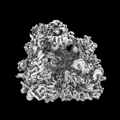 emd_15228.png emd_15228.png | 104.3 KB | ||
| Others | emd_15228_half_map_1.map.gzemd_15228_half_map_2.map.gz | 622.4 MB 622.4 MB | ||
| Archive directory |  http://ftp.pdbj.org/pub/emdb/structures/EMD-15228ftp://ftp.pdbj.org/pub/emdb/structures/EMD-15228 http://ftp.pdbj.org/pub/emdb/structures/EMD-15228ftp://ftp.pdbj.org/pub/emdb/structures/EMD-15228 | HTTPS FTP |
-Related structure data


-Links
| EMDB pages | EMDB (EBI/PDBe) / EMDataResource |
|---|---|
| Related items in Molecule of the Month |

-Map
| File | Download / File: emd_15228.map.gz / Format: CCP4 / Size: 669.9 MB / Type: IMAGE STORED AS FLOATING POINT NUMBER (4 BYTES) | ||||||||||||||||||||||||||||||||||||
|---|---|---|---|---|---|---|---|---|---|---|---|---|---|---|---|---|---|---|---|---|---|---|---|---|---|---|---|---|---|---|---|---|---|---|---|---|---|
| Annotation | refined map of the RQTc1 ribsome | ||||||||||||||||||||||||||||||||||||
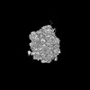
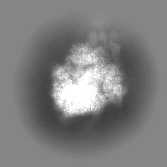
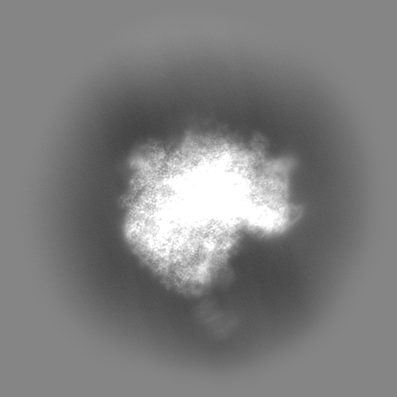
| Projections & slices | Image control
Images are generated by Spider. | ||||||||||||||||||||||||||||||||||||
| Voxel size | X=Y=Z: 1.045 Å | ||||||||||||||||||||||||||||||||||||
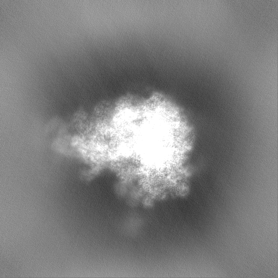
| Density |
| ||||||||||||||||||||||||||||||||||||
| Symmetry | Space group: 1 | ||||||||||||||||||||||||||||||||||||
| Details | EMDB XML:
|
 Z (Sec.)
 Y (Row.)
 X (Col.)


-Supplemental data
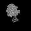
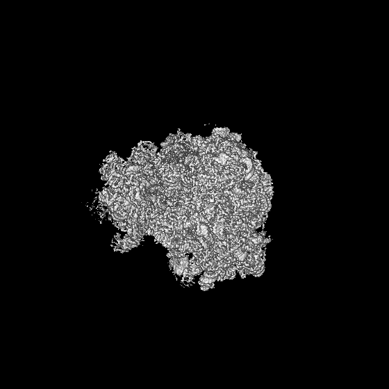
-Half map: halfmap 1
| File | emd_15228_half_map_1.map | ||||||||||||
|---|---|---|---|---|---|---|---|---|---|---|---|---|---|
| Annotation | halfmap 1 | ||||||||||||
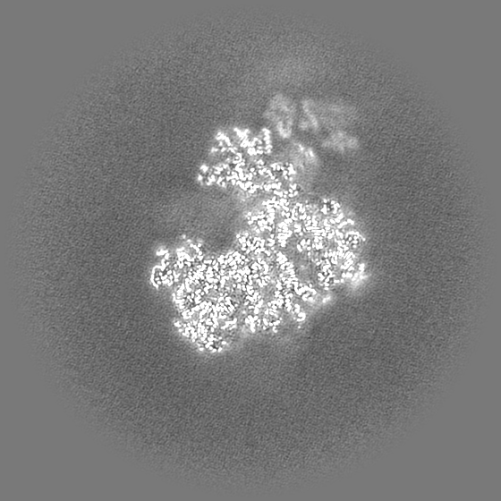
| Projections & Slices |
| ||||||||||||
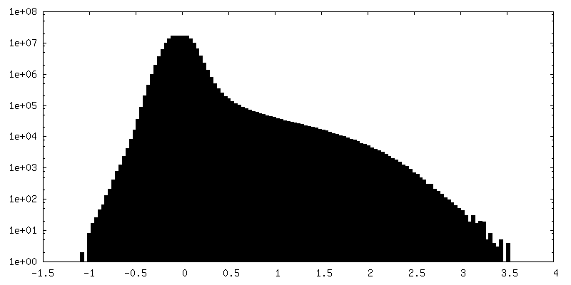
| Density Histograms |


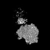
-Half map: halfmap 2
| File | emd_15228_half_map_2.map | ||||||||||||
|---|---|---|---|---|---|---|---|---|---|---|---|---|---|
| Annotation | halfmap 2 | ||||||||||||
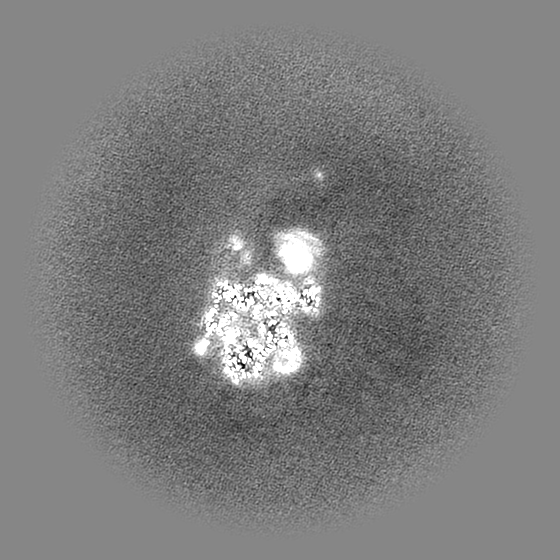
| Projections & Slices |
| ||||||||||||
| Density Histograms |


- Sample components
-Entire : ribosome with bound RQT components (Slh1, Cue3 and Rqt4)
| Entire | Name: ribosome with bound RQT components (Slh1, Cue3 and Rqt4) |
|---|---|
| Components |
|
-Supramolecule #1: ribosome with bound RQT components (Slh1, Cue3 and Rqt4)
| Supramolecule | Name: ribosome with bound RQT components (Slh1, Cue3 and Rqt4) type: complex / ID: 1 / Parent: 0 / Macromolecule list: #1-#82 |
|---|
-Supramolecule #2: RQT complex (Slh1, Cue3 and Rqt4)
| Supramolecule | Name: RQT complex (Slh1, Cue3 and Rqt4) / type: complex / ID: 2 / Parent: 1 / Macromolecule list: #80-#82 |
|---|---|
| Source (natural) | Organism: |
-Supramolecule #3: ribosome
| Supramolecule | Name: ribosome / type: complex / ID: 3 / Parent: 1 / Macromolecule list: #1-#79 |
|---|---|
| Source (natural) | Organism: |
-Experimental details
-Structure determination
| Method | cryo EM |
|---|---|
 Processing Processing | single particle reconstruction |
| Aggregation state | particle |
-Sample preparation
| Buffer | pH: 7.5 |
|---|---|
| Grid | Model: Quantifoil R3/3 / Material: COPPER / Support film - Material: CARBON |
| Vitrification | Cryogen name: ETHANE / Instrument: FEI VITROBOT MARK II |
- Electron microscopy
| Microscope | FEI TITAN KRIOS |
|---|---|
| Image recording | Film or detector model: GATAN K2 SUMMIT (4k x 4k) / Detector mode: COUNTING / Average electron dose: 43.6 e/Å2 |
| Electron beam | Acceleration voltage: 300 kV / Electron source:  FIELD EMISSION GUN FIELD EMISSION GUN |
| Electron optics | Illumination mode: SPOT SCAN / Imaging mode: BRIGHT FIELD / Nominal defocus max: 3.0 µm / Nominal defocus min: 0.5 µm |
| Experimental equipment |  Model: Titan Krios / Image courtesy: FEI Company |

