ムービー
ムービー コントローラー


+ データを開く

- 基本情報
基本情報
| 登録情報 |  | |||||||||
|---|---|---|---|---|---|---|---|---|---|---|
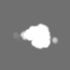
| タイトル | In-cell structure of the actin filament Arp2/3 complex branch junction in lamellipodia of WT B16-F1 mouse melanoma cells | |||||||||
 マップデータ マップデータ | Structure of the Arp2/3 actin branch junction as found in B16-F1 mouse melanoma cell lamellipodia | |||||||||
 試料 試料 |
| |||||||||
| 生物種 |  | |||||||||
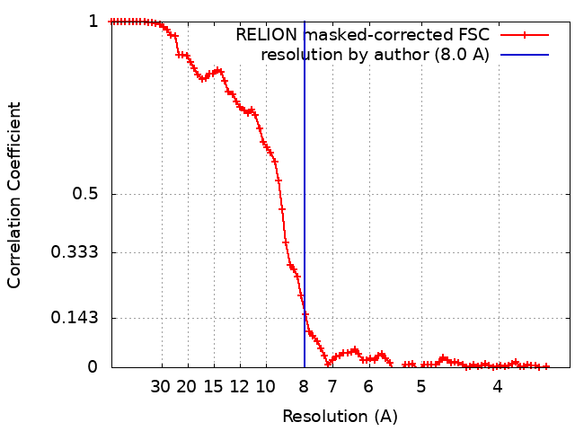
| 手法 | サブトモグラム平均法 / クライオ電子顕微鏡法 / 解像度: 8.0 Å | |||||||||
 データ登録者 データ登録者 | Faessler F / Javoor MG / Datler J / Doering H / Hofer FW / Dimchev G / Hodirnau VV / Rottner K / Schur FKM | |||||||||
| 資金援助 |  オーストリア, 1件 オーストリア, 1件
| |||||||||
 引用 引用 | ジャーナル: Sci Adv / 年: 2023 タイトル: ArpC5 isoforms regulate Arp2/3 complex-dependent protrusion through differential Ena/VASP positioning. 著者: Florian Fäßler / Manjunath G Javoor / Julia Datler / Hermann Döring / Florian W Hofer / Georgi Dimchev / Victor-Valentin Hodirnau / Jan Faix / Klemens Rottner / Florian K M Schur /  要旨: Regulation of the Arp2/3 complex is required for productive nucleation of branched actin networks. An emerging aspect of regulation is the incorporation of subunit isoforms into the Arp2/3 complex. ...Regulation of the Arp2/3 complex is required for productive nucleation of branched actin networks. An emerging aspect of regulation is the incorporation of subunit isoforms into the Arp2/3 complex. Specifically, both ArpC5 subunit isoforms, ArpC5 and ArpC5L, have been reported to fine-tune nucleation activity and branch junction stability. We have combined reverse genetics and cellular structural biology to describe how ArpC5 and ArpC5L differentially affect cell migration. Both define the structural stability of ArpC1 in branch junctions and, in turn, by determining protrusion characteristics, affect protein dynamics and actin network ultrastructure. ArpC5 isoforms also affect the positioning of members of the Ena/Vasodilator-stimulated phosphoprotein (VASP) family of actin filament elongators, which mediate ArpC5 isoform-specific effects on the actin assembly level. Our results suggest that ArpC5 and Ena/VASP proteins are part of a signaling pathway enhancing cell migration. | |||||||||
| 履歴 |
|
- 構造の表示
構造の表示
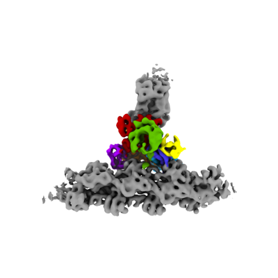
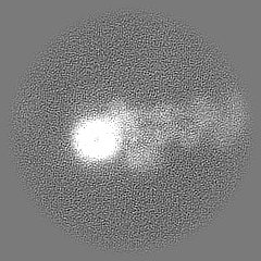
| 添付画像 |
|---|
- ダウンロードとリンク
-EMDBアーカイブ
| マップデータ | emd_15136.map.gz | 49.2 MB |  EMDBマップデータ形式 EMDBマップデータ形式 | |
|---|---|---|---|---|
| ヘッダ (付随情報) | emd-15136-v30.xmlemd-15136.xml | 19.1 KB 19.1 KB | 表示 表示 | EMDBヘッダ |
| FSC (解像度算出) | emd_15136_fsc.xml | 8.6 KB | 表示 | FSCデータファイル |
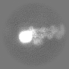
| 画像 |  emd_15136.png emd_15136.png | 43.8 KB | ||
| マスクデータ | emd_15136_msk_1.map | 52.7 MB | マスクマップ | |
| その他 | emd_15136_half_map_1.map.gzemd_15136_half_map_2.map.gz | 27 MB 27 MB | ||
| アーカイブディレクトリ |  http://ftp.pdbj.org/pub/emdb/structures/EMD-15136ftp://ftp.pdbj.org/pub/emdb/structures/EMD-15136 http://ftp.pdbj.org/pub/emdb/structures/EMD-15136ftp://ftp.pdbj.org/pub/emdb/structures/EMD-15136 | HTTPS FTP |
-検証レポート
| 文書・要旨 | emd_15136_validation.pdf.gz | 844.7 KB | 表示 | EMDB検証レポート |
|---|---|---|---|---|
| 文書・詳細版 | emd_15136_full_validation.pdf.gz | 844.2 KB | 表示 | |
| XML形式データ | emd_15136_validation.xml.gz | 14 KB | 表示 | |
| CIF形式データ | emd_15136_validation.cif.gz | 20 KB | 表示 | |
| アーカイブディレクトリ | https://ftp.pdbj.org/pub/emdb/validation_reports/EMD-15136ftp://ftp.pdbj.org/pub/emdb/validation_reports/EMD-15136 | HTTPS FTP |
-関連構造データ


-リンク
| EMDBのページ | EMDB (EBI/PDBe) / EMDataResource |
|---|
-マップ
| ファイル | ダウンロード / ファイル: emd_15136.map.gz / 形式: CCP4 / 大きさ: 52.7 MB / タイプ: IMAGE STORED AS FLOATING POINT NUMBER (4 BYTES) | ||||||||||||||||||||||||||||||||||||
|---|---|---|---|---|---|---|---|---|---|---|---|---|---|---|---|---|---|---|---|---|---|---|---|---|---|---|---|---|---|---|---|---|---|---|---|---|---|
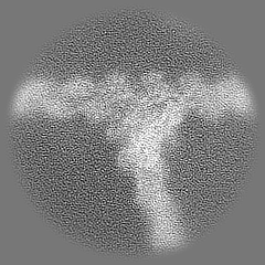
| 注釈 | Structure of the Arp2/3 actin branch junction as found in B16-F1 mouse melanoma cell lamellipodia | ||||||||||||||||||||||||||||||||||||
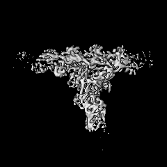
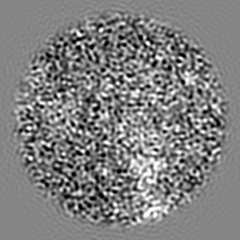
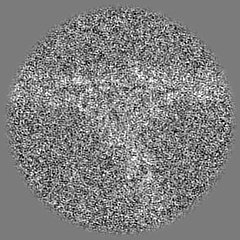
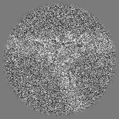
| 投影像・断面図 | 画像のコントロール
画像は Spider により作成 | ||||||||||||||||||||||||||||||||||||
| ボクセルのサイズ | X=Y=Z: 1.693 Å | ||||||||||||||||||||||||||||||||||||
| 密度 |
| ||||||||||||||||||||||||||||||||||||
| 対称性 | 空間群: 1 | ||||||||||||||||||||||||||||||||||||
| 詳細 | EMDB XML:
|
 Z (Sec.)
 Y (Row.)
 X (Col.)


-添付データ
-マスク #1
| ファイル | emd_15136_msk_1.map | ||||||||||||
|---|---|---|---|---|---|---|---|---|---|---|---|---|---|
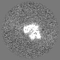
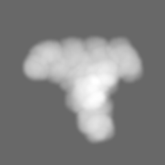
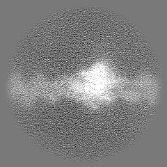
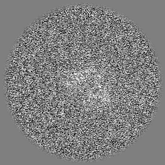
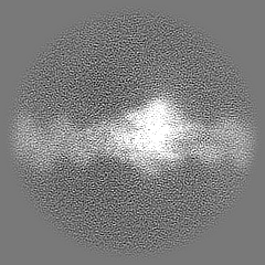
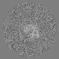
| 投影像・断面図 |
| ||||||||||||
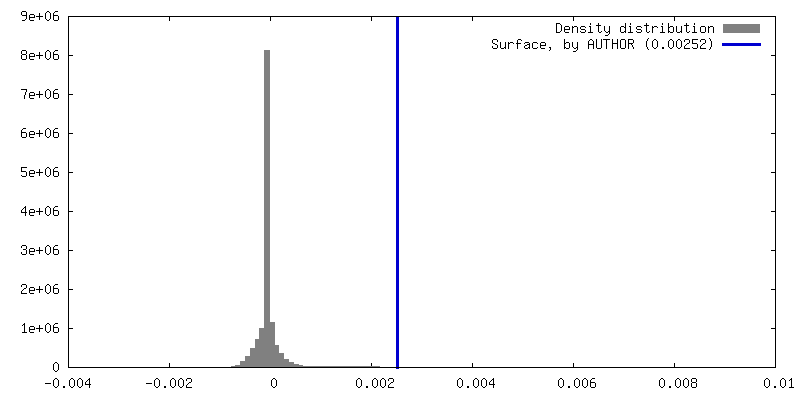
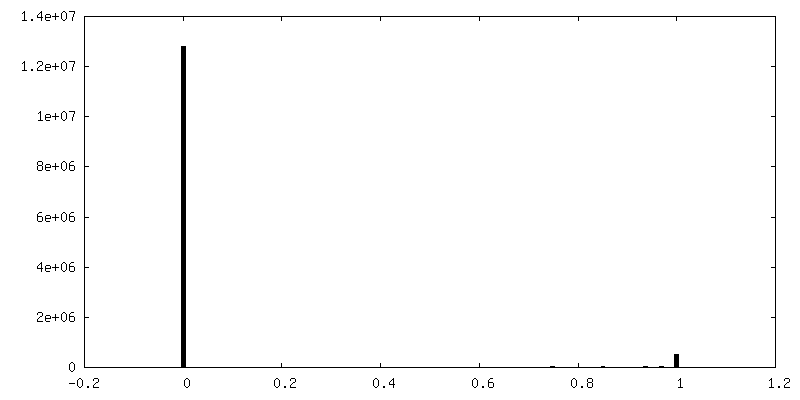
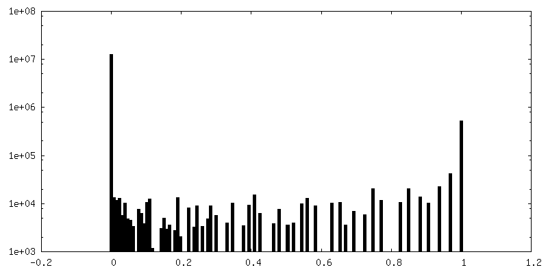
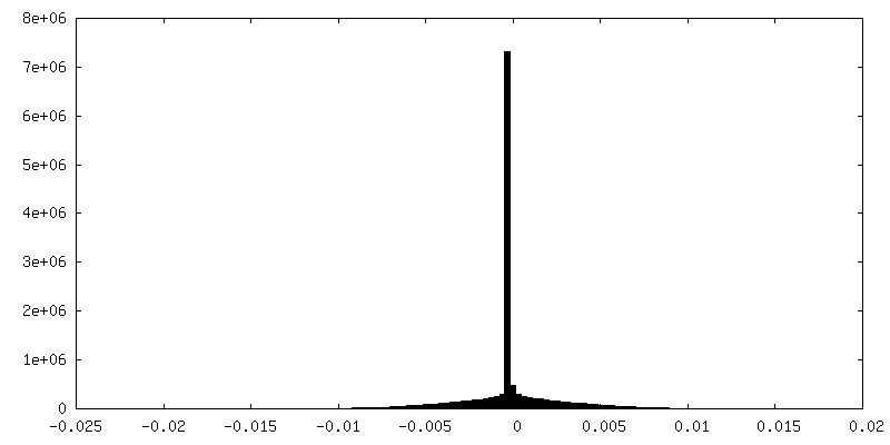
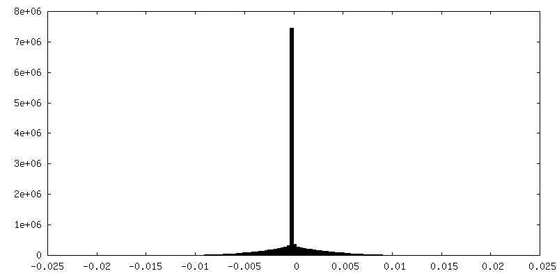
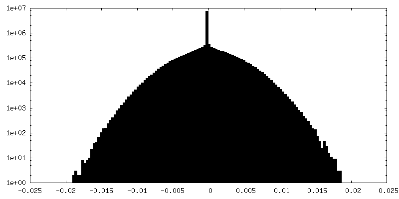
| 密度ヒストグラム |


-ハーフマップ: Halfmap 1 after subtomogram averaging and multi-particle refinemnt
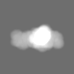
| ファイル | emd_15136_half_map_1.map | ||||||||||||
|---|---|---|---|---|---|---|---|---|---|---|---|---|---|
| 注釈 | Halfmap 1 after subtomogram averaging and multi-particle refinemnt | ||||||||||||
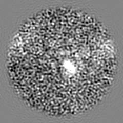
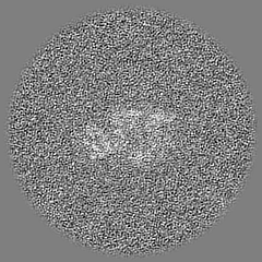
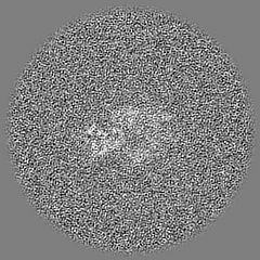
| 投影像・断面図 |
| ||||||||||||
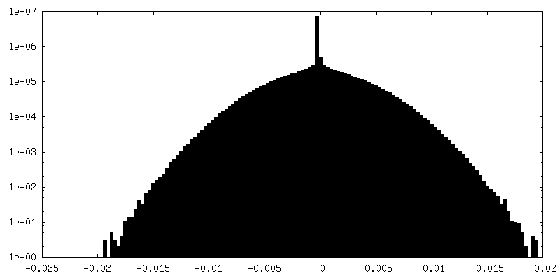
| 密度ヒストグラム |

-ハーフマップ: Halfmap 1 after subtomogram averaging and multi-particle refinemnt
| ファイル | emd_15136_half_map_2.map | ||||||||||||
|---|---|---|---|---|---|---|---|---|---|---|---|---|---|
| 注釈 | Halfmap 1 after subtomogram averaging and multi-particle refinemnt | ||||||||||||
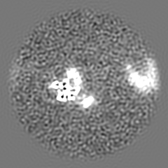
| 投影像・断面図 |
| ||||||||||||
| 密度ヒストグラム |

- 試料の構成要素
-全体 : In-cell structure of the actin filament Arp2/3 complex branch jun...
| 全体 | 名称: In-cell structure of the actin filament Arp2/3 complex branch junction in lamellipodia of WT B16-F1 mouse melanoma cells |
|---|---|
| 要素 |
|
-超分子 #1: In-cell structure of the actin filament Arp2/3 complex branch jun...
| 超分子 | 名称: In-cell structure of the actin filament Arp2/3 complex branch junction in lamellipodia of WT B16-F1 mouse melanoma cells タイプ: complex / ID: 1 / キメラ: Yes / 親要素: 0 |
|---|---|
| 由来(天然) | 生物種: |
-実験情報
-構造解析
| 手法 | クライオ電子顕微鏡法 |
|---|---|
 解析 解析 | サブトモグラム平均法 |
| 試料の集合状態 | cell |
-試料調製
| 緩衝液 | pH: 6.2 構成要素:
詳細: Adjust to pH 6.2 using NaOH Immediately prior to use, add Phalloidin to a final concentration of 1ug/ml | ||||||||||||||
|---|---|---|---|---|---|---|---|---|---|---|---|---|---|---|---|
| グリッド | モデル: Quantifoil R2/2 / 材質: GOLD / メッシュ: 200 / 支持フィルム - 材質: CARBON / 支持フィルム - トポロジー: HOLEY / 前処理 - タイプ: GLOW DISCHARGE / 前処理 - 時間: 120 sec. / 前処理 - 雰囲気: AIR 詳細: The grids were coated with 25ug/ml laminin for 1 hour prior to seeding the cells | ||||||||||||||
| 凍結 | 凍結剤: ETHANE / チャンバー内湿度: 80 % / チャンバー内温度: 277 K / 装置: LEICA EM GP |
- 電子顕微鏡法
| 顕微鏡 | FEI TITAN KRIOS |
|---|---|
| 特殊光学系 | エネルギーフィルター - 名称: GIF Bioquantum / エネルギーフィルター - スリット幅: 20 eV |
| 撮影 | フィルム・検出器のモデル: GATAN K3 (6k x 4k) / 撮影したグリッド数: 1 / 平均電子線量: 2.79 e/Å2 |
| 電子線 | 加速電圧: 300 kV / 電子線源:  FIELD EMISSION GUN FIELD EMISSION GUN |
| 電子光学系 | C2レンズ絞り径: 50.0 µm / 照射モード: FLOOD BEAM / 撮影モード: BRIGHT FIELD / Cs: 2.7 mm / 最大 デフォーカス(公称値): 4.5 µm / 最小 デフォーカス(公称値): 1.5 µm / 倍率(公称値): 53000 |
| 試料ステージ | 試料ホルダーモデル: FEI TITAN KRIOS AUTOGRID HOLDER ホルダー冷却材: NITROGEN |
| 実験機器 |  モデル: Titan Krios / 画像提供: FEI Company |