 ムービー
ムービー コントローラー


+ データを開く

- 基本情報
基本情報
| 登録情報 |  | |||||||||
|---|---|---|---|---|---|---|---|---|---|---|
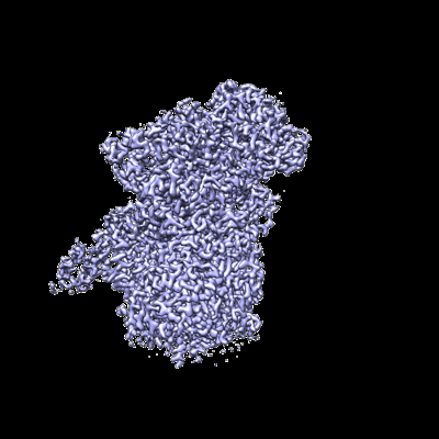
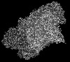
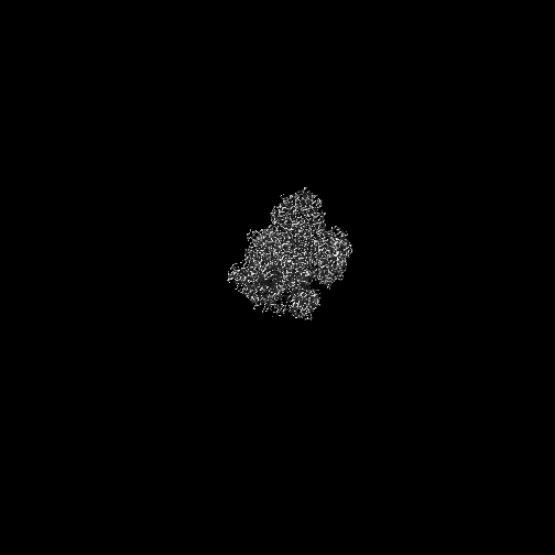
| タイトル | Peripheral arm of Complex I from Ovis aries at pH5.5, Open state | |||||||||
 マップデータ マップデータ | Peripheral arm postprocessed focus refined map | |||||||||
 試料 試料 |
| |||||||||
| 生物種 |  | |||||||||
| 手法 | 単粒子再構成法 / クライオ電子顕微鏡法 / 解像度: 3.2 Å | |||||||||
 データ登録者 データ登録者 | Petrova O / Sazanov L | |||||||||
| 資金援助 | 1件
| |||||||||
 引用 引用 | ジャーナル: Nature / 年: 2022 タイトル: A universal coupling mechanism of respiratory complex I. 著者: Vladyslav Kravchuk / Olga Petrova / Domen Kampjut / Anna Wojciechowska-Bason / Zara Breese / Leonid Sazanov /    要旨: Complex I is the first enzyme in the respiratory chain, which is responsible for energy production in mitochondria and bacteria. Complex I couples the transfer of two electrons from NADH to quinone ...Complex I is the first enzyme in the respiratory chain, which is responsible for energy production in mitochondria and bacteria. Complex I couples the transfer of two electrons from NADH to quinone and the translocation of four protons across the membrane, but the coupling mechanism remains contentious. Here we present cryo-electron microscopy structures of Escherichia coli complex I (EcCI) in different redox states, including catalytic turnover. EcCI exists mostly in the open state, in which the quinone cavity is exposed to the cytosol, allowing access for water molecules, which enable quinone movements. Unlike the mammalian paralogues, EcCI can convert to the closed state only during turnover, showing that closed and open states are genuine turnover intermediates. The open-to-closed transition results in the tightly engulfed quinone cavity being connected to the central axis of the membrane arm, a source of substrate protons. Consistently, the proportion of the closed state increases with increasing pH. We propose a detailed but straightforward and robust mechanism comprising a 'domino effect' series of proton transfers and electrostatic interactions: the forward wave ('dominoes stacking') primes the pump, and the reverse wave ('dominoes falling') results in the ejection of all pumped protons from the distal subunit NuoL. This mechanism explains why protons exit exclusively from the NuoL subunit and is supported by our mutagenesis data. We contend that this is a universal coupling mechanism of complex I and related enzymes. | |||||||||
| 履歴 |
|
- 構造の表示
構造の表示
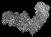
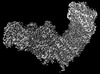
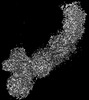
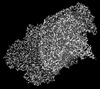
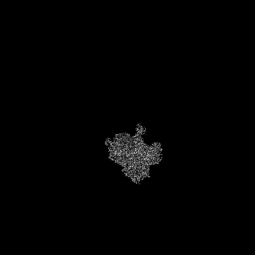
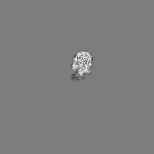
| 添付画像 |
|---|
- ダウンロードとリンク
-EMDBアーカイブ
| マップデータ | emd_15078.map.gz | 26.8 MB |  EMDBマップデータ形式 EMDBマップデータ形式 | |
|---|---|---|---|---|
| ヘッダ (付随情報) | emd-15078-v30.xmlemd-15078.xml | 21.7 KB 21.7 KB | 表示 表示 | EMDBヘッダ |
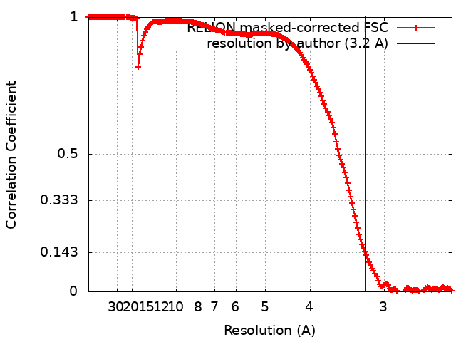
| FSC (解像度算出) | emd_15078_fsc.xml | 17.7 KB | 表示 | FSCデータファイル |
| 画像 |  emd_15078.png emd_15078.png | 100.6 KB | ||
| その他 | emd_15078_additional_1.map.gzemd_15078_half_map_1.map.gzemd_15078_half_map_2.map.gz | 391.5 MB 391.4 MB 391.3 MB | ||
| アーカイブディレクトリ |  http://ftp.pdbj.org/pub/emdb/structures/EMD-15078ftp://ftp.pdbj.org/pub/emdb/structures/EMD-15078 http://ftp.pdbj.org/pub/emdb/structures/EMD-15078ftp://ftp.pdbj.org/pub/emdb/structures/EMD-15078 | HTTPS FTP |
-検証レポート
| 文書・要旨 | emd_15078_validation.pdf.gz | 164.6 KB | 表示 | EMDB検証レポート |
|---|---|---|---|---|
| 文書・詳細版 | emd_15078_full_validation.pdf.gz | 164.1 KB | 表示 | |
| XML形式データ | emd_15078_validation.xml.gz | 497 B | 表示 | |
| CIF形式データ | emd_15078_validation.cif.gz | 373 B | 表示 | |
| アーカイブディレクトリ | https://ftp.pdbj.org/pub/emdb/validation_reports/EMD-15078ftp://ftp.pdbj.org/pub/emdb/validation_reports/EMD-15078 | HTTPS FTP |
-関連構造データ
| 関連構造データ |  7p61C  7p62C  7p63C  7p64C  7p69C  7p7cC  7p7eC  7p7jC  7p7kC  7p7lC  7p7mC  7z7rC  7z7sC 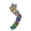 7z7tC  7z7vC  7z80C  7z83C 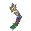 7z84C  7zc5C  7zciC 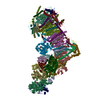 7zd6C  7zdhC 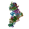 7zdjC 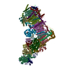 7zdmC 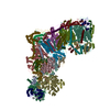 7zdpC 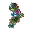 7zebC C: 同じ文献を引用 ( |
|---|


-リンク
| EMDBのページ | EMDB (EBI/PDBe) / EMDataResource |
|---|
-マップ
| ファイル | ダウンロード / ファイル: emd_15078.map.gz / 形式: CCP4 / 大きさ: 488.4 MB / タイプ: IMAGE STORED AS FLOATING POINT NUMBER (4 BYTES) | ||||||||||||||||||||||||||||||||||||
|---|---|---|---|---|---|---|---|---|---|---|---|---|---|---|---|---|---|---|---|---|---|---|---|---|---|---|---|---|---|---|---|---|---|---|---|---|---|
| 注釈 | Peripheral arm postprocessed focus refined map | ||||||||||||||||||||||||||||||||||||
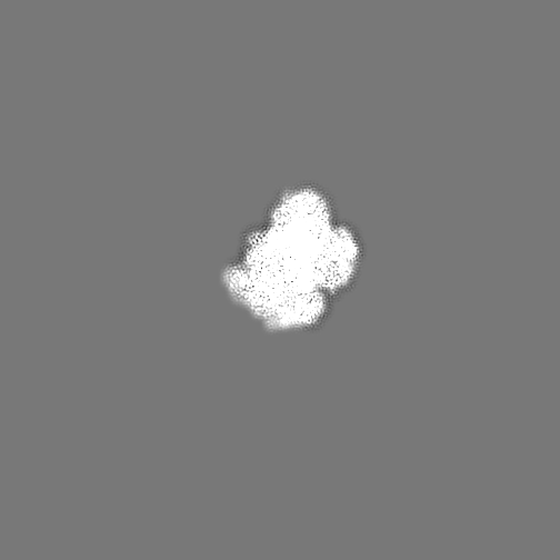
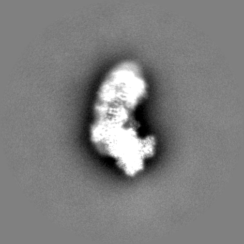
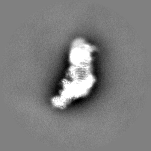
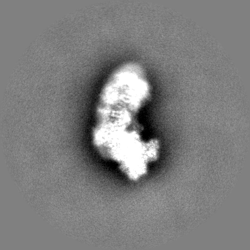
| 投影像・断面図 | 画像のコントロール
画像は Spider により作成 | ||||||||||||||||||||||||||||||||||||
| ボクセルのサイズ | X=Y=Z: 1.22 Å | ||||||||||||||||||||||||||||||||||||
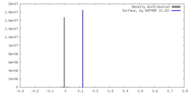
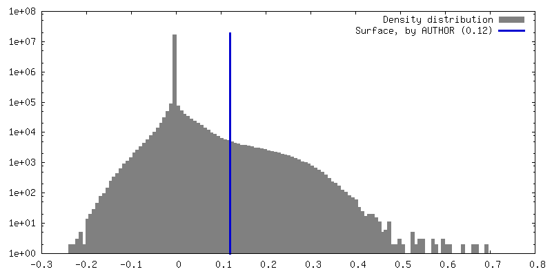
| 密度 |
| ||||||||||||||||||||||||||||||||||||
| 対称性 | 空間群: 1 | ||||||||||||||||||||||||||||||||||||
| 詳細 | EMDB XML:
|
 Z (Sec.)
 Y (Row.)
 X (Col.)


-添付データ
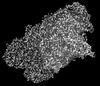
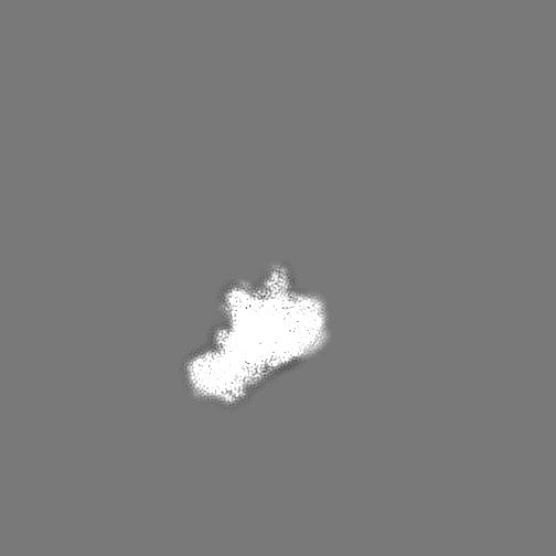
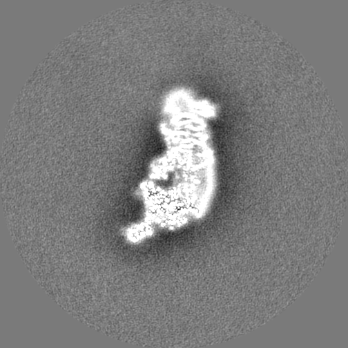
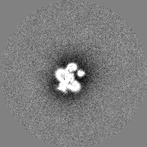
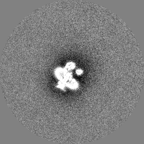
-追加マップ: Peripheral arm focus refined map
| ファイル | emd_15078_additional_1.map | ||||||||||||
|---|---|---|---|---|---|---|---|---|---|---|---|---|---|
| 注釈 | Peripheral arm focus refined map | ||||||||||||
| 投影像・断面図 |
| ||||||||||||
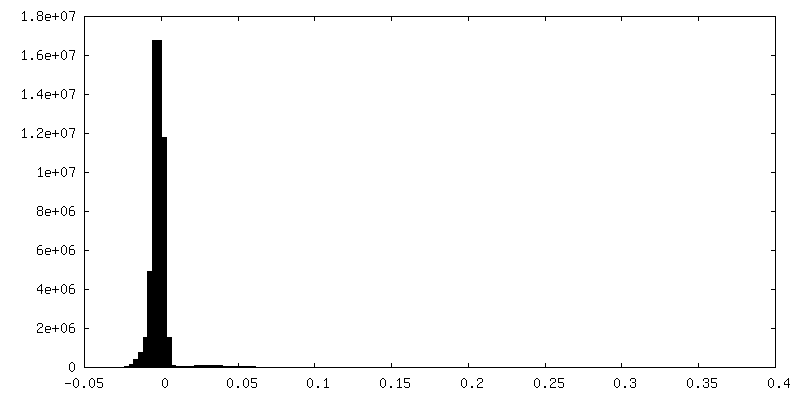
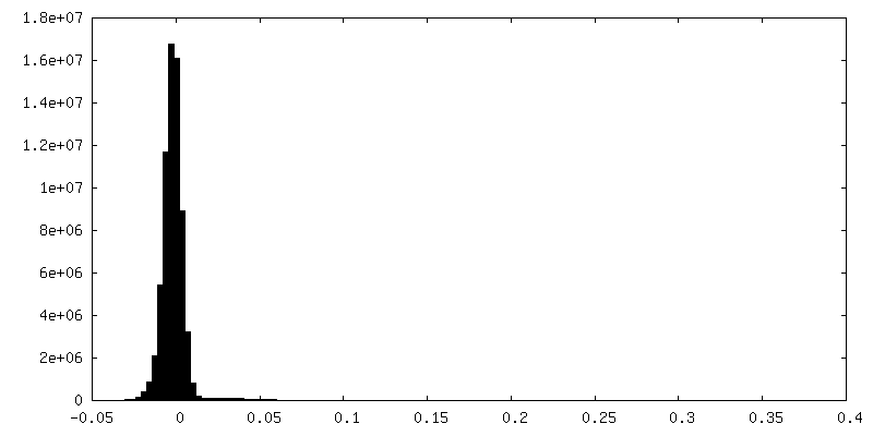
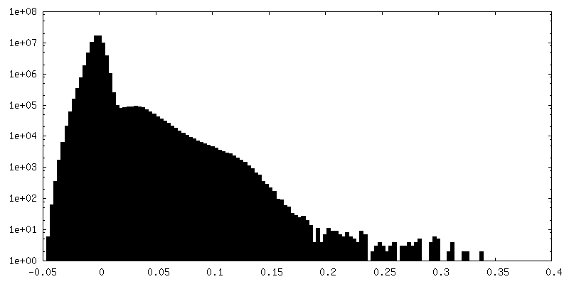
| 密度ヒストグラム |


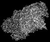
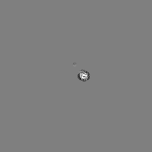
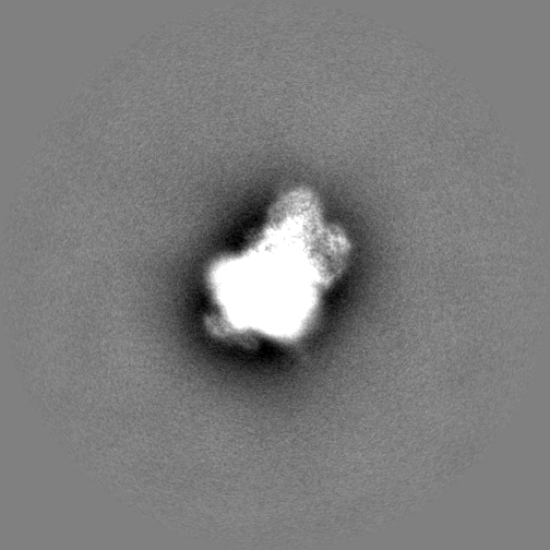
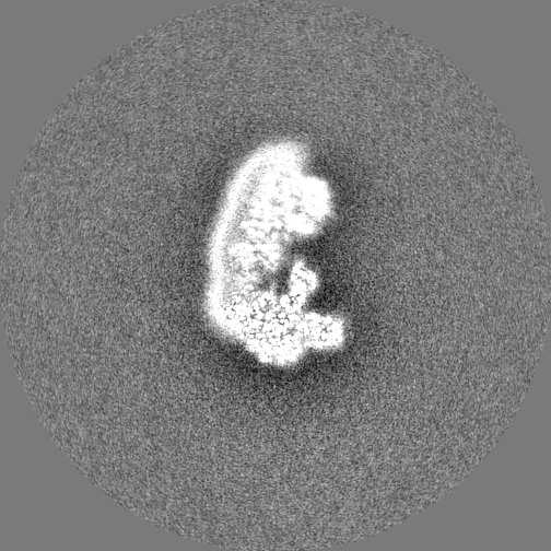
-ハーフマップ: half map 2
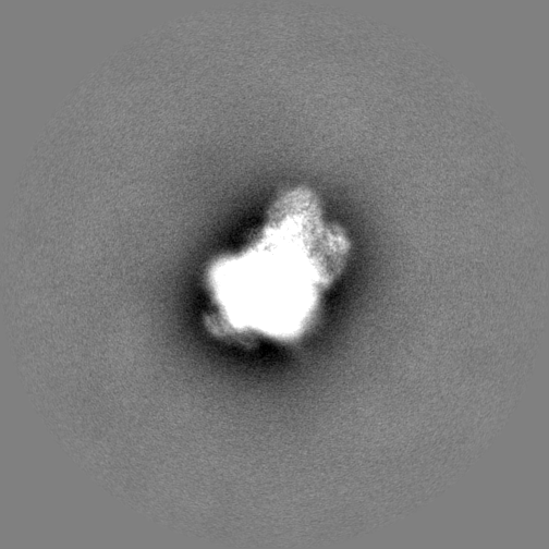
| ファイル | emd_15078_half_map_1.map | ||||||||||||
|---|---|---|---|---|---|---|---|---|---|---|---|---|---|
| 注釈 | half map 2 | ||||||||||||
| 投影像・断面図 |
| ||||||||||||
| 密度ヒストグラム |


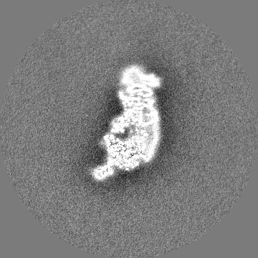
-ハーフマップ: half map 1
| ファイル | emd_15078_half_map_2.map | ||||||||||||
|---|---|---|---|---|---|---|---|---|---|---|---|---|---|
| 注釈 | half map 1 | ||||||||||||
| 投影像・断面図 |
| ||||||||||||
| 密度ヒストグラム |


- 試料の構成要素
-全体 : Complex I from Ovis aries at pH5.5, Open state
| 全体 | 名称: Complex I from Ovis aries at pH5.5, Open state |
|---|---|
| 要素 |
|
-超分子 #1: Complex I from Ovis aries at pH5.5, Open state
| 超分子 | 名称: Complex I from Ovis aries at pH5.5, Open state / タイプ: complex / キメラ: Yes / ID: 1 / 親要素: 0 / 含まれる分子: #1-#44 |
|---|---|
| 由来(天然) | 生物種: |
| 分子量 | 実験値: 1 MDa |
-実験情報
-構造解析
| 手法 | クライオ電子顕微鏡法 |
|---|---|
 解析 解析 | 単粒子再構成法 |
| 試料の集合状態 | particle |
-試料調製
| 濃度 | 3 mg/mL |
|---|---|
| 緩衝液 | pH: 5.5 |
| 凍結 | 凍結剤: ETHANE / チャンバー内湿度: 100 % / チャンバー内温度: 277 K / 装置: FEI VITROBOT MARK IV |
- 電子顕微鏡法
| 顕微鏡 | TFS GLACIOS |
|---|---|
| 撮影 | フィルム・検出器のモデル: FEI FALCON III (4k x 4k) 平均露光時間: 3.3 sec. / 平均電子線量: 90.0 e/Å2 |
| 電子線 | 加速電圧: 200 kV / 電子線源:  FIELD EMISSION GUN FIELD EMISSION GUN |
| 電子光学系 | 照射モード: FLOOD BEAM / 撮影モード: BRIGHT FIELD / 最大 デフォーカス(公称値): 2.0 µm / 最小 デフォーカス(公称値): 1.0 µm / 倍率(公称値): 120000 |
| 試料ステージ | ホルダー冷却材: NITROGEN |
+画像解析

-原子モデル構築 1
| 初期モデル | PDB ID: |
|---|---|
| 精密化 | 空間: REAL / プロトコル: FLEXIBLE FIT / 当てはまり具合の基準: correlation coefficient |
