 ムービー
ムービー コントローラー


+ データを開く

- 基本情報
基本情報
| 登録情報 |  | |||||||||
|---|---|---|---|---|---|---|---|---|---|---|
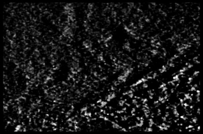
| タイトル | Fibroblast nucleus sub-volume. | |||||||||
 マップデータ マップデータ | sub-volume of EMD-14924. corresponds to Supplementary Movie S2. | |||||||||
 試料 試料 |
| |||||||||
 キーワード キーワード | nucleus / chromatin / STEM / DNA | |||||||||
| 生物種 |  Homo sapiens (ヒト) Homo sapiens (ヒト) | |||||||||
| 手法 | 電子線トモグラフィー法 / クライオ電子顕微鏡法 | |||||||||
 データ登録者 データ登録者 | Sedat J / McDonald A / Elbaum M | |||||||||
| 資金援助 |  イスラエル, 1件 イスラエル, 1件
| |||||||||
 引用 引用 | ジャーナル: Proc Natl Acad Sci U S A / 年: 2022 タイトル: A proposed unified interphase nucleus chromosome structure: Preliminary preponderance of evidence. 著者: John Sedat / Angus McDonald / Hu Cang / Joseph Lucas / Muthuvel Arigovindan / Zvi Kam / Cornelis Murre / Michael Elbaum /   要旨: Cryoelectron tomography of the cell nucleus using scanning transmission electron microscopy and deconvolution processing technology has highlighted a large-scale, 100- to 300-nm interphase chromosome ...Cryoelectron tomography of the cell nucleus using scanning transmission electron microscopy and deconvolution processing technology has highlighted a large-scale, 100- to 300-nm interphase chromosome structure, which is present throughout the nucleus. This study further documents and analyzes these chromosome structures. The paper is divided into four parts: 1) evidence (preliminary) for a unified interphase chromosome structure; 2) a proposed unified interphase chromosome architecture; 3) organization as chromosome territories (e.g., fitting the 46 human chromosomes into a 10-μm-diameter nucleus); and 4) structure unification into a polytene chromosome architecture and lampbrush chromosomes. Finally, the paper concludes with a living light microscopy cell study showing that the G1 nucleus contains very similar structures throughout. The main finding is that this chromosome structure appears to coil the 11-nm nucleosome fiber into a defined hollow structure, analogous to a Slinky helical spring [https://en.wikipedia.org/wiki/Slinky; motif used in Bowerman , 10, e65587 (2021)]. This Slinky architecture can be used to build chromosome territories, extended to the polytene chromosome structure, as well as to the structure of lampbrush chromosomes. | |||||||||
| 履歴 |
|
- 構造の表示
構造の表示
| 添付画像 |
|---|
- ダウンロードとリンク
-EMDBアーカイブ
| マップデータ | emd_15057.map.gz | 49.2 MB |  EMDBマップデータ形式 EMDBマップデータ形式 | |
|---|---|---|---|---|
| ヘッダ (付随情報) | emd-15057-v30.xmlemd-15057.xml | 10.6 KB 10.6 KB | 表示 表示 | EMDBヘッダ |
| 画像 |  emd_15057.png emd_15057.png | 100.2 KB | ||
| Filedesc metadata | emd-15057.cif.gz | 4.2 KB | ||
| アーカイブディレクトリ |  http://ftp.pdbj.org/pub/emdb/structures/EMD-15057ftp://ftp.pdbj.org/pub/emdb/structures/EMD-15057 http://ftp.pdbj.org/pub/emdb/structures/EMD-15057ftp://ftp.pdbj.org/pub/emdb/structures/EMD-15057 | HTTPS FTP |
-検証レポート
| 文書・要旨 | emd_15057_validation.pdf.gz | 410.8 KB | 表示 | EMDB検証レポート |
|---|---|---|---|---|
| 文書・詳細版 | emd_15057_full_validation.pdf.gz | 410.3 KB | 表示 | |
| XML形式データ | emd_15057_validation.xml.gz | 4.9 KB | 表示 | |
| CIF形式データ | emd_15057_validation.cif.gz | 5.4 KB | 表示 | |
| アーカイブディレクトリ | https://ftp.pdbj.org/pub/emdb/validation_reports/EMD-15057ftp://ftp.pdbj.org/pub/emdb/validation_reports/EMD-15057 | HTTPS FTP |
-関連構造データ


-リンク
| EMDBのページ | EMDB (EBI/PDBe) / EMDataResource |
|---|
-マップ
| ファイル | ダウンロード / ファイル: emd_15057.map.gz / 形式: CCP4 / 大きさ: 81.3 MB / タイプ: IMAGE STORED AS FLOATING POINT NUMBER (4 BYTES) | ||||||||||||||||||||
|---|---|---|---|---|---|---|---|---|---|---|---|---|---|---|---|---|---|---|---|---|---|
| 注釈 | sub-volume of EMD-14924. corresponds to Supplementary Movie S2. | ||||||||||||||||||||
| ボクセルのサイズ | X=Y=Z: 33.5 Å | ||||||||||||||||||||
| 密度 |
| ||||||||||||||||||||
| 対称性 | 空間群: 1 | ||||||||||||||||||||
| 詳細 | EMDB XML:
|
-添付データ
- 試料の構成要素
-全体 : WI-38 fibroblast cell
| 全体 | 名称: WI-38 fibroblast cell |
|---|---|
| 要素 |
|
-超分子 #1: WI-38 fibroblast cell
| 超分子 | 名称: WI-38 fibroblast cell / タイプ: cell / ID: 1 / 親要素: 0 / 詳細: cells cultured directly on gold EM grid |
|---|---|
| 由来(天然) | 生物種: Homo sapiens (ヒト) / 器官: lung / 組織: cell culture |
-実験情報
-構造解析
| 手法 | クライオ電子顕微鏡法 |
|---|---|
 解析 解析 | 電子線トモグラフィー法 |
| 試料の集合状態 | cell |
-試料調製
| 緩衝液 | pH: 7.4 詳細: Minimal Essential Medium (Gibco) supplemented with 15% fetal calf serum, L-glutamine, and penicillin/streptomycin. |
|---|---|
| 凍結 | 凍結剤: ETHANE / 装置: LEICA EM GP |
| 詳細 | WI-38 embryonic lung fibroblast cells (obtained from Coriell Institute) were cultured directly on gold EM grids and imaged intact by cryo-STEM tomography (CSTET). |
| 切片作成 | その他: NO SECTIONING |
| 位置合わせマーカー | Manufacturer: obtained from L Duchesne (https://doi.org/10.1021/la802876u ) 直径: 10 nm |
- 電子顕微鏡法
| 顕微鏡 | FEI TECNAI F20 |
|---|---|
| 特殊光学系 | 詳細: STEM data collection in BF mode. |
| 詳細 | NanoProbe STEM acquisition. |
| 撮影 | フィルム・検出器のモデル: OTHER / デジタル化 - サイズ - 横: 2048 pixel / デジタル化 - サイズ - 縦: 2048 pixel / 撮影したグリッド数: 1 / 実像数: 61 / 平均露光時間: 12.6 sec. / 平均電子線量: 3.8 e/Å2 / 詳細: Cryo-STEM tilt series |
| 電子線 | 加速電圧: 200 kV / 電子線源:  FIELD EMISSION GUN FIELD EMISSION GUN |
| 電子光学系 | C2レンズ絞り径: 20.0 µm / 照射モード: SPOT SCAN / 撮影モード: BRIGHT FIELD / Cs: 2.0 mm / 最大 デフォーカス(公称値): 0.0 µm / 最小 デフォーカス(公称値): 0.0 µm |
| 試料ステージ | 試料ホルダーモデル: GATAN 626 SINGLE TILT LIQUID NITROGEN CRYO TRANSFER HOLDER ホルダー冷却材: NITROGEN |
| 実験機器 |  モデル: Tecnai F20 / 画像提供: FEI Company |
-画像解析
| 詳細 | Gatan STEM BF/ADF detector |
|---|---|
| 最終 再構成 | アルゴリズム: BACK PROJECTION / ソフトウェア: (名称: TOMO3D, PRIISM/IVE) 詳細: The original alignment was done using etomo (IMOD), and then optimized using tomoalign as a "thin" specimen. A new aligned stack (.ali) was produced using tomowarpalign (tomoalign) and ...詳細: The original alignment was done using etomo (IMOD), and then optimized using tomoalign as a "thin" specimen. A new aligned stack (.ali) was produced using tomowarpalign (tomoalign) and reconstructed by SIRT using tomo3d. The reconstruction was then binned (by 2) and processed by 3D deconvolution as described in Waugh et al (doi:10.1073/pnas.2000700117) with smoothing parameter 0.1. 使用した粒子像数: 56 |
