 ムービー
ムービー コントローラー


+ データを開く

- 基本情報
基本情報
| 登録情報 | データベース: EMDB / ID: EMD-1498 | |||||||||
|---|---|---|---|---|---|---|---|---|---|---|
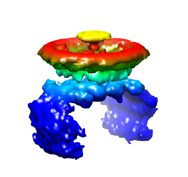
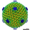
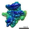
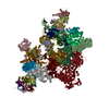
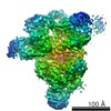
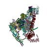
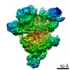
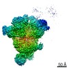
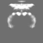
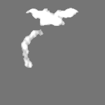
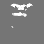
| タイトル | Vertex reconstruction of SH1 spike | |||||||||
 マップデータ マップデータ | spike | |||||||||
 試料 試料 |
| |||||||||
 キーワード キーワード | archaea / virus / SH1 / spike / infection / adsorption | |||||||||
| 生物種 |  Haloarcula phage SH1 (ウイルス) Haloarcula phage SH1 (ウイルス) | |||||||||
| 手法 | 単粒子再構成法 / クライオ電子顕微鏡法 / 解像度: 34.0 Å | |||||||||
 データ登録者 データ登録者 | Jaalinoja HT / Roine E / Laurinmaki P / Kivela HM / Bamford DH / Butcher SJ | |||||||||
 引用 引用 | ジャーナル: Proc Natl Acad Sci U S A / 年: 2008 タイトル: Structure and host-cell interaction of SH1, a membrane-containing, halophilic euryarchaeal virus. 著者: Harri T Jäälinoja / Elina Roine / Pasi Laurinmäki / Hanna M Kivelä / Dennis H Bamford / Sarah J Butcher /  要旨: The Archaea, and the viruses that infect them, are the least well understood of all of the three domains of life. They often grow in extreme conditions such as hypersaline lakes and sulfuric hot ...The Archaea, and the viruses that infect them, are the least well understood of all of the three domains of life. They often grow in extreme conditions such as hypersaline lakes and sulfuric hot springs. Only rare glimpses have been gained into the structures of archaeal viruses. Here, we report the subnanometer resolution structure of a recently isolated, hypersalinic, membrane-containing, euryarchaeal virus, SH1, in which different viral proteins can be localized. The results indicate that SH1 has a complex capsid formed from single beta-barrels, an important missing link in hypotheses on viral capsid protein evolution. Unusual, symmetry-mismatched spikes seem to play a role in host adsorption. They are connected to highly organized membrane proteins providing a platform for capsid assembly and potential machinery for host infection. | |||||||||
| 履歴 |
|
- 構造の表示
構造の表示
| ムービー |
 ムービービューア ムービービューア |
|---|---|
| 構造ビューア | EMマップ: SurfViewMolmilJmol/JSmol |
| 添付画像 |
- ダウンロードとリンク
-EMDBアーカイブ
| マップデータ | emd_1498.map.gz | 874 KB | EMDBマップデータ形式 | |
|---|---|---|---|---|
| ヘッダ (付随情報) | emd-1498-v30.xmlemd-1498.xml | 9.3 KB 9.3 KB | 表示 表示 | EMDBヘッダ |
| 画像 |  1498.png 1498.png | 171.4 KB | ||
| アーカイブディレクトリ |  http://ftp.pdbj.org/pub/emdb/structures/EMD-1498ftp://ftp.pdbj.org/pub/emdb/structures/EMD-1498 http://ftp.pdbj.org/pub/emdb/structures/EMD-1498ftp://ftp.pdbj.org/pub/emdb/structures/EMD-1498 | HTTPS FTP |
-検証レポート
| 文書・要旨 | emd_1498_validation.pdf.gz | 212.8 KB | 表示 | EMDB検証レポート |
|---|---|---|---|---|
| 文書・詳細版 | emd_1498_full_validation.pdf.gz | 211.9 KB | 表示 | |
| XML形式データ | emd_1498_validation.xml.gz | 5.8 KB | 表示 | |
| アーカイブディレクトリ | https://ftp.pdbj.org/pub/emdb/validation_reports/EMD-1498ftp://ftp.pdbj.org/pub/emdb/validation_reports/EMD-1498 | HTTPS FTP |
-関連構造データ
-リンク
| EMDBのページ | EMDB (EBI/PDBe) / EMDataResource |
|---|
-マップ
| ファイル | ダウンロード / ファイル: emd_1498.map.gz / 形式: CCP4 / 大きさ: 12.6 MB / タイプ: IMAGE STORED AS FLOATING POINT NUMBER (4 BYTES) | ||||||||||||||||||||||||||||||||||||||||||||||||||||||||||||||||||||
|---|---|---|---|---|---|---|---|---|---|---|---|---|---|---|---|---|---|---|---|---|---|---|---|---|---|---|---|---|---|---|---|---|---|---|---|---|---|---|---|---|---|---|---|---|---|---|---|---|---|---|---|---|---|---|---|---|---|---|---|---|---|---|---|---|---|---|---|---|---|
| 注釈 | spike | ||||||||||||||||||||||||||||||||||||||||||||||||||||||||||||||||||||
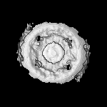
| 投影像・断面図 | 画像のコントロール
画像は Spider により作成 | ||||||||||||||||||||||||||||||||||||||||||||||||||||||||||||||||||||
| ボクセルのサイズ |
| ||||||||||||||||||||||||||||||||||||||||||||||||||||||||||||||||||||
| 密度 |
| ||||||||||||||||||||||||||||||||||||||||||||||||||||||||||||||||||||
| 対称性 | 空間群: 1 | ||||||||||||||||||||||||||||||||||||||||||||||||||||||||||||||||||||
| 詳細 | EMDB XML:
CCP4マップ ヘッダ情報:
| ||||||||||||||||||||||||||||||||||||||||||||||||||||||||||||||||||||
 Z (Sec.)
 Y (Row.)
 X (Col.)


-添付データ
- 試料の構成要素
-全体 : SH1 virus, reconstruction of the non-icosahedrally symmetric spik...
| 全体 | 名称: SH1 virus, reconstruction of the non-icosahedrally symmetric spikes on the vertices |
|---|---|
| 要素 |
|
-超分子 #1000: SH1 virus, reconstruction of the non-icosahedrally symmetric spik...
| 超分子 | 名称: SH1 virus, reconstruction of the non-icosahedrally symmetric spikes on the vertices タイプ: sample / ID: 1000 / Number unique components: 1 |
|---|
-超分子 #1: Haloarcula phage SH1
| 超分子 | 名称: Haloarcula phage SH1 / タイプ: virus / ID: 1 / Name.synonym: SH1 / NCBI-ID: 326574 / 生物種: Haloarcula phage SH1 / ウイルスタイプ: VIRION / ウイルス・単離状態: STRAIN / ウイルス・エンベロープ: No / ウイルス・中空状態: No / Syn species name: SH1 |
|---|---|
| 宿主 | 生物種:  Haloarcula hispanica (好塩性) / 別称: ARCHAEA Haloarcula hispanica (好塩性) / 別称: ARCHAEA |
-実験情報
-構造解析
| 手法 | クライオ電子顕微鏡法 |
|---|---|
 解析 解析 | 単粒子再構成法 |
| 試料の集合状態 | particle |
-試料調製
| 緩衝液 | pH: 7.2 詳細: 150-500 mM NaCl, 10mM MgCl2, 10 M MgSO4, 5 mM KCl, 3 mM CaCl2, 40 mM Tris-HCl pH 7.2 |
|---|---|
| グリッド | 詳細: 400 mesh copper grid, Quantifoil R2/2 holey |
| 凍結 | 凍結剤: ETHANE / チャンバー内温度: 90 K / 装置: HOMEMADE PLUNGER / 詳細: Vitrification instrument: EMBL design 手法: A small vial of ethane is placed inside a larger liquid nitrogen reservoir. The grid holding 3 microliters of the sample is held in place at the bottom of a plunger by the means of fine ...手法: A small vial of ethane is placed inside a larger liquid nitrogen reservoir. The grid holding 3 microliters of the sample is held in place at the bottom of a plunger by the means of fine tweezers. When the liquid ethane is ready, a piece of filter paper is then pressed against the sample to blot off excess buffer, sufficient to leave a thin layer on the grid. The filter paper is removed, and the plunger is allowed to drop into the liquid ethane. Once the grid enters the liquid ethane, the sample is rapidly frozen, and the grid is transferred under liquid nitrogen to a storage box immersed in liquid nitrogen for later use in the microscope. |
- 電子顕微鏡法
| 顕微鏡 | FEI TECNAI F20 |
|---|---|
| 温度 | 最低: 90 K / 最高: 94 K / 平均: 93 K |
| 詳細 | Low dose conditions. |
| 撮影 | カテゴリ: FILM / フィルム・検出器のモデル: KODAK SO-163 FILM / デジタル化 - スキャナー: ZEISS SCAI / デジタル化 - サンプリング間隔: 7 µm / 実像数: 169 |
| 電子線 | 加速電圧: 200 kV / 電子線源:  FIELD EMISSION GUN FIELD EMISSION GUN |
| 電子光学系 | 倍率(補正後): 49300 / 照射モード: FLOOD BEAM / 撮影モード: BRIGHT FIELD / Cs: 2.0 mm / 最大 デフォーカス(公称値): 2.5 µm / 最小 デフォーカス(公称値): 0.7 µm / 倍率(公称値): 50000 |
| 試料ステージ | 試料ホルダー: Side entry liquid nitrogen-cooled cryo specimen holder 試料ホルダーモデル: GATAN LIQUID NITROGEN |
| 実験機器 |  モデル: Tecnai F20 / 画像提供: FEI Company |
-画像解析
| CTF補正 | 詳細: Each micrograph |
|---|---|
| 最終 再構成 | 想定した対称性 - 点群: C2 (2回回転対称) / アルゴリズム: OTHER / 解像度のタイプ: BY AUTHOR / 解像度: 34.0 Å / 解像度の算出法: FSC 0.5 CUT-OFF / ソフトウェア - 名称: IMAGIC, EMAN, BSoft / 詳細: Vertex reconstruction. / 使用した粒子像数: 10196 |
| 最終 2次元分類 | クラス数: 548 |
