 Movie
Movie Controller


+ Open data

- Basic information
| Entry | Database: EMDB / ID: EMD-1498 | |||||||||
|---|---|---|---|---|---|---|---|---|---|---|
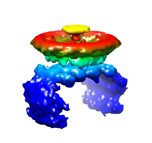
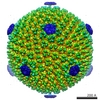
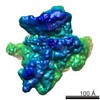
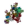
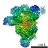
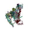
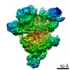
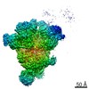
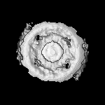
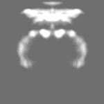
| Title | Vertex reconstruction of SH1 spike | |||||||||
 Map data Map data | spike | |||||||||
 Sample Sample |
| |||||||||
 Keywords Keywords | archaea / virus / SH1 / spike / infection / adsorption | |||||||||
| Biological species |  Haloarcula phage SH1 (virus) Haloarcula phage SH1 (virus) | |||||||||
| Method | single particle reconstruction / cryo EM / Resolution: 34.0 Å | |||||||||
 Authors Authors | Jaalinoja HT / Roine E / Laurinmaki P / Kivela HM / Bamford DH / Butcher SJ | |||||||||
 Citation Citation | Journal: Proc Natl Acad Sci U S A / Year: 2008 Title: Structure and host-cell interaction of SH1, a membrane-containing, halophilic euryarchaeal virus. Authors: Harri T Jäälinoja / Elina Roine / Pasi Laurinmäki / Hanna M Kivelä / Dennis H Bamford / Sarah J Butcher /  Abstract: The Archaea, and the viruses that infect them, are the least well understood of all of the three domains of life. They often grow in extreme conditions such as hypersaline lakes and sulfuric hot ...The Archaea, and the viruses that infect them, are the least well understood of all of the three domains of life. They often grow in extreme conditions such as hypersaline lakes and sulfuric hot springs. Only rare glimpses have been gained into the structures of archaeal viruses. Here, we report the subnanometer resolution structure of a recently isolated, hypersalinic, membrane-containing, euryarchaeal virus, SH1, in which different viral proteins can be localized. The results indicate that SH1 has a complex capsid formed from single beta-barrels, an important missing link in hypotheses on viral capsid protein evolution. Unusual, symmetry-mismatched spikes seem to play a role in host adsorption. They are connected to highly organized membrane proteins providing a platform for capsid assembly and potential machinery for host infection. | |||||||||
| History |
|
- Structure visualization
| Movie |
 Movie viewer Movie viewer |
|---|---|
| Structure viewer | EM map: SurfViewMolmilJmol/JSmol |
| Supplemental images |
- Downloads & links
-EMDB archive
| Map data | emd_1498.map.gz | 874 KB | EMDB map data format | |
|---|---|---|---|---|
| Header (meta data) | emd-1498-v30.xmlemd-1498.xml | 9.3 KB 9.3 KB | Display Display | EMDB header |
| Images |  1498.png 1498.png | 171.4 KB | ||
| Archive directory |  http://ftp.pdbj.org/pub/emdb/structures/EMD-1498ftp://ftp.pdbj.org/pub/emdb/structures/EMD-1498 http://ftp.pdbj.org/pub/emdb/structures/EMD-1498ftp://ftp.pdbj.org/pub/emdb/structures/EMD-1498 | HTTPS FTP |
-Related structure data
-Links
| EMDB pages | EMDB (EBI/PDBe) / EMDataResource |
|---|
-Map
| File | Download / File: emd_1498.map.gz / Format: CCP4 / Size: 12.6 MB / Type: IMAGE STORED AS FLOATING POINT NUMBER (4 BYTES) | ||||||||||||||||||||||||||||||||||||||||||||||||||||||||||||||||||||
|---|---|---|---|---|---|---|---|---|---|---|---|---|---|---|---|---|---|---|---|---|---|---|---|---|---|---|---|---|---|---|---|---|---|---|---|---|---|---|---|---|---|---|---|---|---|---|---|---|---|---|---|---|---|---|---|---|---|---|---|---|---|---|---|---|---|---|---|---|---|
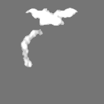
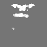
| Annotation | spike | ||||||||||||||||||||||||||||||||||||||||||||||||||||||||||||||||||||
| Projections & slices | Image control
Images are generated by Spider. | ||||||||||||||||||||||||||||||||||||||||||||||||||||||||||||||||||||
| Voxel size |
| ||||||||||||||||||||||||||||||||||||||||||||||||||||||||||||||||||||
| Density |
| ||||||||||||||||||||||||||||||||||||||||||||||||||||||||||||||||||||
| Symmetry | Space group: 1 | ||||||||||||||||||||||||||||||||||||||||||||||||||||||||||||||||||||
| Details | EMDB XML:
CCP4 map header:
| ||||||||||||||||||||||||||||||||||||||||||||||||||||||||||||||||||||
 Z (Sec.)
 Y (Row.)
 X (Col.)


-Supplemental data
- Sample components
-Entire : SH1 virus, reconstruction of the non-icosahedrally symmetric spik...
| Entire | Name: SH1 virus, reconstruction of the non-icosahedrally symmetric spikes on the vertices |
|---|---|
| Components |
|
-Supramolecule #1000: SH1 virus, reconstruction of the non-icosahedrally symmetric spik...
| Supramolecule | Name: SH1 virus, reconstruction of the non-icosahedrally symmetric spikes on the vertices type: sample / ID: 1000 / Number unique components: 1 |
|---|
-Supramolecule #1: Haloarcula phage SH1
| Supramolecule | Name: Haloarcula phage SH1 / type: virus / ID: 1 / Name.synonym: SH1 / NCBI-ID: 326574 / Sci species name: Haloarcula phage SH1 / Virus type: VIRION / Virus isolate: STRAIN / Virus enveloped: No / Virus empty: No / Syn species name: SH1 |
|---|---|
| Host (natural) | Organism:  Haloarcula hispanica (Halophile) / synonym: ARCHAEA Haloarcula hispanica (Halophile) / synonym: ARCHAEA |
-Experimental details
-Structure determination
| Method | cryo EM |
|---|---|
 Processing Processing | single particle reconstruction |
| Aggregation state | particle |
-Sample preparation
| Buffer | pH: 7.2 Details: 150-500 mM NaCl, 10mM MgCl2, 10 M MgSO4, 5 mM KCl, 3 mM CaCl2, 40 mM Tris-HCl pH 7.2 |
|---|---|
| Grid | Details: 400 mesh copper grid, Quantifoil R2/2 holey |
| Vitrification | Cryogen name: ETHANE / Chamber temperature: 90 K / Instrument: HOMEMADE PLUNGER / Details: Vitrification instrument: EMBL design Method: A small vial of ethane is placed inside a larger liquid nitrogen reservoir. The grid holding 3 microliters of the sample is held in place at the bottom of a plunger by the means of fine ...Method: A small vial of ethane is placed inside a larger liquid nitrogen reservoir. The grid holding 3 microliters of the sample is held in place at the bottom of a plunger by the means of fine tweezers. When the liquid ethane is ready, a piece of filter paper is then pressed against the sample to blot off excess buffer, sufficient to leave a thin layer on the grid. The filter paper is removed, and the plunger is allowed to drop into the liquid ethane. Once the grid enters the liquid ethane, the sample is rapidly frozen, and the grid is transferred under liquid nitrogen to a storage box immersed in liquid nitrogen for later use in the microscope. |
- Electron microscopy
| Microscope | FEI TECNAI F20 |
|---|---|
| Temperature | Min: 90 K / Max: 94 K / Average: 93 K |
| Details | Low dose conditions. |
| Image recording | Category: FILM / Film or detector model: KODAK SO-163 FILM / Digitization - Scanner: ZEISS SCAI / Digitization - Sampling interval: 7 µm / Number real images: 169 |
| Electron beam | Acceleration voltage: 200 kV / Electron source:  FIELD EMISSION GUN FIELD EMISSION GUN |
| Electron optics | Calibrated magnification: 49300 / Illumination mode: FLOOD BEAM / Imaging mode: BRIGHT FIELD / Cs: 2.0 mm / Nominal defocus max: 2.5 µm / Nominal defocus min: 0.7 µm / Nominal magnification: 50000 |
| Sample stage | Specimen holder: Side entry liquid nitrogen-cooled cryo specimen holder Specimen holder model: GATAN LIQUID NITROGEN |
| Experimental equipment |  Model: Tecnai F20 / Image courtesy: FEI Company |
-Image processing
| CTF correction | Details: Each micrograph |
|---|---|
| Final reconstruction | Applied symmetry - Point group: C2 (2 fold cyclic) / Algorithm: OTHER / Resolution.type: BY AUTHOR / Resolution: 34.0 Å / Resolution method: FSC 0.5 CUT-OFF / Software - Name: IMAGIC, EMAN, BSoft / Details: Vertex reconstruction. / Number images used: 10196 |
| Final two d classification | Number classes: 548 |
