+ データを開く
- 基本情報
基本情報
| 登録情報 |  | |||||||||
|---|---|---|---|---|---|---|---|---|---|---|
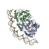
| タイトル | Cryo-EM structure of holo-PdxR from Bacillus clausii bound to its target DNA in the closed conformation, C1 symmetry | |||||||||
 マップデータ マップデータ | Full cryo-EM map of PdxR from Bacillus clausii in complex with its DNA target in closed conformation, symmetry C1 | |||||||||
 試料 試料 |
| |||||||||
 キーワード キーワード | transcription factor / PLP-binding protein / domain-swap homodimer / DNA binding protein | |||||||||
| 機能・相同性 |  機能・相同性情報 機能・相同性情報transaminase activity / pyridoxal phosphate binding / DNA-binding transcription factor activity / DNA binding 類似検索 - 分子機能 | |||||||||
| 生物種 |  Alkalihalobacillus clausii (バクテリア) Alkalihalobacillus clausii (バクテリア) | |||||||||
| 手法 | 単粒子再構成法 / クライオ電子顕微鏡法 / 解像度: 3.9 Å | |||||||||
 データ登録者 データ登録者 | Freda I / Montemiglio LC / Tramonti A / Contestabile R / Vallone B / Exertier C / Savino C / Chaves Sanjuan A / Bolognesi M | |||||||||
| 資金援助 | 1件
| |||||||||
 引用 引用 |  ジャーナル: Nucleic Acids Res / 年: 2023 ジャーナル: Nucleic Acids Res / 年: 2023タイトル: Structural insights into the DNA recognition mechanism by the bacterial transcription factor PdxR. 著者: Ida Freda / Cécile Exertier / Anna Barile / Antonio Chaves-Sanjuan / Mirella Vivoli Vega / Michail N Isupov / Nicholas J Harmer / Elena Gugole / Paolo Swuec / Martino Bolognesi / Anita ...著者: Ida Freda / Cécile Exertier / Anna Barile / Antonio Chaves-Sanjuan / Mirella Vivoli Vega / Michail N Isupov / Nicholas J Harmer / Elena Gugole / Paolo Swuec / Martino Bolognesi / Anita Scipioni / Carmelinda Savino / Martino Luigi Di Salvo / Roberto Contestabile / Beatrice Vallone / Angela Tramonti / Linda Celeste Montemiglio /   要旨: Specificity in protein-DNA recognition arises from the synergy of several factors that stem from the structural and chemical signatures encoded within the targeted DNA molecule. Here, we deciphered ...Specificity in protein-DNA recognition arises from the synergy of several factors that stem from the structural and chemical signatures encoded within the targeted DNA molecule. Here, we deciphered the nature of the interactions driving DNA recognition and binding by the bacterial transcription factor PdxR, a member of the MocR family responsible for the regulation of pyridoxal 5'-phosphate (PLP) biosynthesis. Single particle cryo-EM performed on the PLP-PdxR bound to its target DNA enabled the isolation of three conformers of the complex, which may be considered as snapshots of the binding process. Moreover, the resolution of an apo-PdxR crystallographic structure provided a detailed description of the transition of the effector domain to the holo-PdxR form triggered by the binding of the PLP effector molecule. Binding analyses of mutated DNA sequences using both wild type and PdxR variants revealed a central role of electrostatic interactions and of the intrinsic asymmetric bending of the DNA in allosterically guiding the holo-PdxR-DNA recognition process, from the first encounter through the fully bound state. Our results detail the structure and dynamics of the PdxR-DNA complex, clarifying the mechanism governing the DNA-binding mode of the holo-PdxR and the regulation features of the MocR family of transcription factors. | |||||||||
| 履歴 |
|
- 構造の表示
構造の表示
| 添付画像 |
|---|
- ダウンロードとリンク
-EMDBアーカイブ
| マップデータ |  emd_14852.map.gz emd_14852.map.gz | 47.7 MB |  EMDBマップデータ形式 EMDBマップデータ形式 | |
|---|---|---|---|---|
| ヘッダ (付随情報) |  emd-14852-v30.xml emd-14852-v30.xml emd-14852.xml emd-14852.xml | 21.6 KB 21.6 KB | 表示 表示 |  EMDBヘッダ EMDBヘッダ |
| FSC (解像度算出) |  emd_14852_fsc.xml emd_14852_fsc.xml | 9.2 KB | 表示 |  FSCデータファイル FSCデータファイル |
| 画像 |  emd_14852.png emd_14852.png | 37.1 KB | ||
| Filedesc metadata |  emd-14852.cif.gz emd-14852.cif.gz | 6.9 KB | ||
| その他 |  emd_14852_half_map_1.map.gz emd_14852_half_map_1.map.gz emd_14852_half_map_2.map.gz emd_14852_half_map_2.map.gz | 47 MB 46.8 MB | ||
| アーカイブディレクトリ |  http://ftp.pdbj.org/pub/emdb/structures/EMD-14852 http://ftp.pdbj.org/pub/emdb/structures/EMD-14852 ftp://ftp.pdbj.org/pub/emdb/structures/EMD-14852 ftp://ftp.pdbj.org/pub/emdb/structures/EMD-14852 | HTTPS FTP |
-検証レポート
| 文書・要旨 |  emd_14852_validation.pdf.gz emd_14852_validation.pdf.gz | 865.6 KB | 表示 |  EMDB検証レポート EMDB検証レポート |
|---|---|---|---|---|
| 文書・詳細版 |  emd_14852_full_validation.pdf.gz emd_14852_full_validation.pdf.gz | 865.1 KB | 表示 | |
| XML形式データ |  emd_14852_validation.xml.gz emd_14852_validation.xml.gz | 16.6 KB | 表示 | |
| CIF形式データ |  emd_14852_validation.cif.gz emd_14852_validation.cif.gz | 21.5 KB | 表示 | |
| アーカイブディレクトリ |  https://ftp.pdbj.org/pub/emdb/validation_reports/EMD-14852 https://ftp.pdbj.org/pub/emdb/validation_reports/EMD-14852 ftp://ftp.pdbj.org/pub/emdb/validation_reports/EMD-14852 ftp://ftp.pdbj.org/pub/emdb/validation_reports/EMD-14852 | HTTPS FTP |
-関連構造データ
- リンク
リンク
| EMDBのページ |  EMDB (EBI/PDBe) / EMDB (EBI/PDBe) /  EMDataResource EMDataResource |
|---|
- マップ
マップ
| ファイル |  ダウンロード / ファイル: emd_14852.map.gz / 形式: CCP4 / 大きさ: 64 MB / タイプ: IMAGE STORED AS FLOATING POINT NUMBER (4 BYTES) ダウンロード / ファイル: emd_14852.map.gz / 形式: CCP4 / 大きさ: 64 MB / タイプ: IMAGE STORED AS FLOATING POINT NUMBER (4 BYTES) | ||||||||||||||||||||||||||||||||||||
|---|---|---|---|---|---|---|---|---|---|---|---|---|---|---|---|---|---|---|---|---|---|---|---|---|---|---|---|---|---|---|---|---|---|---|---|---|---|
| 注釈 | Full cryo-EM map of PdxR from Bacillus clausii in complex with its DNA target in closed conformation, symmetry C1 | ||||||||||||||||||||||||||||||||||||
| 投影像・断面図 | 画像のコントロール
画像は Spider により作成 | ||||||||||||||||||||||||||||||||||||
| ボクセルのサイズ | X=Y=Z: 0.889 Å | ||||||||||||||||||||||||||||||||||||
| 密度 |
| ||||||||||||||||||||||||||||||||||||
| 対称性 | 空間群: 1 | ||||||||||||||||||||||||||||||||||||
| 詳細 | EMDB XML:
|
-添付データ
-ハーフマップ: Half cryo-EM map 1 of PdxR from Bacillus...
| ファイル | emd_14852_half_map_1.map | ||||||||||||
|---|---|---|---|---|---|---|---|---|---|---|---|---|---|
| 注釈 | Half cryo-EM map 1 of PdxR from Bacillus clausii in complex with its DNA target in closed conformation, symmetry C1 | ||||||||||||
| 投影像・断面図 |
| ||||||||||||
| 密度ヒストグラム |
-ハーフマップ: Half cryo-EM map 2 of PdxR from Bacillus...
| ファイル | emd_14852_half_map_2.map | ||||||||||||
|---|---|---|---|---|---|---|---|---|---|---|---|---|---|
| 注釈 | Half cryo-EM map 2 of PdxR from Bacillus clausii in complex with its DNA target in closed conformation, symmetry C1 | ||||||||||||
| 投影像・断面図 |
| ||||||||||||
| 密度ヒストグラム |
- 試料の構成要素
-全体 : Binary complex between the PLP-bound homodimeric PdxR and a 48bp ...
| 全体 | 名称: Binary complex between the PLP-bound homodimeric PdxR and a 48bp double strand DNA fragment |
|---|---|
| 要素 |
|
-超分子 #1: Binary complex between the PLP-bound homodimeric PdxR and a 48bp ...
| 超分子 | 名称: Binary complex between the PLP-bound homodimeric PdxR and a 48bp double strand DNA fragment タイプ: complex / ID: 1 / 親要素: 0 / 含まれる分子: all |
|---|---|
| 由来(天然) | 生物種:  Alkalihalobacillus clausii (バクテリア) Alkalihalobacillus clausii (バクテリア) |
| 分子量 | 理論値: 136 KDa |
-分子 #1: PLP-dependent aminotransferase family protein
| 分子 | 名称: PLP-dependent aminotransferase family protein / タイプ: protein_or_peptide / ID: 1 / コピー数: 2 / 光学異性体: LEVO |
|---|---|
| 由来(天然) | 生物種:  Alkalihalobacillus clausii (バクテリア) Alkalihalobacillus clausii (バクテリア) |
| 分子量 | 理論値: 55.499121 KDa |
| 組換発現 | 生物種:  |
| 配列 | 文字列: MELLWCELNR DLPTPLYEQL YAHIKTEITE GRIGYGTKLP SKRKLADSLK LSQNTVEAAY EQLVAEGYVE VIPRKGFYVQ AYEDLEYIR APQAPGDALA TKQDTIRYNF HPTHIDTTSF PFEQWRKYFK QTMCKENHRL LLNGDHQGEA SFRREIAYYL H HSRGVNCT ...文字列: MELLWCELNR DLPTPLYEQL YAHIKTEITE GRIGYGTKLP SKRKLADSLK LSQNTVEAAY EQLVAEGYVE VIPRKGFYVQ AYEDLEYIR APQAPGDALA TKQDTIRYNF HPTHIDTTSF PFEQWRKYFK QTMCKENHRL LLNGDHQGEA SFRREIAYYL H HSRGVNCT PEQVVVGAGV ETLLQQLFLL LGESKVYGIE DPGYQLMRKL LSHYPNDYVP FQVDEEGIDV DSIVRTAVDV VY TTPSRHF PYGSVLSINR RKQLLHWAEA HENRYIIEDD YDSEFRYTGK TIPSLQSMDV HNKVIYLGAF S(LLP)SLIPSVR ISYMVLPAPL AHLYKNKFSY YHSTVSRIDQ QVLTAFMKQG DFEKHLNRMR KIYRRKLEKV LSLLKRYEDK LLIIGERSGL HIVLVVKNG MDEQTLVEKA LAAKAKVYPL SAYSLERAIH PPQIVLGFGS IPEDELEEAI ATVLNAWGFL VPRGSLEHHH H HH UniProtKB: PLP-dependent aminotransferase family protein |
-分子 #2: DNA (48-MER)
| 分子 | 名称: DNA (48-MER) / タイプ: dna / ID: 2 / コピー数: 1 / 分類: DNA |
|---|---|
| 由来(天然) | 生物種:  Alkalihalobacillus clausii (バクテリア) Alkalihalobacillus clausii (バクテリア) |
| 分子量 | 理論値: 14.667447 KDa |
| 配列 | 文字列: (DC)(DT)(DG)(DA)(DC)(DC)(DT)(DC)(DA)(DT) (DC)(DA)(DT)(DT)(DT)(DT)(DC)(DT)(DT)(DA) (DA)(DA)(DA)(DA)(DC)(DT)(DG)(DA)(DC) (DA)(DC)(DT)(DT)(DA)(DC)(DA)(DA)(DT)(DG) (DT) (DG)(DG)(DT)(DC)(DA)(DG)(DT)(DT) |
-分子 #3: DNA (48-MER)
| 分子 | 名称: DNA (48-MER) / タイプ: dna / ID: 3 / コピー数: 1 / 分類: DNA |
|---|---|
| 由来(天然) | 生物種:  Alkalihalobacillus clausii (バクテリア) Alkalihalobacillus clausii (バクテリア) |
| 分子量 | 理論値: 14.894607 KDa |
| 配列 | 文字列: (DA)(DA)(DC)(DT)(DG)(DA)(DC)(DC)(DA)(DC) (DA)(DT)(DT)(DG)(DT)(DA)(DA)(DG)(DT)(DG) (DT)(DC)(DA)(DG)(DT)(DT)(DT)(DT)(DT) (DA)(DA)(DG)(DA)(DA)(DA)(DA)(DT)(DG)(DA) (DT) (DG)(DA)(DG)(DG)(DT)(DC)(DA)(DG) |
-実験情報
-構造解析
| 手法 | クライオ電子顕微鏡法 |
|---|---|
 解析 解析 | 単粒子再構成法 |
| 試料の集合状態 | particle |
- 試料調製
試料調製
| 濃度 | 0.5 mg/mL | ||||||
|---|---|---|---|---|---|---|---|
| 緩衝液 | pH: 7.5 / 構成要素:
| ||||||
| グリッド | モデル: Quantifoil R1.2/1.3 / 材質: COPPER / メッシュ: 300 / 支持フィルム - 材質: CARBON / 支持フィルム - トポロジー: HOLEY / 支持フィルム - Film thickness: 12 / 前処理 - タイプ: GLOW DISCHARGE / 前処理 - 時間: 30 sec. | ||||||
| 凍結 | 凍結剤: ETHANE / チャンバー内湿度: 100 % / チャンバー内温度: 277.15 K / 装置: FEI VITROBOT MARK IV |
- 電子顕微鏡法
| 顕微鏡 | FEI TALOS ARCTICA |
|---|---|
| 撮影 | フィルム・検出器のモデル: FEI FALCON III (4k x 4k) 検出モード: COUNTING / 撮影したグリッド数: 1 / 実像数: 3284 / 平均露光時間: 1.0 sec. / 平均電子線量: 40.0 e/Å2 |
| 電子線 | 加速電圧: 200 kV / 電子線源:  FIELD EMISSION GUN FIELD EMISSION GUN |
| 電子光学系 | 照射モード: FLOOD BEAM / 撮影モード: BRIGHT FIELD / 最大 デフォーカス(公称値): 2.2 µm / 最小 デフォーカス(公称値): 0.8 µm / 倍率(公称値): 120000 |
| 試料ステージ | ホルダー冷却材: NITROGEN |
| 実験機器 |  モデル: Talos Arctica / 画像提供: FEI Company |
+ 画像解析
画像解析
-原子モデル構築 1
| 初期モデル |
| ||||||
|---|---|---|---|---|---|---|---|
| 精密化 | 空間: REAL / プロトコル: RIGID BODY FIT / 温度因子: 142 | ||||||
| 得られたモデル | 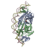 PDB-7zpa: |
 ムービー
ムービー コントローラー


 Z (Sec.)
 Y (Row.)
 X (Col.)


