[English] 日本語
Yorodumi
- EMDB-14736: 3D reconstruction of the cylindrical assembly of DnaJA2 delta G/F... -

+
Open data


ID or keywords:

Loading...

-
Basic information

Entry
Database: EMDB / ID: EMD-14736
Title3D reconstruction of the cylindrical assembly of DnaJA2 delta G/F by imposing D5 symmetry
Map data
Sample
  • Complex: Oligomeric structure of DnaJA2 delta G/F by imposing D5 symmetry
    • Protein or peptide: Ubiquitin-like protein SMT3,DnaJ homolog subfamily A member 2
  • Ligand: ZINC ION
Keywordsoligomer / filaments / helix / holdase / foldase / Hsp40 / DnaJA2 / Hsp70 co-chaperone / CHAPERONE
Function / homology
Function and homology information


SUMO is conjugated to E1 (UBA2:SAE1) / SUMOylation of nuclear envelope proteins / SUMO is transferred from E1 to E2 (UBE2I, UBC9) / SUMO is proteolytically processed / SUMOylation of transcription factors / Postmitotic nuclear pore complex (NPC) reformation / SUMOylation of transcription cofactors / SUMOylation of DNA damage response and repair proteins / SUMOylation of DNA replication proteins / septin ring ...SUMO is conjugated to E1 (UBA2:SAE1) / SUMOylation of nuclear envelope proteins / SUMO is transferred from E1 to E2 (UBE2I, UBC9) / SUMO is proteolytically processed / SUMOylation of transcription factors / Postmitotic nuclear pore complex (NPC) reformation / SUMOylation of transcription cofactors / SUMOylation of DNA damage response and repair proteins / SUMOylation of DNA replication proteins / septin ring / SUMOylation of SUMOylation proteins / Recruitment and ATM-mediated phosphorylation of repair and signaling proteins at DNA double strand breaks / SUMOylation of RNA binding proteins / SUMOylation of chromatin organization proteins / ubiquitin-like protein ligase binding / ATPase activator activity / protein sumoylation / Hsp70 protein binding / HSP90 chaperone cycle for steroid hormone receptors (SHR) in the presence of ligand / condensed nuclear chromosome / PML body / protein tag activity / unfolded protein binding / protein-folding chaperone binding / response to heat / protein refolding / positive regulation of cell population proliferation / extracellular exosome / ATP binding / identical protein binding / membrane / nucleus / metal ion binding / cytoplasm / cytosol
Similarity search - Function
DnaJ homolog subfamily A member 1/2-like / Chaperone DnaJ / DnaJ central domain / Heat shock protein DnaJ, cysteine-rich domain / Zinc finger CR-type profile. / Heat shock protein DnaJ, cysteine-rich domain superfamily / HSP40/DnaJ peptide-binding / Chaperone DnaJ, C-terminal / DnaJ C terminal domain / Nt-dnaJ domain signature. ...DnaJ homolog subfamily A member 1/2-like / Chaperone DnaJ / DnaJ central domain / Heat shock protein DnaJ, cysteine-rich domain / Zinc finger CR-type profile. / Heat shock protein DnaJ, cysteine-rich domain superfamily / HSP40/DnaJ peptide-binding / Chaperone DnaJ, C-terminal / DnaJ C terminal domain / Nt-dnaJ domain signature. / DnaJ domain, conserved site / Rad60/SUMO-like domain / Ubiquitin-2 like Rad60 SUMO-like / DnaJ domain / DnaJ molecular chaperone homology domain / dnaJ domain profile. / Chaperone J-domain superfamily / DnaJ domain / Ubiquitin homologues / Ubiquitin domain profile. / Ubiquitin-like domain / Ubiquitin-like domain superfamily
Similarity search - Domain/homology
DnaJ homolog subfamily A member 2 / Ubiquitin-like protein SMT3
Similarity search - Component
Biological speciesHomo sapiens (human)
Methodsingle particle reconstruction / cryo EM / Resolution: 6.9 Å
AuthorsCuellar J / Velasco-Carneros L / Santiago C / Martin-Benito J / Valpuesta J / Muga A
Funding support Spain, 2 items
OrganizationGrant numberCountry
Spanish Ministry of Science, Innovation, and UniversitiesPID2019-105872GB-I00 Spain
Spanish Ministry of Science, Innovation, and UniversitiesPID2019-111068GB-I00 Spain
Citation
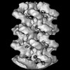
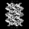
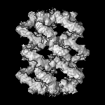
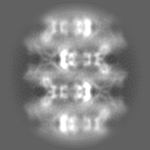
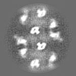
Journal: Nat Commun / Year: 2023
Title: The self-association equilibrium of DNAJA2 regulates its interaction with unfolded substrate proteins and with Hsc70.
Authors: Lorea Velasco-Carneros / Jorge Cuéllar / Leire Dublang / César Santiago / Jean-Didier Maréchal / Jaime Martín-Benito / Moisés Maestro / José Ángel Fernández-Higuero / Natalia Orozco ...Authors: Lorea Velasco-Carneros / Jorge Cuéllar / Leire Dublang / César Santiago / Jean-Didier Maréchal / Jaime Martín-Benito / Moisés Maestro / José Ángel Fernández-Higuero / Natalia Orozco / Fernando Moro / José María Valpuesta / Arturo Muga /
Abstract: J-domain proteins tune the specificity of Hsp70s, engaging them in precise functions. Despite their essential role, the structure and function of many J-domain proteins remain largely unknown. We ...J-domain proteins tune the specificity of Hsp70s, engaging them in precise functions. Despite their essential role, the structure and function of many J-domain proteins remain largely unknown. We explore human DNAJA2, finding that it reversibly forms highly-ordered, tubular structures that can be dissociated by Hsc70, the constitutively expressed Hsp70 isoform. Cryoelectron microscopy and mutational studies reveal that different domains are involved in self-association. Oligomer dissociation into dimers potentiates its interaction with unfolded client proteins. The J-domains are accessible to Hsc70 within the tubular structure. They allow binding of closely spaced Hsc70 molecules that could be transferred to the unfolded substrate for its cooperative remodelling, explaining the efficient recovery of DNAJA2-bound clients. The disordered C-terminal domain, comprising the last 52 residues, regulates its holding activity and productive interaction with Hsc70. These in vitro findings suggest that the association equilibrium of DNAJA2 could regulate its interaction with client proteins and Hsc70.
History
DepositionApr 7, 2022-
Header (metadata) releaseJul 26, 2023-
Map releaseJul 26, 2023-
UpdateJan 24, 2024-
Current statusJan 24, 2024Processing site: PDBe / Status: Released

-
Structure visualization
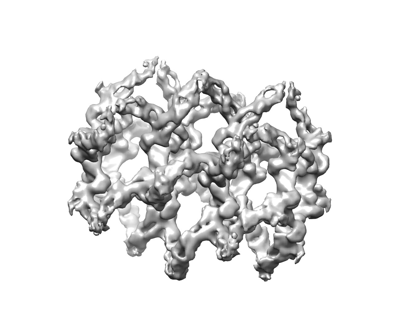
Supplemental images

Downloads & links

-
Map

FileDownload / File: emd_14736.map.gz / Format: CCP4 / Size: 12.9 MB / Type: IMAGE STORED AS FLOATING POINT NUMBER (4 BYTES)
Projections & slices

Image control

Size
Brightness
Contrast
Others
AxesZ (Sec.)Y (Row.)X (Col.)
2.11 Å/pix.
x 150 pix.
= 316.2 Å
2.11 Å/pix.
x 150 pix.
= 316.2 Å
2.11 Å/pix.
x 150 pix.
= 316.2 Å

Surface

Projections

Slices (1/3)

Slices (1/2)

Slices (2/3)

Images are generated by Spider.

Voxel sizeX=Y=Z: 2.108 Å
Density
Contour LevelBy AUTHOR: 0.088
Minimum - Maximum-0.08371178 - 0.44252196
Average (Standard dev.)0.012119809 (±0.040648203)
SymmetrySpace group: 1
Details

EMDB XML:

Map geometry
Axis orderXYZ
Origin000
Dimensions150150150
Spacing150150150
CellA=B=C: 316.2 Å
α=β=γ: 90.0 °

-
Supplemental data

-
Half map: #1

Fileemd_14736_half_map_1.map
Projections & Slices
AxesZYX

Projections

Slices (1/2)
Density Histograms

-
Half map: #2

Fileemd_14736_half_map_2.map
Projections & Slices
AxesZYX

Projections

Slices (1/2)
Density Histograms

-
Sample components

-
Entire : Oligomeric structure of DnaJA2 delta G/F by imposing D5 symmetry

EntireName: Oligomeric structure of DnaJA2 delta G/F by imposing D5 symmetry
Components
  • Complex: Oligomeric structure of DnaJA2 delta G/F by imposing D5 symmetry
    • Protein or peptide: Ubiquitin-like protein SMT3,DnaJ homolog subfamily A member 2
  • Ligand: ZINC ION

-
Supramolecule #1: Oligomeric structure of DnaJA2 delta G/F by imposing D5 symmetry

SupramoleculeName: Oligomeric structure of DnaJA2 delta G/F by imposing D5 symmetry
type: complex / ID: 1 / Parent: 0 / Macromolecule list: #1
Source (natural)Organism: Homo sapiens (human)

-
Macromolecule #1: Ubiquitin-like protein SMT3,DnaJ homolog subfamily A member 2

MacromoleculeName: Ubiquitin-like protein SMT3,DnaJ homolog subfamily A member 2
type: protein_or_peptide / ID: 1 / Number of copies: 40 / Enantiomer: LEVO
Source (natural)Organism: Homo sapiens (human)
Molecular weightTheoretical: 55.980961 KDa
Recombinant expressionOrganism: Escherichia coli (E. coli)
SequenceString: MGHHHHHHGS LQDSEVNQEA KPEVKPEVKP ETHINLKVSD GSSEIFFKIK KTTPLRRLME AFAKRQGKEM DSLRFLYDGI RIQADQAPE DLDMEDNDII EAHREQIGGM ANVADTKLYD ILGVPPGASE NELKKAYRKL AKEYHPDKNP NAGDKFKEIS F AYEVLSNP ...String:
MGHHHHHHGS LQDSEVNQEA KPEVKPEVKP ETHINLKVSD GSSEIFFKIK KTTPLRRLME AFAKRQGKEM DSLRFLYDGI RIQADQAPE DLDMEDNDII EAHREQIGGM ANVADTKLYD ILGVPPGASE NELKKAYRKL AKEYHPDKNP NAGDKFKEIS F AYEVLSNP EKRELYDRYG EQGLREGGNQ SRSRNGRRRG EDMMHPLKVS LEDLYNGKTT KLQLSKNVLC SACSGQGGKS GA VQKCSAC RGRGVRIMIR QLAPGMVQQM QSVCSDCNGE GEVINEKDRC KKCEGKKVIK EVKILEVHVD KGMKHGQRIT FTG EADQAP GVEPGDIVLL LQEKEHEVFQ RDGNDLHMTY KIGLVEALCG FQFTFKHLDG RQIVVKYPPG KVIEPGCVRV VRGE GMPQY RNPFEKGDLY IKFDVQFPEN NWINPDKLSE LEDLLPSRPE VPNIIGETEE VELQEFDSTR GSGGGQRREA YNDSS DEES SSHHGPGVQC AHQ

UniProtKB: Ubiquitin-like protein SMT3, DnaJ homolog subfamily A member 2

-
Macromolecule #2: ZINC ION

MacromoleculeName: ZINC ION / type: ligand / ID: 2 / Number of copies: 80 / Formula: ZN
Molecular weightTheoretical: 65.409 Da

-
Experimental details

-
Structure determination

Methodcryo EM
Processingsingle particle reconstruction
Aggregation statefilament

-
Sample preparation

BufferpH: 7.5
GridModel: Quantifoil R2/2 / Material: COPPER/RHODIUM / Mesh: 300
VitrificationCryogen name: ETHANE / Chamber humidity: 95 % / Chamber temperature: 293 K / Instrument: FEI VITROBOT MARK IV

-
Electron microscopy

MicroscopeFEI TITAN KRIOS
DetailsPreliminary grid screening was performed manually
Image recordingFilm or detector model: GATAN K3 BIOQUANTUM (6k x 4k) / Digitization - Dimensions - Width: 3838 pixel / Digitization - Dimensions - Height: 3710 pixel / Number grids imaged: 1 / Number real images: 10714 / Average exposure time: 2.0 sec. / Average electron dose: 30.0 e/Å2
Electron beamAcceleration voltage: 300 kV / Electron source: FIELD EMISSION GUN
Electron opticsC2 aperture diameter: 100.0 µm / Illumination mode: FLOOD BEAM / Imaging mode: BRIGHT FIELD / Cs: 2.7 mm / Nominal defocus max: 3.0 µm / Nominal defocus min: 1.2 µm
Sample stageSpecimen holder model: FEI TITAN KRIOS AUTOGRID HOLDER / Cooling holder cryogen: NITROGEN
Experimental equipment
Model: Titan Krios / Image courtesy: FEI Company

+
Image processing

Particle selectionNumber selected: 288620
Startup modelType of model: INSILICO MODEL
In silico model: Some of the best 2D classes were used as a template to generate an initial model using RANSAC
Final reconstructionApplied symmetry - Point group: D5 (2x5 fold dihedral) / Resolution.type: BY AUTHOR / Resolution: 6.9 Å / Resolution method: FSC 0.143 CUT-OFF / Software - Name: RELION (ver. 3.0) / Number images used: 47396
Initial angle assignmentType: MAXIMUM LIKELIHOOD / Software - Name: RELION (ver. 3.0)
Final angle assignmentType: PROJECTION MATCHING / Software - Name: RELION (ver. 3.0)
Final 3D classificationNumber classes: 3 / Software - Name: RELION (ver. 3.0)

-
Atomic model buiding 1

RefinementSpace: REAL / Protocol: FLEXIBLE FIT
Output model

PDB-7zhs:
3D reconstruction of the cylindrical assembly of DnaJA2 delta G/F by imposing D5 symmetry

+
About Yorodumi

-
News

-
Feb 9, 2022. New format data for meta-information of EMDB entries

New format data for meta-information of EMDB entries

  • Version 3 of the EMDB header file is now the official format.
  • The previous official version 1.9 will be removed from the archive.

Related info.:EMDB header

External links:wwPDB to switch to version 3 of the EMDB data model

-
Aug 12, 2020. Covid-19 info

Covid-19 info

URL: https://pdbjlvh1.pdbj.org/emnavi/covid19.php

New page: Covid-19 featured information page in EM Navigator.

Related info.:Covid-19 info / Mar 5, 2020. Novel coronavirus structure data

+
Mar 5, 2020. Novel coronavirus structure data

Novel coronavirus structure data

Related info.:Yorodumi Speices / Aug 12, 2020. Covid-19 info

External links:COVID-19 featured content - PDBj / Molecule of the Month (242):Coronavirus Proteases

+
Jan 31, 2019. EMDB accession codes are about to change! (news from PDBe EMDB page)

EMDB accession codes are about to change! (news from PDBe EMDB page)

  • The allocation of 4 digits for EMDB accession codes will soon come to an end. Whilst these codes will remain in use, new EMDB accession codes will include an additional digit and will expand incrementally as the available range of codes is exhausted. The current 4-digit format prefixed with “EMD-” (i.e. EMD-XXXX) will advance to a 5-digit format (i.e. EMD-XXXXX), and so on. It is currently estimated that the 4-digit codes will be depleted around Spring 2019, at which point the 5-digit format will come into force.
  • The EM Navigator/Yorodumi systems omit the EMD- prefix.

Related info.:Q: What is EMD? / ID/Accession-code notation in Yorodumi/EM Navigator

External links:EMDB Accession Codes are Changing Soon! / Contact to PDBj

+
Jul 12, 2017. Major update of PDB

Major update of PDB

  • wwPDB released updated PDB data conforming to the new PDBx/mmCIF dictionary.
  • This is a major update changing the version number from 4 to 5, and with Remediation, in which all the entries are updated.
  • In this update, many items about electron microscopy experimental information are reorganized (e.g. em_software).
  • Now, EM Navigator and Yorodumi are based on the updated data.

External links:wwPDB Remediation / Enriched Model Files Conforming to OneDep Data Standards Now Available in the PDB FTP Archive

-
Yorodumi

Thousand views of thousand structures

  • Yorodumi is a browser for structure data from EMDB, PDB, SASBDB, etc.
  • This page is also the successor to EM Navigator detail page, and also detail information page/front-end page for Omokage search.
  • The word "yorodu" (or yorozu) is an old Japanese word meaning "ten thousand". "mi" (miru) is to see.

Related info.:EMDB / PDB / SASBDB / Comparison of 3 databanks / Yorodumi Search / Aug 31, 2016. New EM Navigator & Yorodumi / Yorodumi Papers / Jmol/JSmol / Function and homology information / Changes in new EM Navigator and Yorodumi

Read more