ジャーナル: Cell / 年: 2008 タイトル: Structural basis of membrane invagination by F-BAR domains. 著者: Adam Frost / Rushika Perera / Aurélien Roux / Krasimir Spasov / Olivier Destaing / Edward H Egelman / Pietro De Camilli / Vinzenz M Unger / 要旨: BAR superfamily domains shape membranes through poorly understood mechanisms. We solved structures of F-BAR modules bound to flat and curved bilayers using electron (cryo)microscopy. We show that ...BAR superfamily domains shape membranes through poorly understood mechanisms. We solved structures of F-BAR modules bound to flat and curved bilayers using electron (cryo)microscopy. We show that membrane tubules form when F-BARs polymerize into helical coats that are held together by lateral and tip-to-tip interactions. On gel-state membranes or after mutation of residues along the lateral interaction surface, F-BARs adsorb onto bilayers via surfaces other than their concave face. We conclude that membrane binding is separable from membrane bending, and that imposition of the module's concave surface forces fluid-phase bilayers to bend locally. Furthermore, exposure of the domain's lateral interaction surface through a change in orientation serves as the crucial trigger for assembly of the helical coat and propagation of bilayer bending. The geometric constraints and sequential assembly of the helical lattice explain how F-BAR and classical BAR domains segregate into distinct microdomains, and provide insight into the spatial regulation of membrane invagination.
全体 : membrane tubule coated with a helical lattice of dimer F-BAR modu...
全体
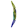
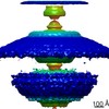
名称: membrane tubule coated with a helical lattice of dimer F-BAR modules from the human protein CIP4
要素
試料: membrane tubule coated with a helical lattice of dimer F-BAR modules from the human protein CIP4
タンパク質・ペプチド: CIP4
-
超分子 #1000: membrane tubule coated with a helical lattice of dimer F-BAR modu...
超分子
名称: membrane tubule coated with a helical lattice of dimer F-BAR modules from the human protein CIP4 タイプ: sample / ID: 1000 詳細: membrane composed of phospholipids and cholesterol. recombinant protein Number unique components: 2
 ムービー
ムービー コントローラー


 データを開く
 基本情報
基本情報 マップデータ
 試料
試料 キーワード
キーワード Homo sapiens (ヒト)
 データ登録者
 引用
引用
 構造の表示
構造の表示 ムービービューア
 ダウンロードとリンク
 1471.gif
 http://ftp.pdbj.org/pub/emdb/structures/EMD-1471
 Z (Sec.)
 Y (Row.)
 X (Col.)


 試料の構成要素
 解析
解析 電子顕微鏡法
 FIELD EMISSION GUN