[English] 日本語
Yorodumi
- EMDB-14678: Cryo-EM structure of the human inward-rectifier potassium 2.1 cha... -

+
Open data


ID or keywords:

Loading...

-
Basic information

Entry
Database: EMDB / ID: EMD-14678
TitleCryo-EM structure of the human inward-rectifier potassium 2.1 channel (Kir2.1)
Map data
Sample
  • Organelle or cellular component: Human inward-rectifier potassium channel 2.1 (Kir2.1)
    • Protein or peptide: Inward rectifier potassium channel 2Inward-rectifier potassium channel
  • Ligand: POTASSIUM IONPotassium
  • Ligand: STRONTIUM ION
Function / homology
Function and homology information


Sensory perception of sour taste / Classical Kir channels / regulation of skeletal muscle contraction via regulation of action potential / relaxation of skeletal muscle / Phase 4 - resting membrane potential / magnesium ion transport / voltage-gated potassium channel activity involved in cardiac muscle cell action potential repolarization / membrane repolarization during action potential / membrane repolarization during cardiac muscle cell action potential / regulation of resting membrane potential ...Sensory perception of sour taste / Classical Kir channels / regulation of skeletal muscle contraction via regulation of action potential / relaxation of skeletal muscle / Phase 4 - resting membrane potential / magnesium ion transport / voltage-gated potassium channel activity involved in cardiac muscle cell action potential repolarization / membrane repolarization during action potential / membrane repolarization during cardiac muscle cell action potential / regulation of resting membrane potential / regulation of membrane repolarization / intracellular potassium ion homeostasis / positive regulation of potassium ion transmembrane transport / membrane depolarization during cardiac muscle cell action potential / inward rectifier potassium channel activity / cardiac muscle cell action potential involved in contraction / regulation of cardiac muscle cell contraction / regulation of monoatomic ion transmembrane transport / relaxation of cardiac muscle / potassium ion import across plasma membrane / regulation of heart rate by cardiac conduction / smooth endoplasmic reticulum / intercalated disc / rough endoplasmic reticulum / voltage-gated potassium channel complex / potassium ion transmembrane transport / phosphatidylinositol-4,5-bisphosphate binding / T-tubule / Activation of G protein gated Potassium channels / Inhibition of voltage gated Ca2+ channels via Gbeta/gamma subunits / potassium ion transport / cellular response to mechanical stimulus / postsynaptic membrane / protein homotetramerization / dendritic spine / neuronal cell body / glutamatergic synapse / Golgi apparatus / membrane / identical protein binding / plasma membrane
Similarity search - Function
Potassium channel, inwardly rectifying, Kir2.1 / Potassium channel, inwardly rectifying, Kir, N-terminal / Inward rectifier potassium channel N-terminal / Potassium channel, inwardly rectifying, transmembrane domain / Inward rectifier potassium channel transmembrane domain / Potassium channel, inwardly rectifying, Kir, cytoplasmic / Potassium channel, inwardly rectifying, Kir / Inward rectifier potassium channel, C-terminal / Inward rectifier potassium channel C-terminal domain / Immunoglobulin E-set
Similarity search - Domain/homology
Inward rectifier potassium channel 2
Similarity search - Component
Biological speciesHomo sapiens (human)
Methodsingle particle reconstruction / cryo EM / Resolution: 4.3 Å
AuthorsFernandes CAH / Venien-Bryan C / Fagnen C / Zuniga D
Funding support France, 3 items
OrganizationGrant numberCountry
Agence Nationale de la Recherche (ANR)ANR-11-EQPX-0008 France
The French Muscular Dystrophy Telethon (AFM-Telethon)23207 France
The French Muscular Dystrophy Telethon (AFM-Telethon)23210 France
Citation
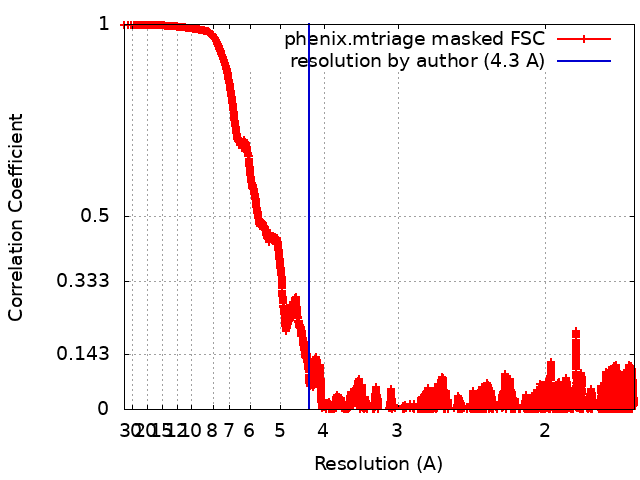
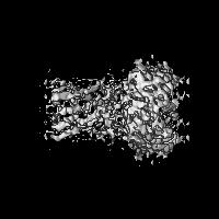
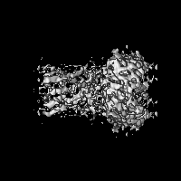
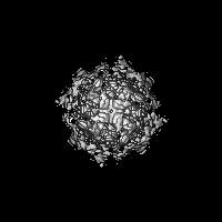
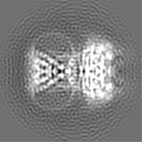
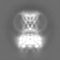
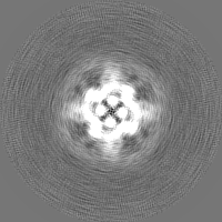
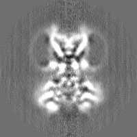
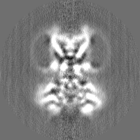
Journal: Sci Adv / Year: 2022
Title: Cryo-electron microscopy unveils unique structural features of the human Kir2.1 channel.
Authors: Carlos A H Fernandes / Dania Zuniga / Charline Fagnen / Valérie Kugler / Rosa Scala / Gérard Péhau-Arnaudet / Renaud Wagner / David Perahia / Saïd Bendahhou / Catherine Vénien-Bryan /
Abstract: We present the first structure of the human Kir2.1 channel containing both transmembrane domain (TMD) and cytoplasmic domain (CTD). Kir2.1 channels are strongly inward-rectifying potassium channels ...We present the first structure of the human Kir2.1 channel containing both transmembrane domain (TMD) and cytoplasmic domain (CTD). Kir2.1 channels are strongly inward-rectifying potassium channels that play a key role in maintaining resting membrane potential. Their gating is modulated by phosphatidylinositol 4,5-bisphosphate (PIP). Genetically inherited defects in Kir2.1 channels are responsible for several rare human diseases, including Andersen's syndrome. The structural analysis (cryo-electron microscopy), surface plasmon resonance, and electrophysiological experiments revealed a well-connected network of interactions between the PIP-binding site and the G-loop through residues R312 and H221. In addition, molecular dynamics simulations and normal mode analysis showed the intrinsic tendency of the CTD to tether to the TMD and a movement of the secondary anionic binding site to the membrane even without PIP. Our results revealed structural features unique to human Kir2.1 and provided insights into the connection between G-loop and gating and the pathological mechanisms associated with this channel.
History
DepositionMar 30, 2022-
Header (metadata) releaseSep 28, 2022-
Map releaseSep 28, 2022-
UpdateOct 5, 2022-
Current statusOct 5, 2022Processing site: PDBe / Status: Released

-
Structure visualization
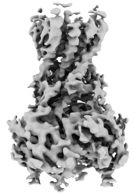
Supplemental images

Downloads & links

-
Map

FileDownload / File: emd_14678.map.gz / Format: CCP4 / Size: 30.5 MB / Type: IMAGE STORED AS FLOATING POINT NUMBER (4 BYTES)
Projections & slices

Image control

Size
Brightness
Contrast
Others
AxesX (Sec.)Y (Row.)Z (Col.)
0.86 Å/pix.
x 200 pix.
= 172. Å
0.86 Å/pix.
x 200 pix.
= 172. Å
0.86 Å/pix.
x 200 pix.
= 172. Å

Surface

Projections

Slices (1/3)

Slices (1/2)

Slices (2/3)

Images are generated by Spider.

Voxel sizeX=Y=Z: 0.86 Å
Density
Contour LevelBy AUTHOR: 3.64
Minimum - Maximum-13.838733 - 21.031654
Average (Standard dev.)-6.3654997e-12 (±1.0)
SymmetrySpace group: 1
Details

EMDB XML:

Map geometry
Axis orderZYX
Origin000
Dimensions200200200
Spacing200200200
CellA=B=C: 172.0 Å
α=β=γ: 90.0 °

-
Supplemental data

-
Half map: #2

Fileemd_14678_half_map_1.map
Projections & Slices
AxesZYX

Projections

Slices (1/2)
Density Histograms

-
Half map: #1

Fileemd_14678_half_map_2.map
Projections & Slices
AxesZYX

Projections

Slices (1/2)
Density Histograms

-
Sample components

-
Entire : Human inward-rectifier potassium channel 2.1 (Kir2.1)

EntireName: Human inward-rectifier potassium channel 2.1 (Kir2.1)
Components
  • Organelle or cellular component: Human inward-rectifier potassium channel 2.1 (Kir2.1)
    • Protein or peptide: Inward rectifier potassium channel 2Inward-rectifier potassium channel
  • Ligand: POTASSIUM IONPotassium
  • Ligand: STRONTIUM ION

-
Supramolecule #1: Human inward-rectifier potassium channel 2.1 (Kir2.1)

SupramoleculeName: Human inward-rectifier potassium channel 2.1 (Kir2.1) / type: organelle_or_cellular_component / ID: 1 / Parent: 0 / Macromolecule list: #1
Source (natural)Organism: Homo sapiens (human)
Molecular weightExperimental: 50 kDa/nm
Recombinant expressionOrganism: Komagataella pastoris (fungus)

-
Macromolecule #1: Inward rectifier potassium channel 2

MacromoleculeName: Inward rectifier potassium channel 2 / type: protein_or_peptide / ID: 1 / Number of copies: 4 / Enantiomer: LEVO
Source (natural)Organism: Homo sapiens (human)
Molecular weightTheoretical: 48.344141 KDa
Recombinant expressionOrganism: Komagataella pastoris (fungus)
SequenceString: MGSVRTNRYS IVSSEEDGMK LATMAVANGF GNGKSKVHTR QQCRSRFVKK DGHCNVQFIN VGEKGQRYLA DIFTTCVDIR WRWMLVIFC LAFVLSWLFF GCVFWLIALL HGDLDASKEG KACVSEVNSF TAAFLFSIET QTTIGYGFRC VTDECPIAVF M VVFQSIVG ...String:
MGSVRTNRYS IVSSEEDGMK LATMAVANGF GNGKSKVHTR QQCRSRFVKK DGHCNVQFIN VGEKGQRYLA DIFTTCVDIR WRWMLVIFC LAFVLSWLFF GCVFWLIALL HGDLDASKEG KACVSEVNSF TAAFLFSIET QTTIGYGFRC VTDECPIAVF M VVFQSIVG CIIDAFIIGA VMAKMAKPKK RNETLVFSHN AVIAMRDGKL CLMWRVGNLR KSHLVEAHVR AQLLKSRITS EG EYIPLDQ IDINVGFDSG IDRIFLVSPI TIVHEIDEDS PLYDLSKQDI DNADFEIVVI LEGMVEATAM TTQCRSSYLA NEI LWGHRY EPVLFEEKHY YKVDYSRFHK TYEVPNTPLC SARDLAEKKY ILSNANSFCY ENEVALTSKE EDDSENGVPE STST DTPPD IDLHNQASVP LEPRPLRRES EI

-
Macromolecule #2: POTASSIUM ION

MacromoleculeName: POTASSIUM ION / type: ligand / ID: 2 / Number of copies: 1 / Formula: K
Molecular weightTheoretical: 39.098 Da

-
Macromolecule #3: STRONTIUM ION

MacromoleculeName: STRONTIUM ION / type: ligand / ID: 3 / Number of copies: 2 / Formula: SR
Molecular weightTheoretical: 87.62 Da
Chemical component information

ChemComp-SR:
STRONTIUM ION / Strontium

-
Experimental details

-
Structure determination

Methodcryo EM
Processingsingle particle reconstruction
Aggregation stateparticle

-
Sample preparation

Concentration0.6 mg/mL
BufferpH: 7.4
Component:
ConcentrationFormulaName
150.0 mMKClPotassium chloride
20.0 mMTris-HClTrisTrizma hydrochloride
0.03 %DDMDodecyl-B-D-maltoside
1.0 mMEDTAEthylenediaminetetraacetic acidEthylenediamine tetraacetic acid
2.0 mMDDTDichlorodiphenyltrichloroethane
GridModel: Quantifoil R1.2/1.3 / Material: COPPER / Mesh: 300 / Support film - Material: CARBON / Support film - topology: HOLEY / Pretreatment - Type: GLOW DISCHARGE
VitrificationCryogen name: ETHANE / Chamber humidity: 100 % / Chamber temperature: 277 K / Instrument: FEI VITROBOT MARK III

-
Electron microscopy

MicroscopeFEI TITAN KRIOS
Electron beamAcceleration voltage: 300 kV / Electron source: FIELD EMISSION GUN
Electron opticsC2 aperture diameter: 100.0 µm / Illumination mode: FLOOD BEAM / Imaging mode: BRIGHT FIELDBright-field microscopy / Cs: 2.7 mm / Nominal defocus max: 2.8000000000000003 µm / Nominal defocus min: 1.2 µm / Nominal magnification: 105000
Sample stageSpecimen holder model: FEI TITAN KRIOS AUTOGRID HOLDER / Cooling holder cryogen: NITROGEN
Image recordingFilm or detector model: FEI FALCON III (4k x 4k) / Detector mode: COUNTING / Digitization - Dimensions - Width: 5760 pixel / Digitization - Dimensions - Height: 4092 pixel / Digitization - Sampling interval: 10.0 µm / Number grids imaged: 1 / Number real images: 9895 / Average exposure time: 4.0 sec. / Average electron dose: 61.7 e/Å2
Experimental equipment
Model: Titan Krios / Image courtesy: FEI Company

-
Image processing

Particle selectionNumber selected: 1031472
CTF correctionSoftware - Name: CTFFIND (ver. 4)
Initial angle assignmentType: MAXIMUM LIKELIHOOD / Software - Name: RELION (ver. 3.0.7)
Final 3D classificationNumber classes: 1 / Software - Name: RELION (ver. 3.0.7)
Final angle assignmentType: MAXIMUM LIKELIHOOD / Software - Name: RELION (ver. 3.0.7)
Final reconstructionNumber classes used: 1 / Applied symmetry - Point group: C4 (4 fold cyclic) / Resolution.type: BY AUTHOR / Resolution: 4.3 Å / Resolution method: FSC 0.143 CUT-OFF / Software - Name: RELION (ver. 3.0.7) / Number images used: 63584
FSC plot (resolution estimation)

-
Atomic model buiding 1

Initial modelPDB ID:

Chain - Residue range: 188-367
DetailsFor structural fitting, it was used dock-in-map (available at PHENIX) that uses both SSM and convolution-based shape searches to find a part of a map that is similar to a model. An initial in silico homology model of human Kir2.1 was generated using I-TASSER using the crystal structure of chicken Kir2.2 channel (PDB ID 3JYC) as a template. For building and refinement of the atomic model, the transmembrane domain (TMD, 55-184 region) of this in silico model was placed into the final sharpened cryo-EM map using the Dock in Map tool available in PHENIX. For the cytoplasmic domain (CTD; 188-367 region), the crystal structure of the CTD from mice Kir2.1 channel (PDB ID 1U4F) was placed into the final cryo-EM map using the same approach. Once the models were placed in the electron density, the loops that connect the two domains (185-187 region) and a N-terminal loop (41-54 region) absent in the in silico model were manually built using Coot.
RefinementSpace: REAL / Protocol: OTHER / Overall B value: 404.73 / Target criteria: Correlation coefficient
Output model

PDB-7zdz:
Cryo-EM structure of the human inward-rectifier potassium 2.1 channel (Kir2.1)

+
About Yorodumi

-
News

-
Feb 9, 2022. New format data for meta-information of EMDB entries

New format data for meta-information of EMDB entries

  • Version 3 of the EMDB header file is now the official format.
  • The previous official version 1.9 will be removed from the archive.

Related info.:EMDB header

External links:wwPDB to switch to version 3 of the EMDB data model

-
Aug 12, 2020. Covid-19 info

Covid-19 info

URL: https://pdbj.org/emnavi/covid19.php

New page: Covid-19 featured information page in EM Navigator.

Related info.:Covid-19 info / Mar 5, 2020. Novel coronavirus structure data

+
Mar 5, 2020. Novel coronavirus structure data

Novel coronavirus structure data

Related info.:Yorodumi Speices / Aug 12, 2020. Covid-19 info

External links:COVID-19 featured content - PDBj / Molecule of the Month (242):Coronavirus Proteases

+
Jan 31, 2019. EMDB accession codes are about to change! (news from PDBe EMDB page)

EMDB accession codes are about to change! (news from PDBe EMDB page)

  • The allocation of 4 digits for EMDB accession codes will soon come to an end. Whilst these codes will remain in use, new EMDB accession codes will include an additional digit and will expand incrementally as the available range of codes is exhausted. The current 4-digit format prefixed with “EMD-” (i.e. EMD-XXXX) will advance to a 5-digit format (i.e. EMD-XXXXX), and so on. It is currently estimated that the 4-digit codes will be depleted around Spring 2019, at which point the 5-digit format will come into force.
  • The EM Navigator/Yorodumi systems omit the EMD- prefix.

Related info.:Q: What is EMD? / ID/Accession-code notation in Yorodumi/EM Navigator

External links:EMDB Accession Codes are Changing Soon! / Contact to PDBj

+
Jul 12, 2017. Major update of PDB

Major update of PDB

  • wwPDB released updated PDB data conforming to the new PDBx/mmCIF dictionary.
  • This is a major update changing the version number from 4 to 5, and with Remediation, in which all the entries are updated.
  • In this update, many items about electron microscopy experimental information are reorganized (e.g. em_software).
  • Now, EM Navigator and Yorodumi are based on the updated data.

External links:wwPDB Remediation / Enriched Model Files Conforming to OneDep Data Standards Now Available in the PDB FTP Archive

-
Yorodumi

Thousand views of thousand structures

  • Yorodumi is a browser for structure data from EMDB, PDB, SASBDB, etc.
  • This page is also the successor to EM Navigator detail page, and also detail information page/front-end page for Omokage search.
  • The word "yorodu" (or yorozu) is an old Japanese word meaning "ten thousand". "mi" (miru) is to see.

Related info.:EMDB / PDB / SASBDB / Comparison of 3 databanks / Yorodumi Search / Aug 31, 2016. New EM Navigator & Yorodumi / Yorodumi Papers / Jmol/JSmol / Function and homology information / Changes in new EM Navigator and Yorodumi

Read more