ムービー
ムービー コントローラー


+ データを開く

- 基本情報
基本情報
| 登録情報 | データベース: EMDB / ID: EMD-1448 | |||||||||
|---|---|---|---|---|---|---|---|---|---|---|
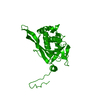
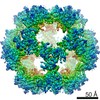
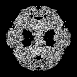
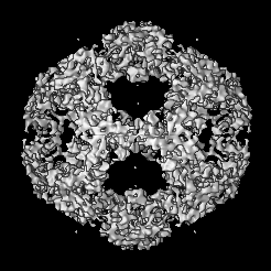
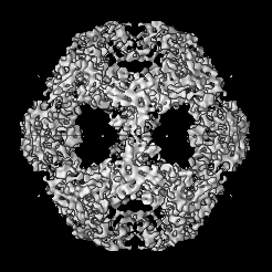
| タイトル | Structures of the human pyruvate dehydrogenase complex cores: a highly conserved catalytic center with flexible N-terminal domains. | |||||||||
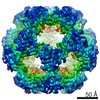
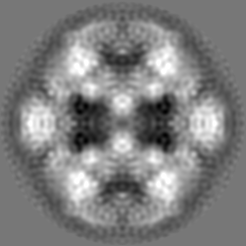
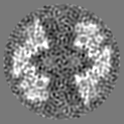
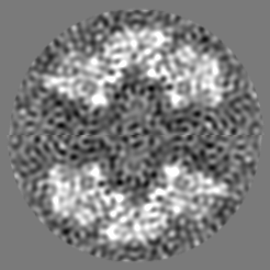
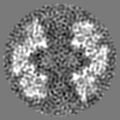
 マップデータ マップデータ | This is the half density map for the truncated human dihydrolipoyl acetyltransferase(E2)dodecahedron | |||||||||
 試料 試料 |
| |||||||||
| 機能・相同性 |  機能・相同性情報 機能・相同性情報PDH complex synthesizes acetyl-CoA from PYR / pyruvate catabolic process / dihydrolipoyllysine-residue acetyltransferase / dihydrolipoyllysine-residue acetyltransferase activity / Regulation of pyruvate dehydrogenase (PDH) complex / Protein lipoylation / pyruvate decarboxylation to acetyl-CoA / pyruvate dehydrogenase complex / Signaling by Retinoic Acid / tricarboxylic acid cycle ...PDH complex synthesizes acetyl-CoA from PYR / pyruvate catabolic process / dihydrolipoyllysine-residue acetyltransferase / dihydrolipoyllysine-residue acetyltransferase activity / Regulation of pyruvate dehydrogenase (PDH) complex / Protein lipoylation / pyruvate decarboxylation to acetyl-CoA / pyruvate dehydrogenase complex / Signaling by Retinoic Acid / tricarboxylic acid cycle / glucose metabolic process / mitochondrial matrix / mitochondrion / identical protein binding 類似検索 - 分子機能 | |||||||||
| 生物種 |  Homo sapiens (ヒト) Homo sapiens (ヒト) | |||||||||
| 手法 | 単粒子再構成法 / クライオ電子顕微鏡法 / 解像度: 8.8 Å | |||||||||
 データ登録者 データ登録者 | Yu X / Hiromasa Y / Tsen H / Stoops JK / Roche TE / Zhou ZH | |||||||||
 引用 引用 | ジャーナル: Structure / 年: 2008 タイトル: Structures of the human pyruvate dehydrogenase complex cores: a highly conserved catalytic center with flexible N-terminal domains. 著者: Xuekui Yu / Yasuaki Hiromasa / Hua Tsen / James K Stoops / Thomas E Roche / Z Hong Zhou /  要旨: Dihydrolipoyl acetyltransferase (E2) is the central component of pyruvate dehydrogenase complex (PDC), which converts pyruvate to acetyl-CoA. Structural comparison by cryo-electron microscopy (cryo- ...Dihydrolipoyl acetyltransferase (E2) is the central component of pyruvate dehydrogenase complex (PDC), which converts pyruvate to acetyl-CoA. Structural comparison by cryo-electron microscopy (cryo-EM) of the human full-length and truncated E2 (tE2) cores revealed flexible linkers emanating from the edges of trimers of the internal catalytic domains. Using the secondary structure constraints revealed in our 8 A cryo-EM reconstruction and the prokaryotic tE2 atomic structure as a template, we derived a pseudo atomic model of human tE2. The active sites are conserved between prokaryotic tE2 and human tE2. However, marked structural differences are apparent in the hairpin domain and in the N-terminal helix connected to the flexible linker. These permutations away from the catalytic center likely impart structures needed to integrate a second component into the inner core and provide a sturdy base for the linker that holds the pyruvate dehydrogenase for access by the E2-bound regulatory kinase/phosphatase components in humans. | |||||||||
| 履歴 |
|
- 構造の表示
構造の表示
| ムービー |
ムービービューア |
|---|---|
| 構造ビューア | EMマップ: SurfViewMolmilJmol/JSmol |
| 添付画像 |

- ダウンロードとリンク
-EMDBアーカイブ
| マップデータ | emd_1448.map.gz | 29.5 MB | EMDBマップデータ形式 | |
|---|---|---|---|---|
| ヘッダ (付随情報) | emd-1448-v30.xmlemd-1448.xml | 9.9 KB 9.9 KB | 表示 表示 | EMDBヘッダ |
| 画像 | 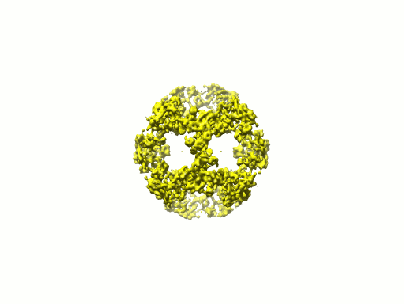 1448.gif 1448.gif | 16.1 KB | ||
| アーカイブディレクトリ |  http://ftp.pdbj.org/pub/emdb/structures/EMD-1448ftp://ftp.pdbj.org/pub/emdb/structures/EMD-1448 http://ftp.pdbj.org/pub/emdb/structures/EMD-1448ftp://ftp.pdbj.org/pub/emdb/structures/EMD-1448 | HTTPS FTP |
-関連構造データ

-リンク
| EMDBのページ | EMDB (EBI/PDBe) / EMDataResource |
|---|---|
| 「今月の分子」の関連する項目 |


-マップ
| ファイル | ダウンロード / ファイル: emd_1448.map.gz / 形式: CCP4 / 大きさ: 55.5 MB / タイプ: IMAGE STORED AS FLOATING POINT NUMBER (4 BYTES) | ||||||||||||||||||||||||||||||||||||||||||||||||||||||||||||||||||||
|---|---|---|---|---|---|---|---|---|---|---|---|---|---|---|---|---|---|---|---|---|---|---|---|---|---|---|---|---|---|---|---|---|---|---|---|---|---|---|---|---|---|---|---|---|---|---|---|---|---|---|---|---|---|---|---|---|---|---|---|---|---|---|---|---|---|---|---|---|---|
| 注釈 | This is the half density map for the truncated human dihydrolipoyl acetyltransferase(E2)dodecahedron | ||||||||||||||||||||||||||||||||||||||||||||||||||||||||||||||||||||
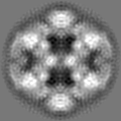
| 投影像・断面図 | 画像のコントロール
画像は Spider により作成 | ||||||||||||||||||||||||||||||||||||||||||||||||||||||||||||||||||||
| ボクセルのサイズ | X=Y=Z: 1.09 Å | ||||||||||||||||||||||||||||||||||||||||||||||||||||||||||||||||||||
| 密度 |
| ||||||||||||||||||||||||||||||||||||||||||||||||||||||||||||||||||||
| 対称性 | 空間群: 1 | ||||||||||||||||||||||||||||||||||||||||||||||||||||||||||||||||||||
| 詳細 | EMDB XML:
CCP4マップ ヘッダ情報:
| ||||||||||||||||||||||||||||||||||||||||||||||||||||||||||||||||||||
 Z (Sec.)
 Y (Row.)
 X (Col.)


-添付データ
- 試料の構成要素
-全体 : the truncated human dihydrolipoyl acetyltransferase
| 全体 | 名称: the truncated human dihydrolipoyl acetyltransferase |
|---|---|
| 要素 |
|
-超分子 #1000: the truncated human dihydrolipoyl acetyltransferase
| 超分子 | 名称: the truncated human dihydrolipoyl acetyltransferase / タイプ: sample / ID: 1000 / 集合状態: dodecahedrial assembly of tE2 / Number unique components: 1 |
|---|---|
| 分子量 | 理論値: 1.6 MDa |
-分子 #1: truncated human dihydrolipoyl acetyltransferase
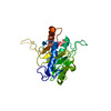
| 分子 | 名称: truncated human dihydrolipoyl acetyltransferase / タイプ: protein_or_peptide / ID: 1 / Name.synonym: tE2 詳細: Human tE2 was prepared from scE2, which contains a PreScission site in the third linker region. Treatment of scE2 with the PreScission protease (Amersham Biosciences) removed the N-terminal 319 amino acids. コピー数: 60 / 集合状態: Dodecahedron / 組換発現: Yes |
|---|---|
| 由来(天然) | 生物種: Homo sapiens (ヒト) / 別称: Human |
| 分子量 | 実験値: 1.6 MDa / 理論値: 1.6 MDa |
| 組換発現 | 生物種:  |
-実験情報
-構造解析
| 手法 | クライオ電子顕微鏡法 |
|---|---|
 解析 解析 | 単粒子再構成法 |
| 試料の集合状態 | particle |
-試料調製
| 濃度 | 0.2 mg/mL |
|---|---|
| 緩衝液 | pH: 7.2 / 詳細: PBS |
| グリッド | 詳細: 200 mesh holey carbon grid |
| 凍結 | 凍結剤: ETHANE / 装置: HOMEMADE PLUNGER / 詳細: Vitrification instrument: lab-made plunger / 手法: Blot for 1 second before plunging |
- 電子顕微鏡法
| 顕微鏡 | JEOL 2010F |
|---|---|
| 温度 | 最低: 100 K / 最高: 100 K / 平均: 100 K |
| 日付 | 2003年10月10日 |
| 撮影 | カテゴリ: CCD / フィルム・検出器のモデル: GENERIC GATAN / 平均電子線量: 12 e/Å2 |
| 電子線 | 加速電圧: 200 kV / 電子線源:  FIELD EMISSION GUN FIELD EMISSION GUN |
| 電子光学系 | 倍率(補正後): 69250 / 照射モード: FLOOD BEAM / 撮影モード: BRIGHT FIELD / Cs: 1.0 mm / 最大 デフォーカス(公称値): 2.1 µm / 最小 デフォーカス(公称値): 0.6 µm / 倍率(公称値): 69250 |
| 試料ステージ | 試料ホルダー: Eucentric / 試料ホルダーモデル: GATAN LIQUID NITROGEN |
-画像解析
| CTF補正 | 詳細: each image |
|---|---|
| 最終 再構成 | 想定した対称性 - 点群: I (正20面体型対称) / アルゴリズム: OTHER / 解像度のタイプ: BY AUTHOR / 解像度: 8.8 Å / 解像度の算出法: FSC 0.5 CUT-OFF / ソフトウェア - 名称: IMIRS / 使用した粒子像数: 2432 |