 ムービー
ムービー コントローラー


+ データを開く

- 基本情報
基本情報
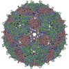
| 登録情報 | データベース: EMDB / ID: EMD-1433 | |||||||||
|---|---|---|---|---|---|---|---|---|---|---|
| タイトル | The three-dimensional structure of genomic RNA in bacteriophage MS2: implications for assembly. | |||||||||
 マップデータ マップデータ | CryoEM map of S2-MS2 | |||||||||
 試料 試料 |
| |||||||||
| 生物種 |  Enterobacterio phage MS2 (ファージ) Enterobacterio phage MS2 (ファージ) | |||||||||
| 手法 | 単粒子再構成法 / クライオ電子顕微鏡法 / 解像度: 17.8 Å | |||||||||
 データ登録者 データ登録者 | Toropova K / Basnak G / Twarock R / Stockley PG / Ranson NA | |||||||||
 引用 引用 | ジャーナル: J Mol Biol / 年: 2008 タイトル: The three-dimensional structure of genomic RNA in bacteriophage MS2: implications for assembly. 著者: Katerina Toropova / Gabriella Basnak / Reidun Twarock / Peter G Stockley / Neil A Ranson /  要旨: Using cryo-electron microscopy, single particle image processing and three-dimensional reconstruction with icosahedral averaging, we have determined the three-dimensional solution structure of ...Using cryo-electron microscopy, single particle image processing and three-dimensional reconstruction with icosahedral averaging, we have determined the three-dimensional solution structure of bacteriophage MS2 capsids reassembled from recombinant protein in the presence of short oligonucleotides. We have also significantly extended the resolution of the previously reported structure of the wild-type MS2 virion. The structures of recombinant MS2 capsids reveal clear density for bound RNA beneath the coat protein binding sites on the inner surface of the T=3 MS2 capsid, and show that a short extension of the minimal assembly initiation sequence that promotes an increase in the efficiency of assembly, interacts with the protein capsid forming a network of bound RNA. The structure of the wild-type MS2 virion at approximately 9 A resolution reveals icosahedrally ordered density encompassing approximately 90% of the single-stranded RNA genome. The genome in the wild-type virion is arranged as two concentric shells of density, connected along the 5-fold symmetry axes of the particle. This novel RNA fold provides new constraints for models of viral assembly. | |||||||||
| 履歴 |
|
- 構造の表示
構造の表示
| ムービー |
 ムービービューア ムービービューア |
|---|---|
| 構造ビューア | EMマップ: SurfViewMolmilJmol/JSmol |
| 添付画像 |

- ダウンロードとリンク
-EMDBアーカイブ
| マップデータ | emd_1433.map.gz | 7.8 MB | EMDBマップデータ形式 | |
|---|---|---|---|---|
| ヘッダ (付随情報) | emd-1433-v30.xmlemd-1433.xml | 7.9 KB 7.9 KB | 表示 表示 | EMDBヘッダ |
| 画像 | 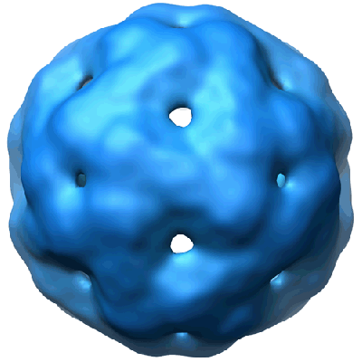 1433.gif 1433.gif | 52.8 KB | ||
| アーカイブディレクトリ |  http://ftp.pdbj.org/pub/emdb/structures/EMD-1433ftp://ftp.pdbj.org/pub/emdb/structures/EMD-1433 http://ftp.pdbj.org/pub/emdb/structures/EMD-1433ftp://ftp.pdbj.org/pub/emdb/structures/EMD-1433 | HTTPS FTP |
-関連構造データ


-リンク
| EMDBのページ | EMDB (EBI/PDBe) / EMDataResource |
|---|
-マップ
| ファイル | ダウンロード / ファイル: emd_1433.map.gz / 形式: CCP4 / 大きさ: 15.3 MB / タイプ: IMAGE STORED AS FLOATING POINT NUMBER (4 BYTES) | ||||||||||||||||||||||||||||||||||||||||||||||||||||||||||||||||||||
|---|---|---|---|---|---|---|---|---|---|---|---|---|---|---|---|---|---|---|---|---|---|---|---|---|---|---|---|---|---|---|---|---|---|---|---|---|---|---|---|---|---|---|---|---|---|---|---|---|---|---|---|---|---|---|---|---|---|---|---|---|---|---|---|---|---|---|---|---|---|
| 注釈 | CryoEM map of S2-MS2 | ||||||||||||||||||||||||||||||||||||||||||||||||||||||||||||||||||||
| 投影像・断面図 | 画像のコントロール
画像は Spider により作成 | ||||||||||||||||||||||||||||||||||||||||||||||||||||||||||||||||||||
| ボクセルのサイズ | X=Y=Z: 2.97 Å | ||||||||||||||||||||||||||||||||||||||||||||||||||||||||||||||||||||
| 密度 |
| ||||||||||||||||||||||||||||||||||||||||||||||||||||||||||||||||||||
| 対称性 | 空間群: 1 | ||||||||||||||||||||||||||||||||||||||||||||||||||||||||||||||||||||
| 詳細 | EMDB XML:
CCP4マップ ヘッダ情報:
| ||||||||||||||||||||||||||||||||||||||||||||||||||||||||||||||||||||
 Z (Sec.)
 Y (Row.)
 X (Col.)


-添付データ
- 試料の構成要素
-全体 : Recombinant MS2 capsids with S2 RNA
| 全体 | 名称: Recombinant MS2 capsids with S2 RNA |
|---|---|
| 要素 |
|
-超分子 #1000: Recombinant MS2 capsids with S2 RNA
| 超分子 | 名称: Recombinant MS2 capsids with S2 RNA / タイプ: sample / ID: 1000 / Number unique components: 2 |
|---|
-分子 #1: MS2 capsid
| 分子 | 名称: MS2 capsid / タイプ: protein_or_peptide / ID: 1 / Name.synonym: MS2 Capsid / 組換発現: No |
|---|---|
| 由来(天然) | 生物種: Enterobacterio phage MS2 (ファージ) / 別称: Bacteriophage MS2 |
-分子 #2: S2 RNA
| 分子 | 名称: S2 RNA / タイプ: rna / ID: 2 / Name.synonym: S2 RNA / 詳細: 31bases / 分類: OTHER / Structure: SINGLE STRANDED / Synthetic?: Yes |
|---|---|
| 由来(天然) | 生物種: Enterobacterio phage MS2 (ファージ) / 別称: Bacteriophage MS2 |
-実験情報
-構造解析
| 手法 | クライオ電子顕微鏡法 |
|---|---|
 解析 解析 | 単粒子再構成法 |
| 試料の集合状態 | particle |
-試料調製
| 凍結 | 凍結剤: ETHANE / チャンバー内湿度: 50 % / チャンバー内温度: 22 K / 装置: HOMEMADE PLUNGER 詳細: Vitrification instrument: double sided pneumatic blotter 手法: 1.4s blot |
|---|
- 電子顕微鏡法
| 顕微鏡 | FEI TECNAI F20 |
|---|---|
| 撮影 | カテゴリ: FILM フィルム・検出器のモデル: GATAN ULTRASCAN 4000 (4k x 4k) デジタル化 - スキャナー: OTHER / 平均電子線量: 15 e/Å2 |
| Tilt angle min | 0 |
| Tilt angle max | 0 |
| 電子線 | 加速電圧: 200 kV / 電子線源:  FIELD EMISSION GUN FIELD EMISSION GUN |
| 電子光学系 | 倍率(補正後): 50500 / 照射モード: FLOOD BEAM / 撮影モード: BRIGHT FIELD / Cs: 2.0 mm |
| 試料ステージ | 試料ホルダー: side entry / 試料ホルダーモデル: GATAN LIQUID NITROGEN |
| 実験機器 |  モデル: Tecnai F20 / 画像提供: FEI Company |
-画像解析
| CTF補正 | 詳細: phase flipping each particle |
|---|---|
| 最終 再構成 | 想定した対称性 - 点群: I (正20面体型対称) / 解像度のタイプ: BY AUTHOR / 解像度: 17.8 Å / 解像度の算出法: FSC 0.5 CUT-OFF / ソフトウェア - 名称: SPIDER |
