- EMDB-14248: Chlamydomonas reinhardtii TSP9 mutant small Photosystem I complex -
+
Open data
ID or keywords:
Loading...
-
Basic information
Entry
Database: EMDB / ID: EMD-14248
Title
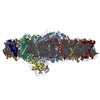
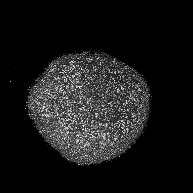
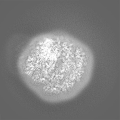
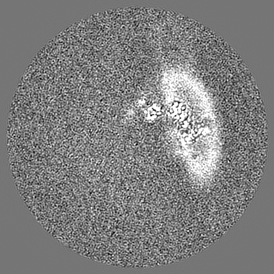
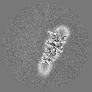
Chlamydomonas reinhardtii TSP9 mutant small Photosystem I complex
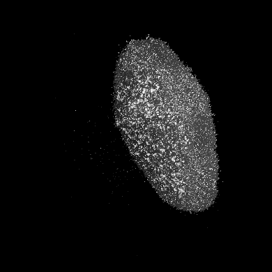
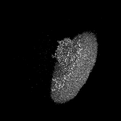
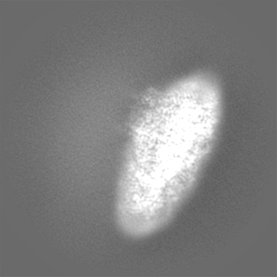
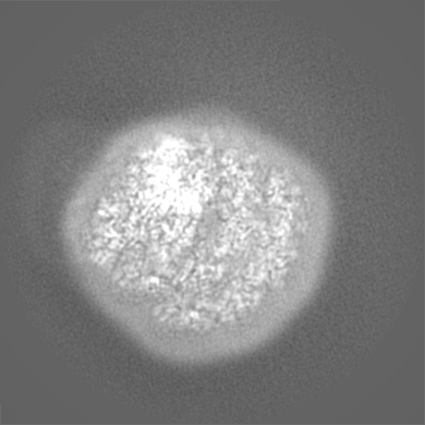
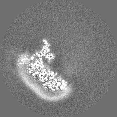
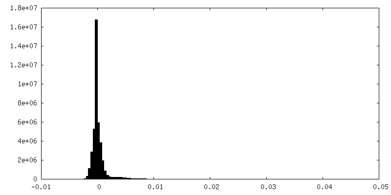
Map data
Sample
Complex: C. reinhardtii Photosystem I supercomplex
Protein or peptide: x 17 types
Ligand: x 22 types
Keywords
Photosystem I / Complex / Green algae / PHOTOSYNTHESIS
Function / homology
Function and homology information
photosynthesis, light harvesting in photosystem I / photosynthesis, light harvesting / chloroplast thylakoid lumen / photosystem I reaction center / photosystem I / photosynthetic electron transport in photosystem I / photosystem I / photosystem II / chlorophyll binding / chloroplast thylakoid membrane ...photosynthesis, light harvesting in photosystem I / photosynthesis, light harvesting / chloroplast thylakoid lumen / photosystem I reaction center / photosystem I / photosynthetic electron transport in photosystem I / photosystem I / photosystem II / chlorophyll binding / chloroplast thylakoid membrane / response to light stimulus / photosynthesis / 4 iron, 4 sulfur cluster binding / electron transfer activity / oxidoreductase activity / magnesium ion binding / metal ion binding Similarity search - Function
Photosystem I reaction center subunit V / Photosystem I reaction center subunit psaK, plant / Photosystem I reaction center subunit V/PsaK, plant / Photosystem I PsaG/PsaK domain, chloroplastic / Photosystem I psaG and psaK proteins signature. / Photosystem I reaction center subunit V/PsaK / Photosystem I psaG / psaK / Photosystem I reaction centre subunit VIII / Photosystem I reaction centre subunit VIII / Chlorophyll A-B binding protein, plant and chromista ...Photosystem I reaction center subunit V / Photosystem I reaction center subunit psaK, plant / Photosystem I reaction center subunit V/PsaK, plant / Photosystem I PsaG/PsaK domain, chloroplastic / Photosystem I psaG and psaK proteins signature. / Photosystem I reaction center subunit V/PsaK / Photosystem I psaG / psaK / Photosystem I reaction centre subunit VIII / Photosystem I reaction centre subunit VIII / Chlorophyll A-B binding protein, plant and chromista / Chlorophyll A-B binding protein / Chlorophyll A-B binding protein / Photosystem I reaction centre subunit VIII superfamily / Photosystem I PsaF, reaction centre subunit III / Photosystem I PsaF, reaction centre subunit III superfamily / Photosystem I reaction centre subunit III / Photosystem I PsaD / Photosystem I, reaction centre subunit PsaD superfamily / PsaD / Photosystem I PsaE, reaction centre subunit IV / Photosystem I reaction centre subunit IV / PsaE / Photosystem I PsaJ, reaction centre subunit IX superfamily / Photosystem I PsaJ, reaction centre subunit IX / Photosystem I reaction centre subunit IX / PsaJ / Photosystem I PsaA / Photosystem I protein PsaC / Photosystem I PsaB / Photosystem I PsaA/PsaB, conserved site / Photosystem I psaA and psaB proteins signature. / : / Photosystem I PsaA/PsaB / Photosystem I PsaA/PsaB superfamily / Photosystem I psaA/psaB protein / Electron transport accessory-like domain superfamily / 4Fe-4S dicluster domain / 4Fe-4S ferredoxin, iron-sulphur binding, conserved site / 4Fe-4S ferredoxin-type iron-sulfur binding region signature. / 4Fe-4S ferredoxin-type iron-sulfur binding domain profile. / 4Fe-4S ferredoxin-type, iron-sulphur binding domain Similarity search - Domain/homology
Photosystem I reaction center subunit VIII / Chlorophyll a-b binding protein, chloroplastic / Photosystem I P700 chlorophyll a apoprotein A2 / Photosystem I P700 chlorophyll a apoprotein A1 / Photosystem I reaction center subunit IV, chloroplastic / Photosystem I reaction center subunit III, chloroplastic / Photosystem I reaction center subunit V, chloroplastic / Photosystem I reaction center subunit psaK, chloroplastic / Photosystem I reaction center subunit IX / Photosystem I iron-sulfur center ...Photosystem I reaction center subunit VIII / Chlorophyll a-b binding protein, chloroplastic / Photosystem I P700 chlorophyll a apoprotein A2 / Photosystem I P700 chlorophyll a apoprotein A1 / Photosystem I reaction center subunit IV, chloroplastic / Photosystem I reaction center subunit III, chloroplastic / Photosystem I reaction center subunit V, chloroplastic / Photosystem I reaction center subunit psaK, chloroplastic / Photosystem I reaction center subunit IX / Photosystem I iron-sulfur center / Chlorophyll a-b binding protein, chloroplastic / Photosystem I reaction center subunit II, chloroplastic / Chlorophyll a-b binding protein, chloroplastic / Chlorophyll a-b binding protein, chloroplastic / Chlorophyll a-b binding protein, chloroplastic / Chlorophyll a-b binding protein, chloroplastic / Chlorophyll a-b binding protein, chloroplastic Similarity search - Component
Biological species
Chlamydomonas reinhardtii (plant)
Method
single particle reconstruction / cryo EM / Resolution: 2.52 Å
Journal: Biomolecules / Year: 2023 Title: Structure of Photosystem I Supercomplex Isolated from a Cytochrome b6f Temperature-Sensitive Mutant. Authors: Tom Schwartz / Mariia Fadeeva / Daniel Klaiman / Nathan Nelson / Abstract: The unicellular green alga, , has been widely used as a model system to study photosynthesis. Its possibility to generate and analyze specific mutants has made it an excellent tool for mechanistic ...The unicellular green alga, , has been widely used as a model system to study photosynthesis. Its possibility to generate and analyze specific mutants has made it an excellent tool for mechanistic and biogenesis studies. Using negative selection of ultraviolet (UV) irradiation-mutated cells, we isolated a mutant (TSP9) with a single amino acid mutation in the Rieske protein of the cytochrome b6f complex. The W143R mutation in the petC gene resulted in total loss of cytochrome b6f complex function at the non-permissive temperature of 37 °C and recovery at the permissive temperature of 25 °C. We then isolated photosystem I (PSI) and photosystem II (PSII) supercomplexes from cells grown at the non-permissive temperature and determined the PSI structure with high-resolution cryogenic electron microscopy. There were several structural alterations compared with the structures obtained from wild-type cells. Our structural data suggest that the mutant responded by excluding the Lhca2, Lhca9, PsaL, and PsaH subunits. This structural alteration prevents state two transition, where LHCII migrates from PSII to bind to the PSI complex. We propose this as a possible response mechanism triggered by the TSP9 phenotype at the non-permissive temperature.
Model: Quantifoil R1.2/1.3 / Material: COPPER / Mesh: 300 / Support film - Material: CARBON / Support film - topology: HOLEY / Support film - Film thickness: 12 / Pretreatment - Type: PLASMA CLEANING / Pretreatment - Time: 40 sec. / Pretreatment - Atmosphere: AIR / Pretreatment - Pressure: 1.0 kPa / Details: Harrick Plasma cleaner PDC-32G-2
Vitrification
Cryogen name: ETHANE / Chamber humidity: 90 % / Chamber temperature: 295 K / Instrument: LEICA EM GP / Details: blot for 3 seconds before plunging.
Details
Chlorophyll concentration was provided
-
Electron microscopy
Microscope
FEI TITAN KRIOS
Image recording
Film or detector model: GATAN K3 BIOQUANTUM (6k x 4k) / Number real images: 13173 / Average exposure time: 2.6 sec. / Average electron dose: 57.74 e/Å2
Electron beam
Acceleration voltage: 300 kV / Electron source: FIELD EMISSION GUN
In the structure databanks used in Yorodumi, some data are registered as the other names, "COVID-19 virus" and "2019-nCoV". Here are the details of the virus and the list of structure data.
Jan 31, 2019. EMDB accession codes are about to change! (news from PDBe EMDB page)
EMDB accession codes are about to change! (news from PDBe EMDB page)
The allocation of 4 digits for EMDB accession codes will soon come to an end. Whilst these codes will remain in use, new EMDB accession codes will include an additional digit and will expand incrementally as the available range of codes is exhausted. The current 4-digit format prefixed with “EMD-” (i.e. EMD-XXXX) will advance to a 5-digit format (i.e. EMD-XXXXX), and so on. It is currently estimated that the 4-digit codes will be depleted around Spring 2019, at which point the 5-digit format will come into force.
The EM Navigator/Yorodumi systems omit the EMD- prefix.
Related info.:Q: What is EMD? / ID/Accession-code notation in Yorodumi/EM Navigator
Yorodumi is a browser for structure data from EMDB, PDB, SASBDB, etc.
This page is also the successor to EM Navigator detail page, and also detail information page/front-end page for Omokage search.
The word "yorodu" (or yorozu) is an old Japanese word meaning "ten thousand". "mi" (miru) is to see.
Related info.:EMDB / PDB / SASBDB / Comparison of 3 databanks / Yorodumi Search / Aug 31, 2016. New EM Navigator & Yorodumi / Yorodumi Papers / Jmol/JSmol / Function and homology information / Changes in new EM Navigator and Yorodumi
 Movie
Movie Controller

 Yorodumi
 Open data
 Basic information
 Map data
 Sample
 Keywords
 Function and homology information
 Chlamydomonas reinhardtii (plant)
 Authors
 Israel, 1 items
  Citation
 Structure visualization
 Downloads & links
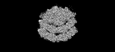 emd_14248.png
 http://ftp.pdbj.org/pub/emdb/structures/EMD-14248


 Z (Sec.)
 Y (Row.)
 X (Col.)


 Sample components


 Processing
 Electron microscopy
 FIELD EMISSION GUN