- EMDB-13798: Cryo-electron tomogram of a podosome at the basal surface of an u... -
+
Open data
ID or keywords:
Loading...
-
Basic information
Entry
Database: EMDB / ID: EMD-13798
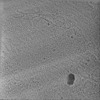
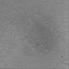
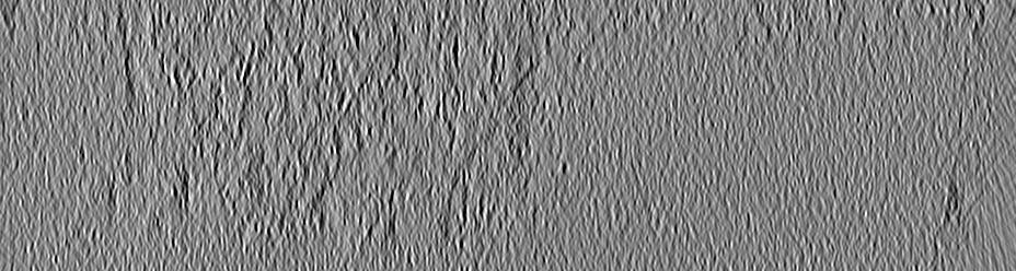
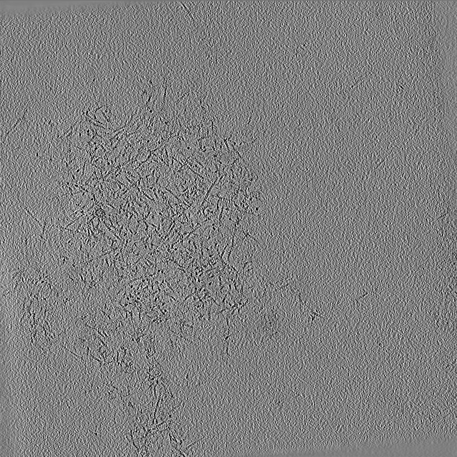
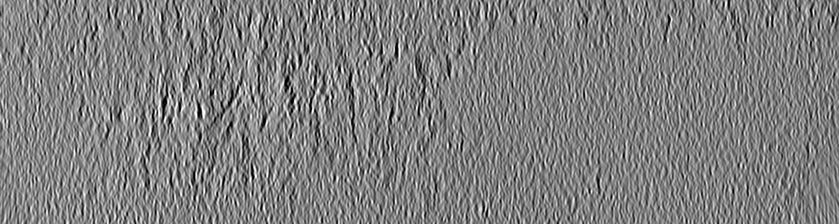
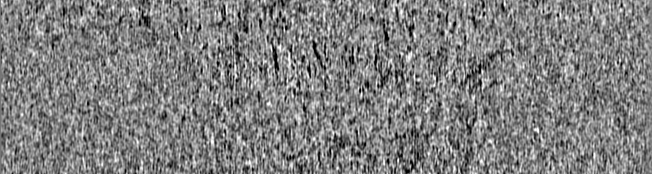
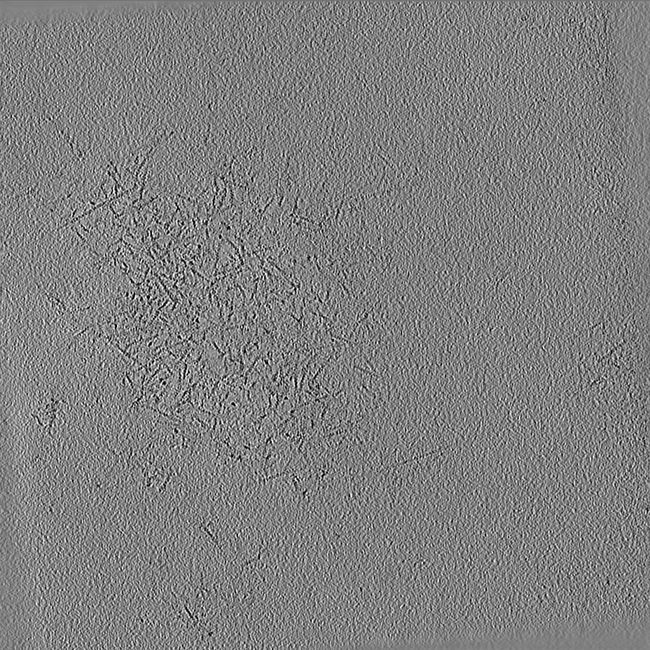
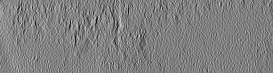
Title
Cryo-electron tomogram of a podosome at the basal surface of an unroofed primary human macrophage treated with cytochalasin D
Map data
Cryo-electron tomogram of a podosome at the basal surface of an unroofed primary human macrophage treated with cytochalasin D
Sample
Organelle or cellular component: Podosome assembled at the basal surface of an unroofed human primary monocyte-derived macrophage treated with cytochalasin D
Journal: Nat Commun / Year: 2022 Title: Elasticity of podosome actin networks produces nanonewton protrusive forces. Authors: Marion Jasnin / Jordan Hervy / Stéphanie Balor / Anaïs Bouissou / Amsha Proag / Raphaël Voituriez / Jonathan Schneider / Thomas Mangeat / Isabelle Maridonneau-Parini / Wolfgang Baumeister ...Authors: Marion Jasnin / Jordan Hervy / Stéphanie Balor / Anaïs Bouissou / Amsha Proag / Raphaël Voituriez / Jonathan Schneider / Thomas Mangeat / Isabelle Maridonneau-Parini / Wolfgang Baumeister / Serge Dmitrieff / Renaud Poincloux / Abstract: Actin filaments assemble into force-generating systems involved in diverse cellular functions, including cell motility, adhesion, contractility and division. It remains unclear how networks of actin ...Actin filaments assemble into force-generating systems involved in diverse cellular functions, including cell motility, adhesion, contractility and division. It remains unclear how networks of actin filaments, which individually generate piconewton forces, can produce forces reaching tens of nanonewtons. Here we use in situ cryo-electron tomography to unveil how the nanoscale architecture of macrophage podosomes enables basal membrane protrusion. We show that the sum of the actin polymerization forces at the membrane is not sufficient to explain podosome protrusive forces. Quantitative analysis of podosome organization demonstrates that the core is composed of a dense network of bent actin filaments storing elastic energy. Theoretical modelling of the network as a spring-loaded elastic material reveals that it exerts forces of a few tens of nanonewtons, in a range similar to that evaluated experimentally. Thus, taking into account not only the interface with the membrane but also the bulk of the network, is crucial to understand force generation by actin machineries. Our integrative approach sheds light on the elastic behavior of dense actin networks and opens new avenues to understand force production inside cells.
A: 12695.04 Å / B: 12695.04 Å / C: 3392.6401 Å α=β=γ: 90.0 °
-
Supplemental data
-
Sample components
-
Entire : Podosome assembled at the basal surface of an unroofed human prim...
Entire
Name: Podosome assembled at the basal surface of an unroofed human primary monocyte-derived macrophage treated with cytochalasin D
Components
Organelle or cellular component: Podosome assembled at the basal surface of an unroofed human primary monocyte-derived macrophage treated with cytochalasin D
-
Supramolecule #1: Podosome assembled at the basal surface of an unroofed human prim...
Supramolecule
Name: Podosome assembled at the basal surface of an unroofed human primary monocyte-derived macrophage treated with cytochalasin D type: organelle_or_cellular_component / ID: 1 / Parent: 0
Cells were treated with cytochalasin D and then unroofed prior to vitrification
Cryo protectant
No
Sectioning
Other: NO SECTIONING
Fiducial marker
Manufacturer: Aurion / Diameter: 10 nm
-
Electron microscopy
Microscope
FEI TITAN KRIOS
Specialist optics
Energy filter - Name: GIF Quantum LS / Energy filter - Slit width: 20 eV
Details
Tilt-series were acquired using a dose-symmetric tilt scheme (Hagen et al. 2017) from -50 to 50 degrees with 2 degree steps.
Image recording
Film or detector model: GATAN K2 SUMMIT (4k x 4k) / Detector mode: COUNTING / Number real images: 51 / Average exposure time: 4.0 sec. / Average electron dose: 3.6 e/Å2
Electron beam
Acceleration voltage: 300 kV / Electron source: FIELD EMISSION GUN
In the structure databanks used in Yorodumi, some data are registered as the other names, "COVID-19 virus" and "2019-nCoV". Here are the details of the virus and the list of structure data.
Jan 31, 2019. EMDB accession codes are about to change! (news from PDBe EMDB page)
EMDB accession codes are about to change! (news from PDBe EMDB page)
The allocation of 4 digits for EMDB accession codes will soon come to an end. Whilst these codes will remain in use, new EMDB accession codes will include an additional digit and will expand incrementally as the available range of codes is exhausted. The current 4-digit format prefixed with “EMD-” (i.e. EMD-XXXX) will advance to a 5-digit format (i.e. EMD-XXXXX), and so on. It is currently estimated that the 4-digit codes will be depleted around Spring 2019, at which point the 5-digit format will come into force.
The EM Navigator/Yorodumi systems omit the EMD- prefix.
Related info.:Q: What is EMD? / ID/Accession-code notation in Yorodumi/EM Navigator
Yorodumi is a browser for structure data from EMDB, PDB, SASBDB, etc.
This page is also the successor to EM Navigator detail page, and also detail information page/front-end page for Omokage search.
The word "yorodu" (or yorozu) is an old Japanese word meaning "ten thousand". "mi" (miru) is to see.
Related info.:EMDB / PDB / SASBDB / Comparison of 3 databanks / Yorodumi Search / Aug 31, 2016. New EM Navigator & Yorodumi / Yorodumi Papers / Jmol/JSmol / Function and homology information / Changes in new EM Navigator and Yorodumi
 Movie
Movie Controller

 Yorodumi
 Open data
 Basic information
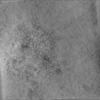
 Map data
 Sample
 Homo sapiens (human)
 Authors
 Germany, 3 items
  Citation
 Structure visualization
 Downloads & links
 EMDB map data format
 emd_13798.png
 http://ftp.pdbj.org/pub/emdb/structures/EMD-13798
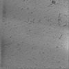
 Z (Sec.)
 Y (Row.)
 X (Col.)


 Sample components
 Processing
 Electron microscopy
 FIELD EMISSION GUN
